History of presentation
A female neonate with prenatal diagnosis of dextrocardia, double-outlet right ventricle with normally related great arteries, and a subaortic ventricular septal defect was born at 28 weeks gestational age. She was found to be cyanotic, hypotonic, and apneic at delivery prompting intubation and surfactant administration. She was mechanically ventilated and an umbilical venous catheter was placed for continued venous access. She was transferred to our centre for further care.
Investigations
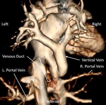
Postnatal echocardiography confirmed the findings from prenatal imaging, with the additional finding of infradiaphragmatic total anomalous pulmonary venous return. CT imaging to further delineate the course of the vertical vein (Fig 1) demonstrated an uncommon variant of infradiaphragmatic total anomalous pulmonary venous return in which the vertical vein inserted directly into the venous duct. Due to the difficulty of surgical repair in patients of this size, a strategy of stenting of the venous duct as a temporising measure with the goal of surgical repair at a larger size was pursued.
Three-dimensional reconstruction of total anomalous pulmonary venous return. Three-dimensional CT reconstruction of the vertical vein, venous duct, and portal veins viewed from posterior. The vertical vein is seen draining directly into the venous duct. ICV = inferior caval vein.

Management
The umbilical venous catheter was left in place to help maintain patency of the venous duct while awaiting intervention. On day of life 6 (weight 940g), she underwent cardiac catheterisation and stenting of the venous duct. Special considerations in the cardiac catheterisation lab were undertaken given the infant’s extremely low birthweight and prematurity: the catheterisation suite was kept at the highest possible ambient temperature to minimise heat loss from the patient during the procedure. Ultrasound guidance was used for all access. Judicious use of flushes was mantained throughout the procedure to minimise volume overload. Care was taken to limit the number of contrast angiograms to protect the premature kidneys. Finally, great care was taken to limit radiation exposure from fluoroscopy throughout the case, given this patient’s likely need for multiple interventional procedures over the course of her lifetime.
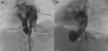
We obtained access by exchanging the existing umbilical venous catheter for a 4-French Brite Tip™ (Cordis, Switzerland) sheath. Angiography through the umbilical venous sheath again demonstrated the unusual anatomy of the vertical vein entering the venous duct directly. There was focal narrowing noted at the entry of the vertical vein into the venous duct (Fig 2a, Online Video 1). Given the need to stent not only the vertical vein but also the insertion point of the vertical vein into the venous duct, additional access was obtained in the left internal jugular vein and a 4-French Brite Tip™ (Cordis, Switzerland) sheath was placed. Angioplasty of the vertical vein and the stenotic entrance of the vertical vein into the venous duct was performed using a 5 mm x 12 mm Quantum Apex™ balloon (Boston Scientific, Marlborough, MA); the stenotic lesion at the vertical vein–venous duct junction was observed to be very compliant with minimal response to balloon angioplasty. A 5-mm Zotarolimus-eluting Resolute Onyx™ stent (Medtronic, Minneapolis, MN) was therefore deployed across the stenotic lesion from the left internal jugular venous access sheath, with no angiographic appearance of obstruction following stent deployment.
Angiography of the Vertical Vein. (a) Angiogram of the vertical vein demonstrates focal narrowing (*) at the site of entry into the venous duct. See Video 1. (b) Angiogram of the stented venous duct at 2 months of age demonstrates continued ductal patency. See Video 2.

Discussion
Total anomalous pulmonary venous return can be classified according to the path of abnormal pulmonary venous return: supracardiac (to systemic veins superior to the heart), cardiac (to the coronary sinus), infracardiac (to systemic or portal veins inferior to the heart), or mixed (a combination of several means of anomalous drainage). By far, the most common site of infracardiac total anomalous pulmonary venous return drainage is to the portal veinsReference Brown, Geva, Allen, Shaddy, Penny, Feltes and Cetta1. Less common sites of anomalous drainage include the hepatic veins, inferior vena cava, or the venous duct. Drainage directly to the venous duct (seen in our case) directly represents a rare entity, with only a handful of cases described in large historical case series of total anomalous pulmonary venous returnReference Burroughs and Edwards2,Reference Duff, Nihill and McNamara3 . While stenting the venous duct alone may be sufficient for other forms of infradiaphragmatic total anomalous pulmonary venous return, in our case, stenting of the vertical vein and venous duct was necessary to treat pulmonary venous obstruction. Given that the venous duct normally closes shortly after birth, leaving the umbilical venous catheter in place until the time of intervention to temporarily maintain vessel patency was life-saving in this patient and facilitates the intervention. Initial intervention was desired within the first week of life to minimise infection risk associated with prolonged umbilical venous catheter placement.
Surgical repair of obstructed total anomalous pulmonary venous return may be technically challenging due to small patient size and without prompt intervention, and the prognosis is usually poor. Venous duct stenting in neonates has been reportedReference Higaki, Yamamoto and Nakano4-Reference Oonishi, Hayabuchi and Sakata7 as a means of delaying surgical repair. Careful delineation of the insertion of the vertical vein must be determined, as this greatly influences the approach needed for stent placement. Surgical repair remains the definitive management for total anomalous pulmonary venous return, with short-term 3-year survival rates of 85% reported in a recent large international registryReference Seale, Uemura and Webber8. Surveillance for later development of obstruction of the repaired pulmonary venous confluence is essential, as this significant source of late morbidity and mortality occurs in approximately 15% of patientsReference Seale, Uemura and Webber8.
Follow-up
The patient was stable following her intervention, and a repeat catheterisation for stent dilation at 2 months of age demonstrated a widely patent stent (Fig 2b, Online Video 2). She underwent surgical repair of total anomalous pulmonary venous return at 3 months of age (weight 2.7 kg) with additional surgical revision of the pulmonary venous confluence at 5 months of age. The residual iatrogenic portosystemic shunt caudal to the stent spontaneously closed. She is now 17 months of age and otherwise healthy, growing, and developing well.
Conclusions
Stenting of the venous duct as a bridge to later surgical repair is technically feasible and can be accomplished even in neonates <1 kg in weight. Special considerations in the cardiac catheterisation lab are essential for safe procedures in premature neonates.
Supplementary material
To view supplementary material for this article, please visit https://doi.org/10.1017/S1047951120002188
Acknowledgments
We would like to thank the entire Heart Center at Texas Children’s Hospital who aided in the care of this patient. We would also like to especially thank our colleagues in the Neonatal Intensive Care Unit (Dr. AnnaMarie Arias-Shah, Dr. Leah Elizondo, and Dr. Danielle Rios) who were so instrumental in the initial resuscitation and management of this patient.
Financial support
This research received no specific grant from any funding agency, commercial or not-for-profit sectors.
Conflicts of interest
None.
Ethical standards
Not applicable.