Implications
Protein biomarkers are tools to design tenderness prediction tests for the beef industry. This study showed that the expression of these markers is influenced by muscle and animal type, which are two factors important in beef production in France. Rearing systems are characterised by different production factors, capable of modifying the validity of tenderness prediction tests, notably by changes in protein expression profiles. The knowledge brought by this study is a first step in understanding these effects on protein biomarker expression. Finally, this type of knowledge could lead to improved management of beef tenderness during the animal's life.
Introduction
Beef is an important economic value in different countries such as the United States, Australia and France (Veysset et al., Reference Veysset, Bebin and Lherm2005). Consumers express an interest in high organoleptic meat quality, especially tenderness (Geay et al., Reference Geay, Bauchart, Hocquette and Culioli2001). This quality is highly variable (Dransfield et al., Reference Dransfield, Martin, Bauchart, Abouelkaram, Lepetit, Culioli, Jurie and Picard2003; Picard et al., Reference Picard, Jurie, Bauchart, Dransfield, Ouali, Martin, Jailler, Lepetit and Culioli2007), which is a problem for the industry. The French beef industry is interested in meat tenderness tests that are suitable for the majority of production systems used in France. These production systems differ notably by animal type (cows, heifers, steers, calves, young bulls) and the breed used (beef, hardy, dairy breeds). For a few years now, several research programmes have identified some potential biomarkers of beef tenderness, at the protein (Picard et al., Reference Picard, Berri, Lefaucheur, Molette, Sayd and Terlouw2010), mRNA (Bernard et al., Reference Bernard, Cassar-Malek, LeCunff, Dubroeucq, Renand and Hocquette2007) and DNA (Hocquette et al., Reference Hocquette, Lehnert, Barendse, Cassar-Malek and Picard2007) levels, in different production systems.
Several studies have shown that the abundance of some gene transcripts and/or proteins can be affected by the production system, animal and muscle type (Hocquette et al., Reference Hocquette, Lehnert, Barendse, Cassar-Malek and Picard2007). The study by Cassar-Malek et al. (Reference Cassar-Malek, Jurie, Bernard, Barnola, Micol and Hocquette2009) showed that the production system (grass feeding of animals v. maize) lead to an oxidative switch in muscle metabolism. In the same way, different breeds exhibit differences in their muscle characteristics, as described in Jurie et al. (Reference Jurie, Picard, Hocquette, Dransfield, Micol and Listrat2007). Schreurs et al.'s (Reference Schreurs, Garcia, Jurie, Agabriel, Micol, Bauchart, Listrat and Picard2008) study showed that the development of muscle characteristics differs between muscle types because of difference in testosterone levels in bulls and steers.
Therefore, the expression of some potential tenderness protein biomarkers can be influenced by factors such as animal and/or muscle type. The knowledge of these influences could be exploited to manage the abundance of different tenderness biomarkers in beef production in order to introduce management methods that would enhance the meat quality for the beef industry.
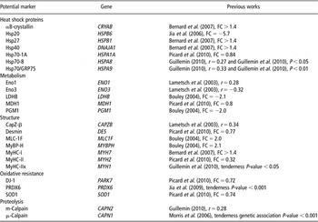
The objective of this study was to analyse the effect of animal and muscle type on the expression of beef tenderness potential protein markers, previously identified in some genomic programmes as reported in Table 1. The proteins belong to five families according to their biological functions: heat shock proteins (HSPs), metabolism, structure, oxidative resistance and proteolysis.
Potential tenderness markers identified by previous works

MyHC-I = myosins of heavy chains I; PRDX6 = peroxiredoxin 6; SOD1 = superoxide dismutase 1.
FC = protein fold change associated with tenderness according to cited literature; r = correlation coefficient associated with tenderness measurement according to cited literature.
Material and methods
Animals and samples
This study was conducted on 67 Charolais young bulls and 44 Charolais steers (castrated at 3 months of age) from the INRA programme MUGENE. The young bulls were slaughtered at 17 months of age on average and the steers at 30 months of age on average at the INRA experimental slaughterhouse in compliance with current ethical guidelines for animal welfare. Muscle samples from the Longissimus thoracis (LT; fast oxido-glycolytic with 25% of myosins of heavy chains I (MyHC-I), 62% of MyHC-IIa and 13% of MyHC-IIx in our experiment by electrophoresis (Picard et al., Reference Picard, Barboiron, Duris, Gagniere, Jurie and Geay1999), and in accordance with Jurie et al. (Reference Jurie, Robelin, Picard, Renand and Geay1995, Reference Jurie, Picard, Hocquette, Dransfield, Micol and Listrat2007) in bovine) and Semitendinosus (ST; fast glycolytic with 11% of MyHC-I, 24% of MyHC-IIa and 66% of MyHC-IIx in our experiment, and in accordance with Schreurs et al. (Reference Schreurs, Garcia, Jurie, Agabriel, Micol, Bauchart, Listrat and Picard2008) in bovine) muscles were excised for each animal within 15 min after slaughter. Muscle samples were immediately frozen in liquid nitrogen and stored at −80°C until protein extraction. Approximately 100 mg of muscle samples were used for protein extraction.
Total protein extractions were performed according to Bouley et al. (Reference Bouley, Chambon and Picard2004) in a denaturation/extraction buffer (8.3 M urea, 2 M thiourea, 1% DiThioThreitol, 2% CHAPS) and stored at −20°C until use. All chemical reagents were from Sigma (St. Louis, MO, USA). The protein concentration was determined by spectrophotometry with the Bradford assay (Bradford, Reference Bradford1976).
Immunological protein quantification
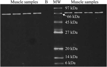
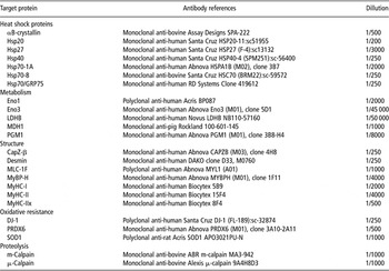
The conditions for use and specificity of primary antibodies against the 24 proteins analysed on bovine muscle were assessed by western blotting according the methodology described by Guillemin et al. (Reference Guillemin, Meunier, Jurie, Cassar-Malek, Hocquette, Levéziel and Picard2009a). Briefly, western blots were used in order to check the specificity of all the antibodies. An antibody was considered specific against the studied protein when one band at the expected molecular weight was detected by western blot. BLAST analyses were done between the 24 studied bovine protein sequences against human protein databases, to assure that proteins share more than 90% of identity. An example of the human Hsp70-1A primary antibody validation is illustrated in Figure 1 and it shows that this human antibody binds to a bovine protein of the Hsp70-1A theoretical molecular weight. All the other 23 primary antibodies were tested for their specificity as described (data not shown). Optimal dilution ratios were determined in the same time, from suppliers’ conditions and adapted to bovine muscle samples. Conditions used and suppliers for all primary antibodies are reported in Table 2.
Primary antibody validation: Hsp70-1A (B = blank; MW = molecular weight, Hsp70-1A theoretical molecular weight = 70.22 kDa).

Suppliers and conditions for each primary antibody used in this study

MyHC-I = myosins of heavy chains I; PRDX6 = peroxiredoxin 6; SOD1 = superoxide dismutase 1.
Secondary fluorescent-conjugated IRDye 800CW antibodies (anti-mouse, anti-sheep, anti-rabbit) were supplied by LI-COR Biosciences (Lincoln, NE, USA) and used at 1/20 000.
Protein quantifications with the validated antibodies were done by Dot blot, as described by Guillemin et al. (Reference Guillemin, Meunier, Jurie, Cassar-Malek, Hocquette, Levéziel and Picard2009a). Dot blot is a technique 15 times faster than western blot, characterised by the same technical variations (10%). This characteristic allowed us to analyse these 24 protein markers in less time than with western blot (6 months for Dot blot and 53 for western blot). Briefly, protein samples were spotted in quadruplicate on a nitrocellulose membrane with the Minifold I Dot blot from Schleicher & Schuell Biosciences (Dassel, Germany) and hybridised with the specific antibody of each protein, with conditions defined by western blot. Then membranes were scanned by the scanner Odyssey (LI-COR Biosciences) at 800 nm. Protein abundance for each sample, given in arbitrary units, was normalised according to a mix of different samples, used as a reference.
Statistical analysis
Analysis of variance was performed using the GLM procedure of SAS version 9.1. The effects of animal type (bull v. steer), muscle (LT v. ST), animal-type × muscle-type interaction and animal tested within animal type were introduced into the model. The animal variation was used as the error term for the animal-type effect. When significant effects were detected, differences were evaluated by the PDIFF option of SAS.
Results
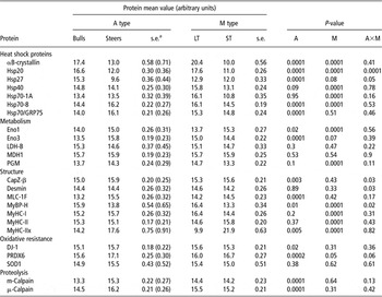
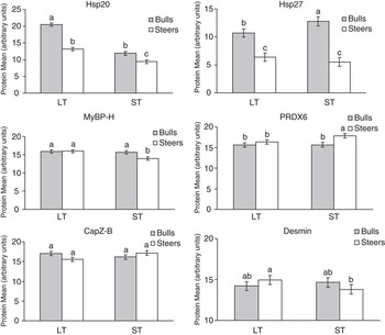
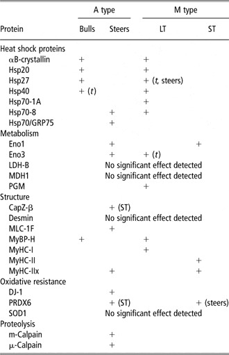
There were effects of animal and/or muscle type on 20 of the 24 proteins studied (Table 3). Figure 2 illustrates the levels of the six proteins, which have significant interactions between animal- and muscle-type effects.
Effect of A and M type on potential protein biomarkers of tenderness

A = animal; M = muscle; LT = Longissimus thoracis; ST = Semitendinosus; MyHC-I = myosins of heavy chains I; PRDX6 = peroxiredoxin 6; SOD1 = superoxide dismutase 1.
as.e. for bulls; s.e. for steers are given in brackets.
Means and significant differences of protein expression with animal- and muscle-type effect interactions. Protein expressions were quantified in arbitrary units.

HSPs
Whereas abundance of αB-crystallin and Hsp27 was significantly higher in bulls than in steers (P = 0.0001), amounts of Hsp70-8 and Hsp70/GRP75 were significantly higher in steers than bulls (P = 0.0001). Hsp40 and Hsp70-1A were not different between bulls and steers.
The LT muscle exhibited significantly higher abundance of αB-crystallin (P < 0.001), Hsp40 (P < 0.001), Hsp70-1A (P < 0.001) and Hsp70-8 (P = 0.001) than the ST muscle. Abundance of Hsp70/GRP75 was not different between muscles.
Significant animal-type × muscle-type interactions were observed for Hsp20 (P < 0.001) and Hsp27 (P = 0.05). Both Hsp were higher in bulls than in steers, and were higher in LT than in ST muscle with a greater difference between bulls and steers in LT muscle than in ST muscle for Hsp20, and only in steers for Hsp27 (Figure 2).
Proteins of energy metabolism
Abundance of Eno1 and Eno3 (glycolytic enzymes) was significantly higher in steers than in bulls (P = 0.02 and P< 0.001, respectively). The levels of three other proteins (LDH-B, PGM (glycolytic enzymes) and MDH1 (oxidative enzyme)) were not significantly different between bulls and steers.
The ST muscle exhibited a significantly higher abundance of Eno1 (P < 0.001) than the LT muscle. Conversely, abundance of PGM was higher in LT than in ST muscle (P < 0.001). No significant muscle effect was detected for Eno3, LDH-B and MDH1.
Structural and contractile proteins
Abundance of MLC-1F and MyHC-IIx was significantly higher in steers than in bulls (P < 0.001 and P = 0.005, respectively). The bulls exhibited significantly higher abundance of MyBP-H (P = 0.01) than steers. There was no animal-type effect for Desmin, MyHC-I and MyHC-II.
Abundance of MyBP-H and MyHC-I was significantly higher in LT muscle than in ST muscle (P < 0.001 and P < 0.001, respectively). The interaction detected (P = 0.02) for MyBP-H amount did not change this difference. Whereas abundance of MyHC-I was higher in LT than in ST muscle, that of the MyHC-II (IIa + IIx) and MyHC-IIx was significantly higher in ST muscle than in LT (P < 0.001). No significant muscle effect was detected for CapZ-β, Desmin and MLC-1F.
Significant animal-type × muscle-type interactions were observed for CapZ-B, desmin and MyBP-H (P < 0.05; Table 2). Whereas no significant difference was observed between bulls and steers in LT muscle, in ST muscle, desmin and MyBP-H were more abundant in bulls than in steers and CapZ-B was more abundant in steers than in bulls.
Proteins of oxidative resistance
Abundance of DJ-1 and peroxiredoxin 6 (PRDX6) was significantly higher in steers than in bulls (P = 0.02 and P< 0.001, respectively). Concerning PRDX6, this difference was only observed in ST muscle (animal-type × muscle-type interaction effect, P = 0.06; Figure 2). No significant muscle effect was detected for these two proteins.
Abundance of superoxide dismutase 1 (SOD1) was not different between animal types and between muscle types.
Proteins involved in proteolysis
Whereas no significant differences were observed between muscles for m-calpain and μ-calpain, abundance in these two proteins was significantly higher in steers than in bulls (P < 0.001; Table 3).
Discussion
This study describes the effect of important factors in meat production (e.g. muscle and animal type) on the expression of some protein biomarkers of beef tenderness. The originality of this study is to provide new data about 24 proteins, for which there is very little information in the literature on bovine species. There are only data on their function and regulation, and for some of these mainly in humans. All the results described above are summarised in Table 4. We detected muscle-type effects on 14 of the 24 analysed proteins. This effect concerns mainly the Hsp family (excepted Hsp70/GRP75), the contractile proteins and some metabolic proteins. We identified an animal effect on 15 proteins of the 24 analysed. The animal effect assessed in this study was the difference between young bulls and steers. These two types of animals are characterised by some differences at the muscular, cellular and proteins levels (Cassar-Malek et al., Reference Cassar-Malek, Picard, Bernard and Hocquette2008), which likely explain the animal-type effects found in this study.
Summary of the A- and M-type effects detected or not for the 24 proteins

A = animal; M = muscle; LT = Longissimus thoracis; ST = Semitendinosus; MyHC-I = myosins of heavy chains I; PRDX6 = peroxiredoxin 6; SOD1 = superoxide dismutase 1.
An over-expression is indicated by a +.
The context specificity (A or M type) of the detected effect is indicated in brackets when necessary.
When the detected effect was not significant but showed a tendency, the letter t is added in brackets.
Protein of energy metabolism
It is well documented that the two muscles analysed in this study, the LT and the ST, have different characteristics in terms of muscle fibre composition (Totland and Kryvi, Reference Totland and Kryvi1991). According to the literature data, the ST muscle may exhibit a lower oxidative activity (Jurie et al., Reference Jurie, Picard, Hocquette, Dransfield, Micol and Listrat2007) and a strong higher glycolytic activity notably a higher LDH activity compared to the LT (Jurie et al., Reference Jurie, Robelin, Picard, Renand and Geay1995). The LT muscle that contains a higher proportion of slow fibres and higher oxidative metabolism than ST in young bulls is also characterised by a smaller fibre cross-sectional areas compared to the ST muscle (Schreurs et al., Reference Schreurs, Garcia, Jurie, Agabriel, Micol, Bauchart, Listrat and Picard2008).
In this study, we observed a muscle-type effect for only two of the glycolytic enzymes quantified: PGM and Eno1. Eno1, the α-enolase, is a glycolytic enzyme that catalyses the conversion of 2-phospho-d-glycerate to phosphoenolpyruvate. This enolase is also involved in the immunoglobulin response and is a transcriptional regulator. Eno3, the β-enolase, is involved in the development and regeneration of muscle. In striated muscle, there are homodimers of β-enolase or heterodimers of α/β-enolase. Here, Eno1 was more abundant in ST muscle in accordance with the literature data. Moreover, it was more abundant in steers as Eno3. As muscles of steers are more glycolytic than bulls (Brandstetter et al., Reference Brandstetter, Picard and Geay1998), this result is consistent with the literature data.
These two proteins, PGM and Eno1, were the only ones showing an animal-type effect among the metabolic proteins analysed in our study. PGM, which was more abundant in LT in our study, is involved in the first steps of glycolysis by catalysing the transformation of glucose-1-phosphate in glucose-6-phosphate. LDH-B, another glycolytic enzyme, catalyses the later steps of glycolysis with the pyruvate conversion to lactate. Their involvement at different stages of glycolysis could explain the differences in muscle-type effect observed. MDH1, an oxidative enzyme catalysing the transformation of malate to oxalo-actetate, was no different between muscles.
In the literature, data obtained for metabolism were mainly provided by enzyme activity assays. They showed, for example, that the LDH activity was higher in steers than in bulls (Brandstetter et al., Reference Brandstetter, Sauerwein, Veerkamp, Geay and Hocquette2002). In this study, we did not find any differences in LDH-B expression (glycolytic metabolism) between muscles, as for MDH1 (oxidative metabolism). This is in contradiction to the enzyme activity differences characterising the LT and ST muscles according to the literature. We can hypothesise that enzymes activity differences between muscles are mainly due to mechanisms of activation/inhibition rather than regulation of enzyme quantity. This would explain why there were no effects detected for LDH-B and MDH1 for either animal or muscle type.
Structural and contractile proteins
In accordance with the literature data, we observed a lower proportion of slow MyHC-I and higher proportions of fast (IIa and IIx) MyHCs in ST muscle. These MyHC isoforms are specific to the slow-twitch fibre type I, the fast-twitch oxido-glycolytic fibre IIa and the fast-twitch glycolytic fibre IIx, respectively (Lefaucheur, Reference Lefaucheur2010). Myosins are hexameric proteins composed of two heavy chains (MyHC) and four light chains (MLC). The MLC-1F quantified in our study showed no differences between the two muscles. However, this isoform is associated with fast-twitch fibres IIa and IIx (Choi and Kim, Reference Choi and Kim2009), which are very different between the two muscles. Myosins are motor proteins able to convert nutrients into energy for muscle contraction. MyHC isoforms have an ATPase activity, which defines the speed of fibre contraction. They are therefore very important for determining the contractile type. MLC isoforms have a regulatory role in contraction and seem to be less sensitive to the muscle type. However, they showed a high animal-type effect with a higher proportion of MLC-1F in steers than in bulls. This was in accordance with the higher proportion of the fast MyHC-IIx as MLC-1F makes a fine adjustment to myosin motor activity and is expressed more in fast-twitch and glycolytic fibre (Choi and Kim, Reference Choi and Kim2009). MyHC-IIx is the only MyHC different between the two types of animals in our study, but the effect was lower than that of muscle type. This result concurs with the literature data. For example, Brandstetter et al. (Reference Brandstetter, Picard and Geay1998) showed that MyHC-IIx was over-expressed in steers more than in bulls. This study by Brandstetter et al. found that only the MyHC-IIx was over-expressed in steers, at the expense of MyHC-IIa, thereby showing a balance of these two MyHCs between steers and bulls. In our study, the antibody used against fast MyHC recognises MyHC-IIa and MyHC-IIx. As no animal effect was detected on MyHC-II, we can conclude that the total amount of MyHC-II is constant in steers and bulls. Hence, this balance of MyHC-II is dependent upon the regulation of animal type (e.g. age and sexual hormones) between amounts of MyHC-IIa and MyHC-IIx. Brandstetter et al.'s (Reference Brandstetter, Picard and Geay1998) study also showed that the quantity of the MyHC-I was constant between steers and bulls, which is in accordance with the results of this study.
In our study, the MyBP-H showed a highly significant effect of muscle type with a higher proportion in LT muscle (more oxidative), which concurs with the animal-type effect observed with greater abundance in young bulls (more oxidative according to the literature). However, this is not consistent with the literature data on this protein. It is a protein of the thick myosin filament that binds to the myosins (Clark et al., Reference Clark, McElhinny, Beckerle and Gregorio2002), controlling the polymerisation and the assembly of the thick filament. This protein participates in the regulation of muscle contraction by inhibiting the myosin ATPase activity. It is considered to be associated with fast-twitch fibres (Lee et al., Reference Lee, McArdle and Griffiths2007). However, its roles as yet are unknown. Gilbert et al. (Reference Gilbert, Cohen, Pardo, Basu and Fischman1999) hypothesise that MyBP-H plays a role in the regulation of the synthesis of the thick filament, which could concur with our data.
The abundance of the two structural proteins, desmin and CapZ-β was no different between the two muscles. CapZ-β, a capping protein of the thin actin filament at the barbed and pointed ends, has a role in the organisation of the thin filament and its anchor to the Z-line (Clark et al., Reference Clark, McElhinny, Beckerle and Gregorio2002). The CapZ protein is a heterodimer of CapZ-α and CapZ-β. The proportion of CapZ-β was no different between the two muscles, so the abundance of the protein does not seem to be associated with the contractile and metabolic properties of muscles. However, it was significantly higher in the ST of steers illustrating a hormonal effect on its expression. The data in the literature show that the ST muscle shows strong anatomical differences between males, castrated males and females (Brandstetter et al., Reference Brandstetter, Picard and Geay1998). This higher reactivity to androgens compared to LT could explain that differences in CapZ-β abundance were observed only in ST.
Desmin is a structural protein of the intermediate filament (Paulin and Li, Reference Paulin and Li2004). This intermediate filament maintains the cytoarchitecture of muscle fibre and connects the contractile apparatus to the sarcolemma and the cytoskeleton (Herrmann et al., Reference Herrmann, Bar, Kreplak, Strelkov and Aebi2007). In our study, desmin was one of the proteins showing no effect on muscle and animal types. The human desmin gene is regulated by a combination of different transcription factors. Some of these could regulate the quantity of desmin according the physiological characteristics of muscles. Our data suggest that in bovines, hormonal signals, metabolism and contractile activities do not influence this protein essential for cell integrity.
HSPs
The proteins of the HSP family quantified in our study showed very large differences between muscle type and animal type in accordance with the literature data showing high modifications of their expression under several physiological conditions (Kiang and Tsokos, Reference Kiang and Tsokos1998).
HSPs are mostly chaperones, essential for normal cell function, and also enable cells to resist stress (for review: Fink, Reference Fink1999). They stabilise myofibrillar proteins under stress conditions and prevent them from losing function. This family is composed of several proteins, which can be classified into two main groups. The group of small HSP comprises 10 known members, among them the proteins Hsp20, Hsp27 and αB-crystallin, quantified in our study. These three proteins are present in great abundance in muscle tissue. According to Vicart et al. (Reference Vicart, Caron, Guicheney, Li, Prevost, Faure, Chateau, Chapon, Tome, Dupret, Paulin and Fardeau1998), their high expression level would allow them to exert an intermediate protective effect in response to stress conditions with no lag time necessary for protein synthesis. They constitute a dynamic complex (Sun and MacRae, Reference Sun and MacRae2005). This complex is implicated in the control of phosphorylation of myosin light chains, control of translation via the factor eIF4G, regulation of the apoptotic pathway by sequestration of Bcl-X and Bax, protection against aggregation of actin, desmin, titin and myosin and finally protection against heat shock of the citrate synthase and the malate dehydrogenase enzymes of the oxidative metabolism. Therefore, this complex plays an important role in the cell, which is greatly influenced by such environmental factors as cell signals or stresses.
In our study, the three small HSPs showed a large effect of muscle and animal type, with an interaction between the two effects for Hsp20 and 27. These three HSPs were more abundant in LT than in ST and in young bulls compared to steers. These results are consistent with the study of Golenhofen et al. (Reference Golenhofen, Perng, Quinlan and Drenckhahn2004) in rats, showing a higher abundance of HSPs in soleus (slow oxidative) compared to extensor digitorum longus (fast glycolytic) muscle. In cattle, LT contains a lower proportion of fast glycolytic fibres and higher proportion of oxidative fibres (I and IIa) compared with ST (Jurie et al., Reference Jurie, Picard, Hocquette, Dransfield, Micol and Listrat2007). Moreover, steers are characterised by a lower proportion of oxidative fibres and a higher proportion of fast glycolytic fibres (Brandstetter et al., Reference Brandstetter, Picard and Geay1998). There are very few data in the literature on differences in HSP expression according to the muscle type. Our results in cattle with those of Golenhofen et al. (Reference Golenhofen, Perng, Quinlan and Drenckhahn2004) in rats, show that HSPs form a complex stress–response system in skeletal muscle with some common and some distinct functions between different muscle types. Golenhofen et al. (Reference Golenhofen, Perng, Quinlan and Drenckhahn2004), using immunostaining, showed a higher intensity with an anti-αB-crystallin antibody in type I fibres compared to II fibres. Moreover, they observed some fast fibres displaying no staining and others with a strong staining, suggesting a differential expression between IIa and IIx/IIb fibres. We could suppose that in cattle the same differential expression is observed, given that the HSP family has a much conserved expression. Further analysis will be conducted on our samples by immunohistochemistry with the anti-HSP antibodies used in this study in order to validate this hypothesis. We can suppose that the LT muscle, which is more oxidative, needs protection against oxidative stress and the consequences of reactive oxygen species (ROS) formation in the cell.
The other group of HSP is made up of the HSP 70 family: Hsp70-1A, Hsp70-8 and Hsp70-GRP75. They function in the anti-apoptotic pathway, in the inhibition of ROS formation as well as a chaperone activity (Laufen et al., Reference Laufen, Mayer, Beisel, Klostermeier, Mogk, Reinstein and Bukau1999). Moreover, Hsp70-GRP75 controls cell proliferation and is used by the cell in case of glucose starvation to avoid ROS formation and cell aging (Kaul et al., Reference Kaul, Deocaris and Wadhwa2007). The two other HSPs (namely Hsp70-1A and Hsp70-8) bring chaperone activity with Hsp40 to ensure a good functioning of the muscle under constitutive oxidative stress conditions. In our study, we showed a high muscle-type effect on these HSP, except for Hsp70/GRP75. On the contrary, this HSP presents as high an animal-type effect as for Hsp70-8. For Hsp40, our results concerning muscle-type effect are in accordance with those of Cassar-Malek et al. (Reference Cassar-Malek, Guillemin, Hocquette, Micol, Bauchart and Picard2010) showing a higher expression in oxidative muscles of cattle as we observed in LT compared to ST muscle. The animal-type effect observed for Hsp70-8 and GRP75 is the result of physiological factors combining the age of the animal and the testosterone effect (Voss et al., Reference Voss, Stallone, Li, Cornelussen, Knuefermann and Knowlton2003; Lee et al., Reference Lee, McArdle and Griffiths2007). These authors demonstrated that oestrogen was positively correlated with the level of Hsp70-8 and Hsp70-GRP75. Levels of expression of the latter protein is also positively correlated with levels of thyroid hormones (Kaul et al., Reference Kaul, Deocaris and Wadhwa2007), which are known to be less abundant in young bulls than in steers. This is in accordance with the results of our study, which show that the muscles of steers have a greater abundance of Hsp70-GRP75 and also of Hsp70-8.
Finally, our results allow us to conclude that, despite their common function, for example anti-apoptotic activity and protection against cellular stresses, HSPs seem to be used specifically according to animal and muscle type. This is evident for the small HSP family complex, which protects the enzymes of the oxidative metabolism. This function is necessary in an oxidative metabolism context, which can explain why these proteins were more expressed in young bulls than in steers. Higher level of Hsp70 proteins in steers than bulls could reflect a more global role in chaperone activity on cell proteins, as the small HSP family complex is under-expressed in a less androgenic context than in steers.
Protein involved in oxidative resistance
The two proteins, DJ-1 and PRDX6, act against oxidative stress: DJ-1 as a quencher for ROS (Junn et al., Reference Junn, Taniguchi, Jeong, Zhao, Ichijo and Mouradian2005) and PRDX6 as a catalyser of the H2O2 reduction (Wang et al., Reference Wang, Phelan, Forsman-Semb, Taylor, Petros, Brown, Lerner and Paigen2003). ROS-driven oxidative stress influences many biological processes, including cell survival and death. Higher levels of ROS generated during muscle metabolic activities induce numerous physiological reactions and can cause significant damage. Accordingly, cells have evolved antioxidant defence systems including antioxidant enzymes such as SOD and PRDX. These last constitute a new family of antioxidants functioning in concert to detoxify ROS while playing an important role in cellular signalling by limiting ROS levels. Kubo et al. (Reference Kubo, Hasanova, Tanaka, Fatma, Takamura, Singh and Akagi2010), using Prdx6 knockout mice, have described the ability of PRDX6 to abolish ROS-driven oxidative stress.
In our study, the most variable protein according to muscle or animal type was PRDX6, with an interaction between the two factors. Its abundance was higher in steers’ muscles. We can hypothesise that the higher proportions of fast glycolytic fibres in steers require an active mechanism to counter the effects of oxidative stress, due to the lower quantity of HSPs. Therefore, PRDX6 and DJ-1 are important for steers in the case of oxidative stress. The specific difference for PRDX6, expressed more in ST muscle in steers, shows that this protein typically acts on glycolytic fibres. This could be a specific requirement of the ST glycolytic muscle in steers, which needs an active mechanism against ROS, like H2O2 reduction by PRDX6.
SOD1 is an enzyme that dismutes the superoxide anion, and is therefore involved in antioxidant defences (Moradas-Ferreira et al., Reference Moradas-Ferreira, Costa, Piper and Mager1996). We observed no effect of both animal and muscle type in our study. We can therefore hypothesise that there is no difference in terms of ROS production between steers and bulls, or between LT and ST, under our experimental conditions.
Proteolytic proteins
The two proteins, m- and μ-calpain, from the calcium-dependent proteolytic system, were no different between the two muscles, but showed very large differences between animal types. They were more abundant in steers than in young bulls. These two enzymes are mainly responsible for protein degradation in muscle. Our results are in accordance with previous studies showing the influence of animal type on muscle proteolysis. For example, cortisol concentrations, higher in steers, increases the proteolysis rate (Fritsche and Steinhart, Reference Fritsche and Steinhart1998). Moreover, testosterone concentrations, lower in steers, decreases this proteolysis rate (Lee et al., Reference Lee, McArdle and Griffiths2007). We hypothesise that in our conditions, animal-type effects were mainly the result of sex hormone action, but other hormones conditioning muscle properties of animals came into play.
Conclusion
This study provides some original insight into the effect of muscle and animal type on the abundance of 24 proteins considered as biomarkers of beef tenderness. It showed that animal and muscle types are characterised mainly by a differential expression of heat shock proteins and oxidative resistance proteins, depending on the glycolytic/oxidative potential of fibres, and contractile muscle type. The small HSP family is expressed in the more oxidative LT muscle, to protect proteins against ROS. It also uses Hsp70-1A and Hsp70-8 to refold proteins. In young bulls, the Hsp20 complex is also used, but with no other Hsp70-enhanced activity. This property is used by steers, where the abundance of Hsp20 complex is low. Steers use the protein DJ-1 to actively protect their cells. The glycolytic ST muscle has none of the oxidative protection system. We hypothesise that in case of cell stress, a strong and active pathway is engaged in the ST muscle to protect cells against ROS, by the way of SOD1 and PRDX6.
One of the aims of beef tenderness research is to bring new practices to manage tenderness in living animals. This aim requires knowledge of meat quality markers, notably the factors that optimise their expression. The results obtained by this study could bring new rearing practices for managing beef tenderness. These original data could be used in order to choose the best rearing conditions for a strong expression of tenderness biomarkers. This study, done on 222 different samples and on 24 proteins, provides the first knowledge of bovine biomarker expression according to rearing practices in order to manage beef tenderness in the future (Guillemin et al., Reference Guillemin, Cassar-Malek, Hocquette, Jurie, Micol, Listrat, Levéziel, Renand and Picard2009b).
Acknowledgements
The PhD grant of N. Guillemin is provided by INRA and the regional council of Auvergne in France. This study was funded by the National Agency of Research and APIS-GENE for the MUGENE programme (through the AGENAE/GENANIMAL call). The authors thank Christiane Barboiron for her skilled technical assistance, David Chadeyron for determining enzymatic activities, the INRA experimental farms of Bourges for animal breeding and the experimental slaughterhouse of our Unit for animal slaughtering and muscle sampling.