We herein report a surgical case in which precise preoperative simulation was very useful using a three-dimensional model in a small neonate having transposition of the great arteries (TGA), coarctation of the aorta (CoA), and low take-off of the right pulmonary artery (PA).
Clinical summary
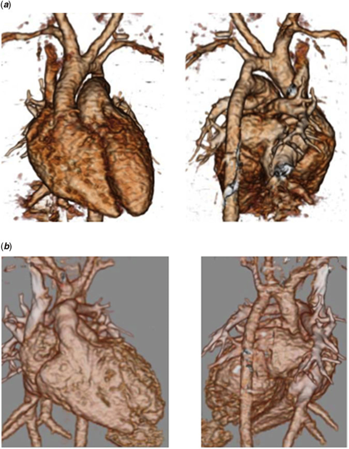
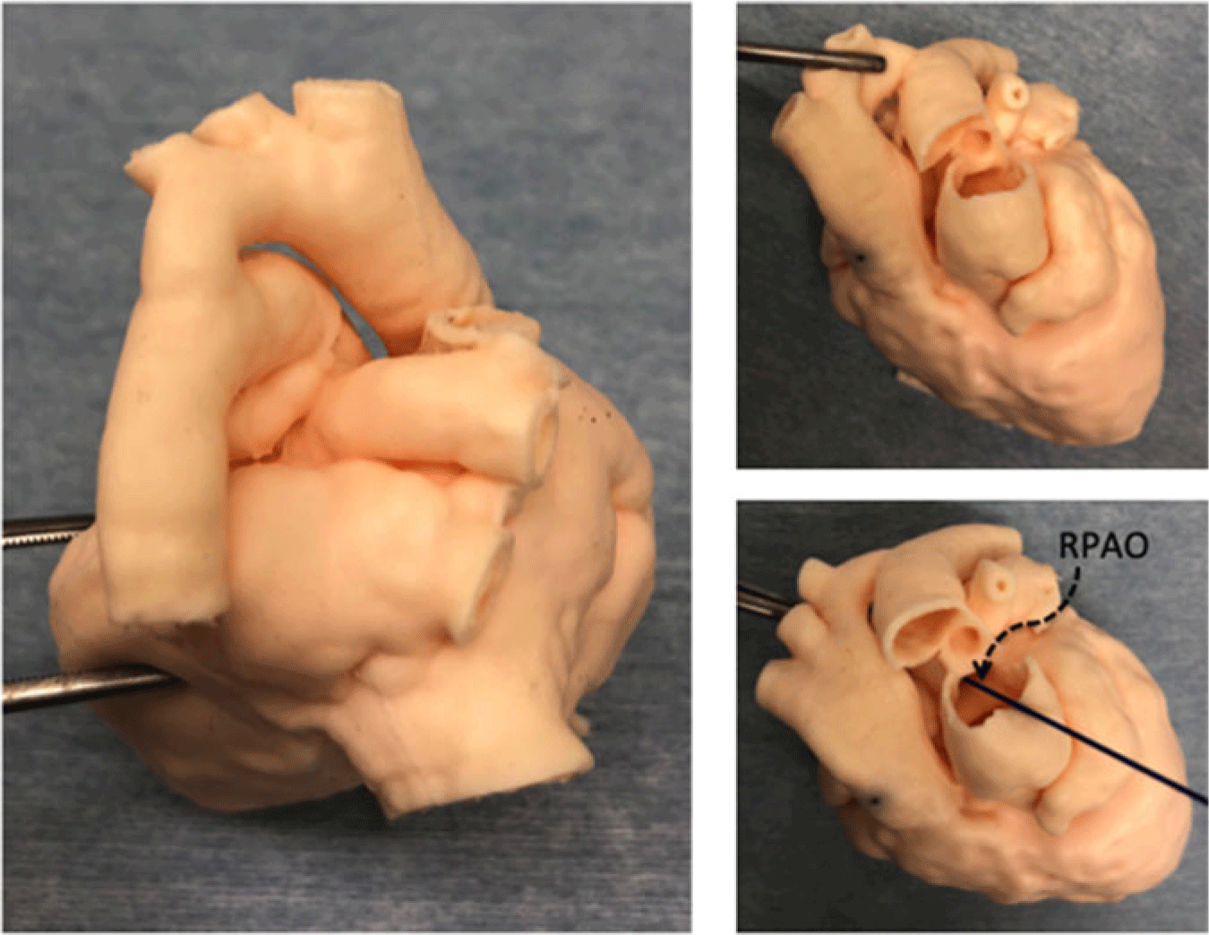
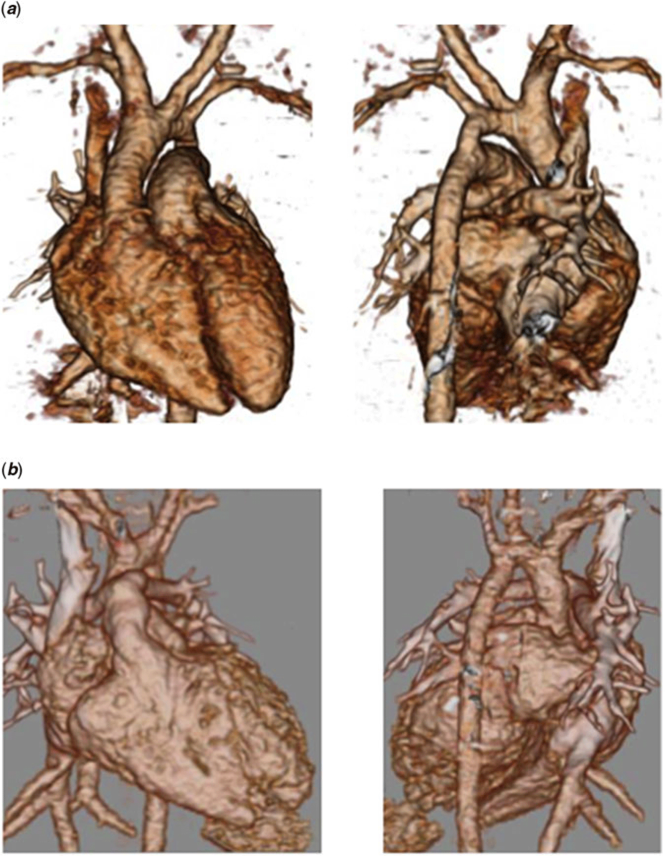
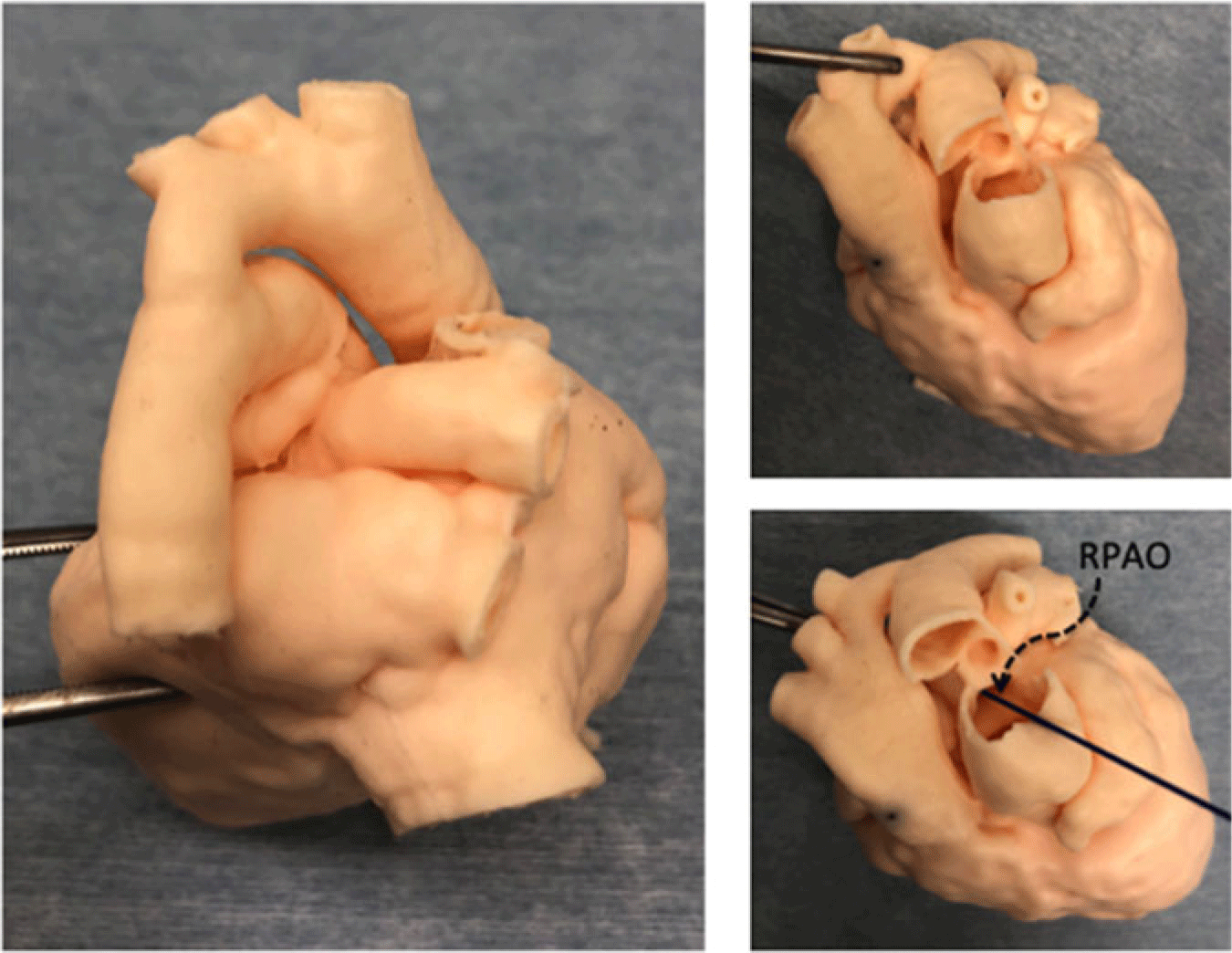
A female infant was born at a local hospital (gestational age 38 weeks and 1 day, the Caesarean section, body weight 2.2 kg) without antenatal diagnosis. She was blue, and eventually diagnosed as having TGA associated with severe CoA on echocardiography. She was in a mild shock status because of restrictive persistent ductus arteriosus on arrival to our institution. Prostaglandin infusion was started immediately and her circulation could be stabilised. A laid-back coronary arteriography illustrated the right-hand facing sinus of the aorta giving rise to the right coronary artery together with the left anterior descending artery while the left circumflex artery coming off from the left-hand facing sinus. Furthermore, three-dimensional computed tomography (CT) demonstrated low take-off of the right PA and counter-clockwise rotation around the PA bifurcation (Fig 1a). Precise assessment was established using a patient-specific three-dimensional model created on the basis of her data set (Fig 2). Because of those combined anatomical features, we considered that primary repair should be pertinent, rather than initial palliative surgery such as CoA repair or bilateral PA banding, even in a neonate less than 2.5 kg.
(a) Preoperative three-dimensional computed tomography. (b) Postoperative three-dimensional computed tomography.
A patient-specific three-dimensional model was developed using data based on routine preoperative scanning of computed tomography (1 mm-thick slices). RPAO; right pulmonary arterial orifice.
Surgical technique
Surgical repair was carried out at the age of 16 days. The aortic arch was reconstructed by means of the extended end-to-end anastomosis in a standard fashion. The ascending aorta was transected 6 mm above the aortic valve. The orifice for the left circumflex artery was extremely small – far smaller than considered prior to surgery. The PA trunk was divided at the bifurcation level, leaving a triangular sleeve of the posterior wall tissue of the right PA to the pulmonary trunk as a flap. This flap was fixed so as to augment the defect related to the right PA originating just above the sinus of the pulmonary valve. Thus, the neo-aortic root provided an unobstructed channel with the pulmonary valve intact.Reference Asada, Yamagishi, Itatani and Yaku1 A pair of the coronary arterial (CA) cuffs was translocated into the left-hand facing sinus when viewed from the neo-aortic root because commissural mismatch was obvious between the aortic and pulmonary valves. The cuff for the greater CA orifice was implanted in a trap-door fashion, and the other by simple translocation anastomosis. The defects where the CA cuffs were harvested were reconstructed using a glutaraldehyde-treated autologous pericardium. The central PA was placed anteriorly (the so-called Lecompte manoeuvre); initial counter-clockwise twist around the PA bifurcation was naturally relieved. An unobstructed neo-pulmonary tract was readily reconstructed using a fresh autologous pericardial patch.
Three-dimensional CT performed at 1 month subsequent to repair showed an excellent result (Fig 1b). Transthoracic echocardiography showed no stenosis at each orifice of transplanted CA, and no flow acceleration across the distal arch or the bifurcation of the PA.
Discussion
When TGA is complicated with co-existing CoA and unusual PA orientation, surgical strategy and reparative techniques become somehow challenging.Reference Hirata, Chen, Quaegebeur and Mosca2, Reference Stoica, Carpenter and Campbell3 In particular, residua or sequelas would be more frequent in small infants weighting less than 2.5 kg.Reference Fricke, Bulstra and Loyer4 In our patient, preoperative simulation using a model appeared greatly helpful to design incision lines.Reference Yoo, Spray, Austin, Yun and van Arsdell5 The chosen method turned out to be appropriate for reconstruction of the pathways across the great arteries.
On the contrary, resolution was not adequately fine for the CA in this size of the heart. Preoperative CT was carried out by the paediatric cardiologist purposing to visualise the aortic arch and the site of CoA in a routine way. Accordingly, CT slices were set at 1 mm thick. We could have obtained a finer image if we had arranged scanning width as thin as 0.5 mm or even thinner aiming at construction of a more detailed three-dimensional model from the beginning. Even with 1-mm-thick slices, however, the data set became rather large – to say nothing of finer resolution. Furthermore, total irradiation for ultra-thin slices should have been greater than that for ordinary diagnosing purpose. Resolution and irradiation issue need to be considered sensibly.
The heart cast created in the present study was made of flexible urethane – sufficiently soft to manipulate. The structural features can be examined precisely. The surgeon is actually able to put incisions for surgical simulation. These are benefits. It costed 200,000 JPY. According to the company, the cast can be created within 100 hours. Technology might make these conditions more reasonable in the future.
Acknowledgements
We greatly appreciate our colleagues who participated in taking care of this particular patient, in particular Dr T. Hayashi, chief of paediatric cardiologist at the neonatal intensive care unit, Nara Medical University.
Financial Support
The three-dimensional model used in this study was produced with a financial support from Japan Agency for Medical Research and Development.
The 3D-printed model was outsourced to Cross Medical Ltd, Japan.
Conflict of Interest
The authors have no conflicts of interest to disclose.
Ethical Standards
The authors assert that all procedures contributing to this work comply with the ethical standards of the relevant national guidelines on human medical regulations and with the Helsinki Declaration of 1975, as revised in 2008. An individual ethical approval for this study was waived according to the policy (case report in an anonymous style) of the Nara Medical University review board.