Apical hypertrophic cardiomyopathy is an uncommon phenotypic variant of hypertrophic cardiomyopathy, characterised by large negative precordial T-waves on electrocardioram and spade-like configuration of the left ventricular cavity. Reference Hughes, Knott and Malcomoson1 Symptoms typically occur in the 4th decade, with only a few cases described in subjects younger than 18 years. We report a 17-year-old boy complaining about episodes of dyspnea and chest pain. Using cardiovascular magnetic resonance imaging, the diagnosis of apical hypertrophic cardiomyopathy with apical subendocardial late gadolinium enhancement was made.
Case report
A 17-year-old boy was referred to our department. The patient complained of dyspnoea attacks occurring at variable frequency during the previous 6 months, not exacerbated by exertion and presented at rest. Limitation in ordinary physical and sports activities was denied. Furthermore, the patient reported episodes of chest pressure in the previous 2 years, occurring irregularly. No fever and travelling to tropical regions were reported.
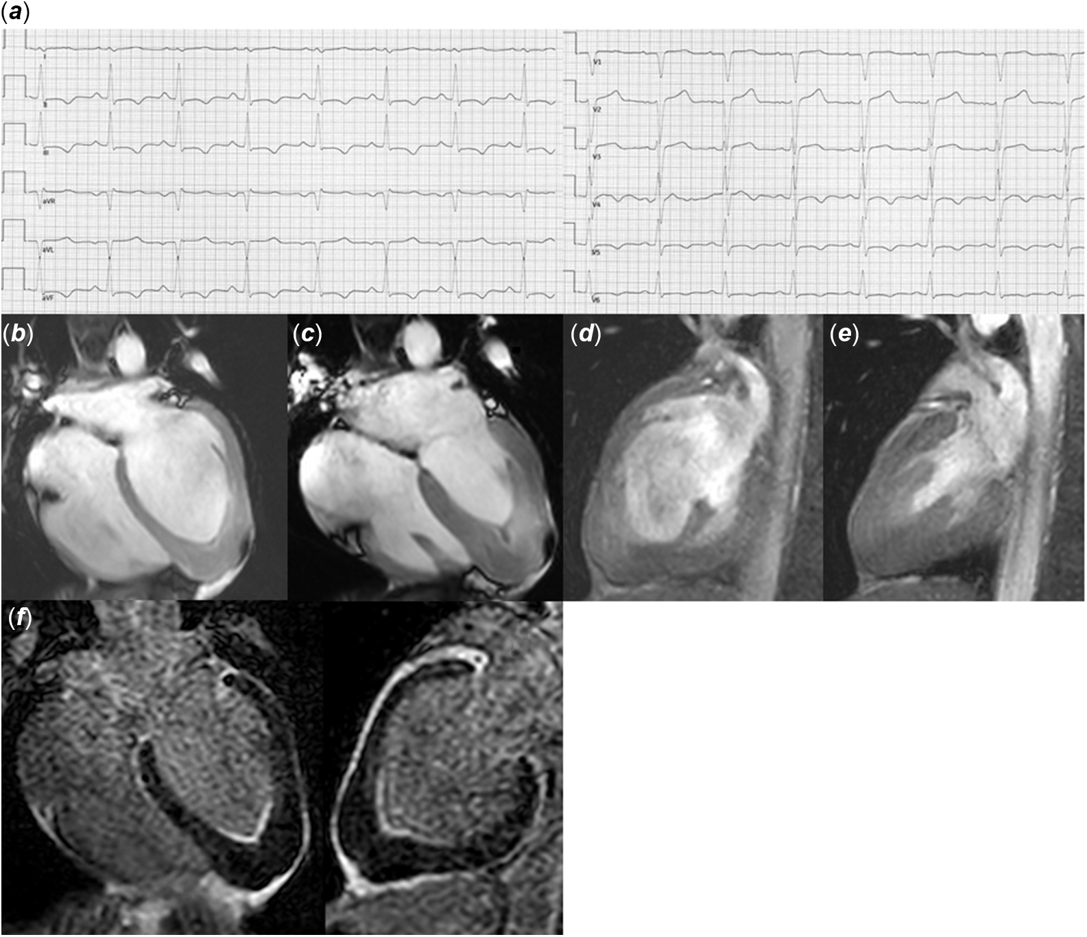
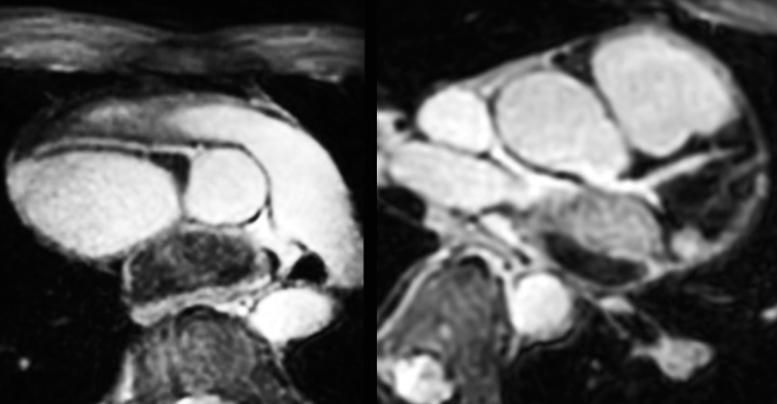
On admission, the patient was asymptomatic, afebrile, with blood pressure of 118/74 mmHg, heart rate of 96 bpm, and oxygen saturation of 100%. Physical examination was unremarkable except for a deformity of the thoracic wall consistent with pectus excavatum. Electrocardiogram showed T-wave inversion in the limb leads, consistent with inferior myocardial ischaemia (Fig 1a). Echocardiography showed normal biventricular systolic and diastolic function. There was no valve dysfunction and no evidence of coronary artery origin abnormalities or cardiomyopathy, although being technically limited due to the above-described funnel chest configuration. Laboratory data revealed slightly elevated high-sensitive troponin T (14.4 ng/L, normal reference range < 14 ng/L) and creatine kinase-MB values (30.8 U/L, normal values <25 U/L), stable at serial controls, and mildly increased N-terminal-proB-type brain natriuretic peptide (NT-pro-BNP) levels (199 ng/L, normal reference range <109 ng/L). Blood count, leucocytes, and inflammatory markers were within normal limits. For further investigation, the patient was admitted to our paediatric cardiology department and a second line imaging assessment using cardiovascular magnetic resonance imaging was performed. Cardiovascular magnetic resonance imaging revealed a spade-like configuration of the left ventricular cavity at end-diastole, best appreciated on ventricular longitudinal axis views (Fig 1b) and an increased apical myocardial wall thickness (17–18 mm) with loss of the typically detectable progressive tapering of myocardial wall thickness towards the apex. No evidence of an apical diverticulum was found. The left ventricle was normal in size and function. In order to assess potential areas of myocardial scarring, late gadolinium enhancement was performed, showing subendocardial enhancement in the hypertrophied apical region, with extension to the mid ventricle (Fig 1c). The origin and proximal course of the coronary arteries were normal (Fig 2). A second echocardiography focusing on the apical myocardium was unable to identify the apical myocardial hypertrophy due to the described chest wall deformity. After discharge, the patient was followed-up in our outpatient department and was referred to our human genetics department. Cardiopulmonary exercise testing was performed, demonstrating normal exercise capacity (VO2 max 42 ml/kg/minute) without induction of symptoms or arrhythmias, electrocardiogram repolarisation abnormalities. 24-hour electrocardiogram monitoring did not demonstrate pathological rhythm disturbances. As there was no relevant intra-ventricular gradient, we decided to not start the patient on beta-blockers. Genetic testing covering the most commonly altered genes in patients with hypertrophic cardiomyopathy and relevant cardiac conditions did not show a causal genetic variant. The patient was therefore subjected to exome sequencing with results still pending.
(a) Electrocardiogram showing T-wave inversion in the inferior leads, (b & c) four-chamber view at diastole and systole demonstrating the presence of apical hypertrophy, (d & e) two-chamber view at diastole and systole, (f) cardiovascular magnetic resonance imaging showing apical subendocardial late gadolinium enhancement.

Electrocardiogram gated mDixon angiography illustrating normal origin and proximal course of the coronary arteries.

Discussion
Apical hypertrophic cardiomyopathy is a phenotypic variant of hypertrophic cardiomyopathy, characterised by left ventricular hypertrophy limited to the distal apex. Initially described in Japan, where it accounts for approximately 25% of all cases of hypertrophic cardiomyopathy, it is a relatively rare finding outside this population, with a reported prevalence of 1–3%. Reference Kitaoka, Doi, Casey, Hitomi, Furuno and Maron2 To date, only very few cases of apical hypetrophic cardiomyopathy in subjects younger than 18 years have been described, Reference Miyamoto, Horigome, Kawano and Sumazaki3–Reference Maurizi, Passantino and Spaziani6 of whom only one was reported to be symptomatic, presenting with exertional chest pain. Reference Zhao, He and Liu4 Due to its rare presentation in childhood and adolescence and to intrinsic limitations of echocardiography in displaying left ventricular apex, particularly in instances of poor echogenic windows, high grade of awareness is required when approaching this cohort of patients. The diagnosis of apical hypertrophic cardiomyopathy may frequently be missed despite consistent symptoms and electrocardiogram changes if the initial echocardiographic evaluation does not provide evidence of cardiomyopathy. In the presence of otherwise unexplained symptoms and/or electrocardiogram findings and persistent clinical suspicion of structural heart disease, it is therefore essential to refer such patients to a second line imaging assessment. As it overcomes the technical limitations imposed to echocardiography, cardiovascular magnetic resonance imaging has established itself as the gold standard for the diagnosis and previous studies have shown that echocardiography can miss the diagnosis in 40% of later by cardiovascular magnetic resonance imaging-detected apical hypertrophic cardiomyopathies. Reference Hughes, Knott and Malcomoson1 In our case, identifying apical hypertrophy was only possible referring the patient to cardiovascular magnetic resonance imaging, which was partly due to the pectus excavatum. Besides to accurate estimation, of biventricular morphology and function, cardiovascular magnetic resonance imaging allows the assessment of tissue fibrosis by evaluating late gadolinium enhancement. In our report, subendocardial distribution of gadolinium was observed in the left ventricular apical region. In apical hypertrophic cardiomyopathy, apical and subendocardial late gadolinium enhancement patterns are typically reported, Reference Kim, Kim and Hwang7 supporting the hypothesis that apical ischaemia due to a mismatch between blood supply and increased oxygen demand in the hypertrophied myocardium might play a pathogenic role in the formation of fibrotic tissue. After careful review of literature, we found only one patient under 18 years, in whom cardiovascular magnetic resonance imaging – late gadolinium enhancement sequences were performed, documenting the apical distribution of the contrast agent. Although data regarding the paediatric population are lacking, hypertrophic cardiomyopathy registry data including 2755 middle-aged patients documented late gadolinium enhancement in 45.8% of subjects presenting the apical variant of the disease. Reference Neubauer, Kolm and Ho8 Apical hypertrophic cardiomyopathy patients demonstrating late gadolinium enhancement on the magnetic resonance imaging were reported to experience more severe symptoms and more episodes of non-sustained ventricular tachycardia on Holter electrocardiogram monitoring compared to those without late gadolinium enhancement, suggesting that areas of fibrosis might represent potential arrhythmogenic substrates. Reference Kebed, Al Adham and Bishu9,Reference Amano, Tacheda and Tachy10
As it typically presents with apical obliteration and subendocardial distribution of late gadolinium enhancement, endomyocardial fibrosis may potentially mimic apical hypertrophic cardiomyopathy, making it challenging, to differentiate between these two pathological entities. Although being endemic in Africa, endomyocardial fibrosis was also described in Europe and affects predominantly children and young adults with a bimodal distribution peaking at 10 and 30 years of age. Clinical presentation and presence of eosinophilia are variable and to date no specific laboratory test for the diagnosis of endomyocardial fibrosis is available. As it provides optimal visualisation of the ventricular apex, cardiovascular magnetic resonance imaging is a fruitful tool. In our patient, obliteration was only observed in systole, whereas in endomyocardial fibrosis, being related to the presence of mural thrombus, it is constant during the entire cardiac cycle. The triple-layered pattern of late gadolinium enhancement typically described in endomyocardial fibrosis (V sign) was not documented in our case. Taking in account also the absence of other imaging features consistent with endomyocardial fibrosis (atrioventricular valve dysfunction, restrictive pattern across atrioventricular valves) and the negative history for fever and travelling to tropical regions abroad, we excluded this diagnosis. Reference Dato11
Conclusion
With this case presentation, we aim to increase awareness of apical hypertrophic cardiomyopathy, which is rarely described in the pediatric population. Typical electrocardiogram alterations and symptoms can be frequently interpreted secondary to coronary artery anomalies and/or myocardial ischaemia in these patients, leading to a missed or delayed diagnosis. As it overcomes the limitations presented by echocardiography, cardiovascular magnetic resonance imaging is an excellent tool to establish the correct diagnosis.
Learning objects
Apical hypertrophic cardiomyopathy is an uncommon phenothypic variant of hypertrophic cardiomyopathy, rarely described in the paediatric population.
Due to intrinsic limitations of echocardiography in displaying left ventricular apex, high grade of awareness is required when approaching patients with apical hypertrophic cardiomyopathy. As it overcomes the technical limitations imposed to echocardiography, cardiovascular magnetic resonance imaging has established itself as the gold standard for the diagnosis.
Acknowledgement
None.
Financial support
This research received no specific grant from any funding agency, commercial or not-for-profit sectors.
Conflicts of interest
None.
Ethical standards
All procedures performed in the study were in accordance with the ethical standards of the institutional research committee and with the Helsinki declaration of 1975, as revised in 2008.