Introduction
The Cichlidae family is considered the most numerous non-Ostariophysi fish family with an estimation of 1900 species (Reis et al., Reference Reis, Kullander and Ferraris2003). These were known to belong to the Order Perciformes, however Wiley & Johnson (Reference Wiley, Johnson, Nelson, Schultze and Wilson2010) split this Order into many other Orders, and the Cichlidae family was inserted into the Labriformes.
Ornamental fish production is considered at present to be one of the most profitable agribusiness in the world, and most potential species are cichlid (Porto, Reference Porto2008). Laetacara araguaiae is one of these. This South American cichlid described in the Araguaia Basin River, Goiás, Brazil by Ottoni & Costa (Reference Ottoni and Costa2009) can be found in quite degraded streams. Several studies involving this species have been performed, such as one monitoring programme for the fish fauna (Casatti et al., Reference Casatti, Langeani and Ferreira2006; Araújo & Tejerina-Garro, Reference Araújo and Tejerina-Garro2007; Graça & Pavanelli, Reference Graça and Pavanelli2007; Perez & Garavello, Reference Perez and Garavello2007), and an environmental trophic characterization (Luz et al., Reference Luz, Abujanra, Agostinho and Gomes2001).
Teresa & Gonçalves-de-Freitas (Reference Teresa and Gonçalves-de-Freitas2011) described the reproductive behaviour of L. araguaiae, which is characterized by cooperation between males and females during mating, nest building, territory defense, followed by sharing of parental care. However, nothing is known about the reproductive biology of this species and data related to gonadal development with emphasis in germ cells morphology and oocytes maturation throughout oogenesis do not exist.
Oocyte development patterns are very similar, being a preserved process among the different Teleostei groups (França et al., Reference França, Grier and Quagio-Grassioto2010; Grier, Reference Grier2000; Grier, Reference Grier2002; Grier et al., Reference Grier, Uribe and De La Rosa-Cruz2005; Ishiba et al., Reference Ishiba, Quagio-Grassiotto and França2010; Mazzoni et al., Reference Mazzoni, Grier and Quagio-Grassiotto2010; Martins et al., Reference Martins, Moura, Santos, Rizzo and Bazzoli2010; Quagio-Grassiotto et al., Reference Quagio-Grassiotto, Grier, Mazzoni, Nóbrega and Amorim2011; Wildner et al., Reference Wildner, Grier and Quagio-Grassiotto2013). However, histological characterization of the dynamics of this process, which is called oogenesis, allows the comprehension and determination of the maturational processes, spawning type and fertilization during the reproductive period from each species (Selman & Wallace, Reference Selman and Wallace1989; Lima et al., Reference Lima, Bernardino, Val-Sella, Fava-de-Moraes and Borella1991; Ishiba et al., Reference Ishiba, Quagio-Grassiotto and França2010).
Thus, due to the scarcity of information about L. araguaiae oogenesis and the necessity of studies that provide basic answers to reproduction questions, conservation and management, the present study was undertaken to provide histological and ultrastructural analysis of the oogenesis of this ornamental species.
Materials and methods
Eight female Laetacara araguaiae with standard sizes ranging from 2.0–5.9 cm were sampled monthly between April 2012 and March 2013 from the Dois Córregos River, Mato Grosso do Sul State, Brazil (20º25′00.5′′S, 51º28′38.4′′W. Alt: 329 m) using a sieve with a net mesh of approximately 8 mm. Specimens were anesthetized with 0.5% benzocaine and sacrificed according to the institutional animal care protocols and approval (007/2012/CEUA-FEIS/UNESP). Then, total length (Lt, cm) and standard length (Ls, cm); body weight (Wb, g), and gonad weight (Wg, g) were measured. Gonadosomatic index (GSI) was calculated with the formula: GSI = (Wg/Wb)*100 according to Vazzoler (Reference Vazzoler1996). Ovaries were removed, fragmented and fixed overnight in 4% paraformaldehyde and 2% glutaraldehyde in Sorensen phosphate buffer, pH 7.4.
For light microscopy, samples were dehydrated in alcoholic solution in increasing concentration, embedded in glycol–metacrylate historesin (Technovit 7100), sectioned at 3.0 μm on a microtome equipped with a glass blade (LEICA RM 2245) and stained with hematoxylin and eosin. All material was examined microscopically (ZEISS–Scope.A1) and photographed with an AxioCam MRc 5.
Samples for transmission electron microscopy (TEM) were post-fixed for 2 h at 4°C in 4% osmium tetroxide, and dehydrated through an acetone series. They were subsequently dehydrated in a critical point dryer Pelco CPD 2 (Ted Pella, Redding, CA). Posteriorly, coated with gold under vacuum with a Coating Unit E5100 (Polaron Equipment, Watford, Hertfordshire, UK) and evaluated for morphological parameters using a Philips–CM 100 TEM.
Results
Ovarian structure
Laetacara araguaiae ovaries are paired organs, dorsally located in the abdominal cavity. They generally are yellowish in colour, changing according to oocytes growth. They show an elongated sac-like form that merges caudally to form one unique duct, which passes through the urogenital papilla. Septa from the surrounding tunica albuginea form the ovigerous lamellae into the gonads. The place of germinal epithelium development, the ovigerous lamellae is formed by germ line cells surrounded by somatic cells. In the germinal epithelium, the oogonia, which is the primordial germ cell from female gametogenesis, undergoes several mitotic divisions before starting folliculogenesis and the oocytes undergo different development stages ending L. araguaiae oogenesis.
Folliculogenesis
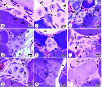
Three types of oogonia are observed in L. araguaiae germinal epithelium: type A-undifferentiated, A-differentiated and B-oogonia. Type A-undifferentiated oogonia are elongated showing one basophilic and irregular nucleus, presenting one small evident nucleolus, which is initially found isolated (Fig. 1 a). These cells proliferate by successive mitosis to form a type A-differentiated oogonia nest. This spermatogonial type is round, with a spherical, voluminous and less basophilic nuclei, showing visible nuages and a voluminous nucleolus (Fig. 1 b). They are separated by pre-follicular cells remaining quiescent in the epithelium or committed to the fate of oogenesis by mitotic divisions (Fig. 1 c), giving origin to the B-oogonia nest. Type B-oogonia are smaller than the previous cells, showing one more basophilic nucleus that contains surrounding nuages. They present one or more nucleolus (Fig. 1 c). This cell type enters meiosis becoming prophasic oocytes that are enclosed inside cysts situated in the ovarian stroma, which remains surrounded by pre-follicular cells.
Light microscopy folliculogenesis in L. araguaiae. (a) Type A undifferentiated oogonia (aug); (b) type A differentiated oogonia nests (adg) (c) B oogonia nest (bo), mitotic division oogonia (dm); (d) oocytes in leptotene cysts (ol); (e) oocytes in zygotene (oz); (f) oocytes in pachytene (op); (g) oocytes in late pachytene (opt); (h) diplotene (do); (i) ovarian follicles (fo). Abbreviations: (pf) pre-follicular cells; (lu) lumen; (bm) basal membrane; (cs) synaptonemal complex. Stain: haematoxylin and eosin.

Initial oocyte cysts in L. araguaiae can be distinguished by chromatin organization according to the meiotic prophase stage. Leptotene oocytes show a basophilic nucleus with decondensed chromatin and one evident nucleolus. (Fig. 1 d). In zygotene oocytes, chromatin is condensed into small granules (Fig. 1 e). Pachytene stages show strong chromosome condensation, due to synaptonemal complex formation and pairing of homologous chromosomes (Fig. 1 f). In late pachytene, the synaptonemal complex undergoes disorganization, homologous chromosomes are retracted, and pre-follicular cells start the isolation of oocytes into the cysts (Fig. 1 g). Diplotene oocytes, which are individualized, undergo cell volume increase, both the nucleus and nucleolus are more voluminous, the cytoplasm is less basophilic and the secretion of basement membrane by pre-follicular cells starts (Fig. 1 h). Oocytes in diplotene are totally surrounded and individualized by follicular cells, forming the ovarian follicle and ending the folliculogenesis stage (Fig. 1 i).
Primary growth
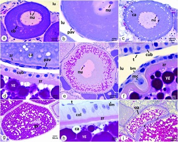
Primary growth starts with initial pre-vitellogenic oocytes showing a more basophilic cytoplasm and a central nucleus with few dispersed nucleolus. Posteriorly many nucleoli are observed in the perinuclear region (Fig. 2 a, b). The Balbiani corpuscle and several ‘nuages’, whose material came from nucleus, are observed surrounding the nucleus (Figs 2 a and 3 a). Follicular cells proliferate giving origin to a follicular epithelium, which is initially formed by squamous cells, evidenced by their flattened nucleus, which rests on the basement membrane (Figs 2 b and 3b).
Light microscopy oogenesis in L. araguaiae. (a–c) Primary growth oocytes: (a) pre-vitellogenic oocytes with perinuclear nucleolus (nu); (b) smooth follicular cell (pav); (c) oocyte with cortical alveoli (ca) positioned in the periphery. (d) Comparison of the zona radiata (zr) thickness and follicular cell shape between cortical alveoli and vitellogenic oocyte. (e, f) Secondary growth oocyte in: (e) yolk granules accumulation (yg) at the oocytes periphery; (f) micropyle detail (mi) with the presence of micropylar cell (mc). (g, h) Oocyte in maturation: (g) germinal vesicle migration (gv) to the periphery of the nucleus; (h) follicular complex details: thick zona radiate (zr), follicular columnar layer (col), basal membrane (bm) and theca (t). (i) Atresic oocyte (oa). Abbreviations: (cb) Balbiani's corpuscle; (nu) nucleolus; (ca) cortical alveoli; (cub) cuboid follicular layer; (lu) lumen. Stain: haematoxylin and eosin.

Transmission electron micrographs of oogenesis in L. araguaiae. (a–c) Primary growth oocyte: (a) pre-vitellogenic oocyte with nuages (ng) around the nucleus; (b) smooth follicular cells (pav); (c) cortical alveoli oocyte, with the first zona radiata layer (zr). (d–i) Secondary growth oocytes: (d) yolk granules accumulation (yg); (e) oocytes general view; (f) microvilli (mc) provided from both oocyte and follicular layers forming the outer zona radiata layer; (g) internal (it) and external theca (et); (h) tri-laminar zona radiata detail: internal (zi), intermediate (zm) and external layers (ze); mitochondria (m) are observed in the cytoplasm; (i) cuboid follicular layer (cub), advanced stage of vitellogenesis. (j–l) Oocyte in maturation: (j) follicular columnar layer (col) filled with secretory vesicles (sv); (k) basal membrane separating theca and follicular layers; (l) zona radiata pores (po). Abbreviations: (nu) nucleolus; (ca) cortical alveoli (lu) lumen; (bm) basal membrane; (rer) rough endoplasmic reticulum; and (bv) blood vessels.

At the end of primary growth, the oocytes are not basophilic anymore and many membranous organelles, such as mitochondria and rough endoplasmic reticulum can be observed in the cytoplasm. Cortical alveoli are concentrated at the cell periphery (Fig. 3 c). At this moment, the follicular layer shows cytoplasmic extensions projected in the direction of the oocyte, which is involved by one narrow electron-dense layer, starting the zona radiata formation (Figs 2 d and 3 c). Moreover, the first layer of theca cells, which are characterized by flattened nuclei, is positioned externally to the basement membrane (Fig. 2 d).
Finishing this stage, the follicular complex is organized as follows: externally is observed the first layer of theca cells, following by a basement membrane, a layer of flattened follicular cells and one narrow zona radiata surrounding the oocyte.
Secondary growth
Oocytes in secondary growth or vitellogenesis are characterized by the initial deposition of yolk microgranules. They cross the follicular complex via intercellular spaces, pass through the pores in zona radiata layer and finally reach the oocyte cytoplasm. The yolk is intensively incorporated at the beginning of this stage (Fig. 3 d). Cortical alveoli go forward in the direction towards the nucleus. They gradually spread in the cytoplasm and mix into the recently aggregated yolk granules (Fig. 4 d). A second layer of theca cells, also showing a flattened nucleus, is observed; blood vessels are spreading throughout this layer (Fig. 3 g).
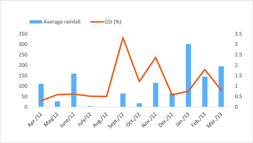
Mean values of GSI in Laetacara araguaiae females and pluviometric precipitation (mm). Source: clima.feis.unesp.br.

The cells from follicular layer undergo some changes, acquiring a cuboid shape with one oval and centralized nucleus. Organelles such as mitochondria, rough endoplasmic reticulum and ribosomes are spread throughout their cytoplasm (Fig. 3 g, h).
At this stage, the zona radiata layer is thick, quite evident and shows three morphologically distinguishable layers: an external one, showing little electron-dense material; the intermediate, which is thin and very electron-dense. This is the first layer to be formed. The internal one that is also electron-dense, presenting a multilamellar shape. This is the thicker and the last layer to be formed (Fig. 3 h). The layers have pores that work as communication channels. These pores, also named channel pores, allow the passage of microvilli that support the contact between the follicular layer and oocyte (Fig. 3 f). At this stage, the oocyte micropile presents an invagination in the zona radiata that is fuelled by one micropylar cell (Fig. 2 f).
Atresic oocytes can occur at any moment during oogenesis, however most of these are observed at the vitellogenesis stage. They are characterized by an irregular shape, the presence of yolk granules, blood vessels, follicular cells in degeneration and numerous associated melano-macrophages (Fig. 2 i).
Final maturation
At this stage, oocytes show the cytoplasm filled by yolk granules and cortical alveoli of different sizes (Fig. 2 g). Follicular layer cells take a columnar shape with one round nucleus on the base and numerous secretory vesicles in the cytoplasm (Figs 2 h and 3 j, k). Cells from the theca layer show the same shape, and stands out from follicular layer by the basement membrane. The zona radiata reaches the maximum thickness and is observed at the external part of microvilli channels opening (Fig. 3 l). The event that best characterizes this stage is the nucleolus (here named germinal vesicle) migration to the oocyte periphery in the direction of the micropyle, also named the fertilization cone.
Reproductive period: gonadosomatic index and pluviometric data
L. araguaiae females presented three reproductive peaks throughout the year from Dois Córregos River, Mato Grosso do Sul State, Brazil. The highest GSI values were observed in September and November 2012 and in February 2013 (Fig. 4). They coincided with the most elevated rainfall levels during the sampling period (Fig. 4).
Discussion
The study of oogenesis in fish is a basic step to better understand the following development phases as, after oogenesis, oocytes are able to enter primary growth and continue the process. The description of folliculogenesis in different oogonia types in L. araguaiae (A-undifferentiated oogonia, A-differentiated and B-oogonia) allows a detailed understanding of the oogenesis initial process, such as mitotic proliferation of these cells giving rise to nests and clusters of oogonia types A and B, as well as prophasic oocyte development until they achieve the diplotene phase, which involves ovarian follicle formation.
The different types of oogonia observed in the germinal epithelium of the Labriformes L. araguaiae were also stated to occur in other fish groups, such as the Siluriformes Pimelodus maculatus (Quagio-Grassiotto et al., Reference Quagio-Grassiotto, Grier, Mazzoni, Nóbrega and Amorim2011) and the Gymnotiformes Gymnotus sylvius (França et al., Reference França, Grier and Quagio-Grassioto2010), showing that this characteristic is probably well preserved in fish.
The involvement of somatic cells in ovarian follicle formation, undergoing diverse changes to differentiate into follicular cells was first described by Grier et al. (Reference Grier, Uribe-Aranzábal and Parenti2007) as a characteristic commonly observed in Teleostei. This condition is also described for L. araguaiae. In ovarian follicle formation of sexually mature animals, pre-follicle cells progressively infiltrate into the cysts surrounding each oocyte to make them individuals (Grier et al., Reference Grier, Uribe-Aranzábal, Patiño and Jamieson2009), as can be observed in L. araguaiae and in Gymnotus sylvius (França et al., Reference França, Grier and Quagio-Grassioto2010) in which pre-follicle cell elongation progressively isolates the oocytes, starting the late pachytene stage.
In L. araguaiae, as in other Teleostei, primary growth starts before full follicle formation or before the oocyte is completely surrounded by the follicular complex and starts primary growth, when meiosis stagnates in diplotene (Patiño & Sullivan, Reference Patiño and Sullivan2002).
During oocyte primary growth an intense synthesis of RNA takes place (Selman & Wallace, Reference Selman and Wallace1989), resulting in a cytoplasmic basophilia at the end of the diplotene stage (Grier et al., Reference Grier, Uribe-Aranzábal, Patiño and Jamieson2009), which can be histologically established, as described in L. araguaiae. In these initial oocytes, Balbiani corpuscles show fissure aspects, due to an intense proliferation of cell membranous organelles, observed in most studied fish (Wallace & Selman, Reference Wallace and Selman1981; França et al., Reference França, Grier and Quagio-Grassioto2010; Quagio-Grassiotto et al., Reference Quagio-Grassiotto, Grier, Mazzoni, Nóbrega and Amorim2011). Nuages observed in oogonia at this phase in L. araguaiae were also reported in Fundulus heteroclitus, on that occasion the authors named them as ribonucleoprotein electron-dense particles (Wallace & Selman, Reference Wallace and Selman1981).
The beginning of zona radiata formation at the end of primary growth, as well as its aspects throughout development, follows a similar pattern to that observed in other fish species (França et al., Reference França, Grier and Quagio-Grassioto2010; Shabanipour & Hossayni, Reference Shabanipour and Hossayni2010; Ma et al., Reference Ma, Zhu, Zhou and Yang2012). In L. araguaiae, the zona radiata was also observed as a single layer of electron-dense material and, posteriorly in vitellogenesis, as a tri-laminar structure with pores and microvilli presence, responsible for substances transport. Grier (Reference Grier2012) observed the same structure in Sciaenops ocellatus, which was formed from both the oocyte and follicular cells. According to Suzuki et al. (Reference Suzuki, Agostinho and Winemiller2000) the pores are responsible for maintenance of abrasion resistance, while the follicular layer is related to adhesiveness. Furthermore, the zona radiata is involved in vitellogenin transfer during vitellogenesis (Shabanipour & Heidari, Reference Shabanipour and Heidari2004). Vitellogenesis, oocyte maturation and incubation are very complex processes that need active synthesis and a lot of protein and lipid transportation (Cakici & Ucuncu, Reference Cakici and Ucuncu2007).
Follicular layer changes along L. araguaiae oogenesis are related to the polymorphism of the follicular cells as a result of their active movement during oocyte envelopment, admitting other shapes through oocyte development. In the secondary growth, cells from oocytes follicular layer in L. araguaiae become cuboid shaped and, as observed in Serrasalmus spilopleura (Quagio-Grassiotto & Guimarães, Reference Quagio-Grassiotto and Guimarães2003), this occurs at this stage because the follicular cells acquire typical characteristics of glycoprotein production cells, showing dilated intercellular space.
In L. araguaiae, the maturation stage was related to germinal vesicle migration (nucleus) in the direction of the animal pole, the place of the micropyle. However, differently from some Perciformes, yolk hydration and liquefaction were not observed (Grier, Reference Grier2012; Wildner et al., Reference Wildner, Grier and Quagio-Grassiotto2013). At the end of this stage, meiosis is recovered and will be finished by oocyte fertilization time. Ovulation occurs at the basement membrane, shared by the ovarian follicle and lamellae epithelium (Patiño & Sullivan, Reference Patiño and Sullivan2002; Quagio-Grassiotto et al., Reference Quagio-Grassiotto, Wilder and Ishiba2013).
It is during the final phases of the gonadal development that is possible to verify an increase in the volume and weight of the ovaries; the GSI is the one index usually indicative of the reproductive period of the species (Vazzoler, Reference Vazzoler1996). Some studies developed with female fish generally analyse the GSI data by season of the year, monthly or bimonthly data and, in this way, identify the reproductive peak of the species, which can occur more than once throughout the year (Barbieri, Reference Barbieri1989; Querol et al., Reference Querol, Querol and Gomes2002; Romagosa et al. Reference Romagosa, Paiva, Andrade-Talmelli and Godinho2003; Wildner et al., Reference Wildner, Grier and Quagio-Grassiotto2013), as observed in L. araguaiae female. In the process of gonadal maturation, an increase in the index values occurs in which the maturation peak coincides with advantage stages of maturation (Navarro, et al., Reference Navarro, Silva, Ribeiro Filho, Calado, Rezende, Silva and Santos2006).
The increase in the values of the river and pluviometric precipitation levels seems to be the ecologic factor that is important in the phenomenon of tropical fish reproduction, such as in Pseudoplatystoma fasciatum (Romagosa et al., Reference Romagosa, Paiva, Andrade-Talmelli and Godinho2003); Prochilodus brevis (Chellappa et al., Reference Chellappa, Bueno, Chellappa, Chellappa and Val2009); Heros efasciatus (Favero et al., Reference Favero, Pompeu and Prado-Valladares2010); Cichlasoma orientale (Gurgel, et al., Reference Gurgel, Verani, Câmara, Barros and Chellappa2011); and Cichla kelberi (Siqueira-Silva et al., Reference Siqueira-Silva, Vicentini, Veríssimo-Silveira and Ninhaus-Silveira2013), acting as a decisive factor in the final maturation of the gamete and in spawning (Baldisserotto, Reference Baldisserotto2002).
This fact was also observed for L. araguaiae in which the reproductive peaks of the species coincides with the beginning of the rainy periods and consequently the elevation of the river water level. According to Vazzoler (Reference Vazzoler1996), this occurs because, in this period, there is higher oxygen availability and higher food quantities in the environment that will contribute to egg hatching and offspring survival, respectively.
Laetacara araguaiae oocytes undergoes different development stages, primary and secondary growth or vitellogenesis and maturation. In the end they are able to spawn three times during the year. This information is important as detailed histological studies that describe oogenesis in ornamental cichlid species are rare and also contribute to management and planning of species conservation in the wild, as its capture can be controlled based on the reproduction periods described here.
Acknowledgements
We thank the Fundação de Amparo à Pesquisa do Estado de São Paulo (FAPESP) (process no. 2012/13413-9), for financial support, Laboratório de Ictiologia Neotropical (LINEO), Univ. Estadual Paulista of UNESP and Electron Microscopy Center of IBB-UNESP. The Laboratory of Hydraulics and Irrigation, Faculty of Ilha Solteira SP/BR for providing the pluviometric date. We also thank Jumma Miranda Araújo Chagas and Douglas Castro Ribeiro for their help during specimen sampling.