Implication
These data demonstrated the involvement of metabolic hormones and mitogen-activated protein kinase (MAPK)-dependent intracellular mechanisms in the control of oocyte nuclear maturation. This expands the existing knowledge concerning hormonal mechanisms (metabolic hormones leptin, ghrelin, obestatin and IGF-I) mediating the effect of nutrition and MAPK-dependent intracellular mechanism in the control of oocyte maturation. The knowledge concerning the effect of hormones could be used for control of oocyte maturation, while expression of MAPK could be potentially useful for assessment of oocyte quality in assisted reproduction and animal embryo production.
Introduction
Nutrition is an important factor affecting fecundity, but the mechanism of this effect remains to be studied. There is growing evidence that the effect of nutrition and metabolism on reproductive processes can be mediated by metabolic hormones – insulin-like growth factor I (IGF-I), leptin, ghrelin and obestatin. Furthermore, these hormones could be useful for regulation of oocyte maturation (meiotic divisions of chromosomes and cytoplasmic maturation enabling oocyte fertilization and further development (Nebreda and Ferby, Reference Nebreda and Ferby2000) in assisted reproduction and biotechnology. IGF-I was reported to be a promoter of porcine oocyte nuclear maturation/meiosis (Xia et al., Reference Xia, Tekpetey and Armstrong1994; Illera et al., Reference Illera, Lorenco, Illera and Petters1998; Sirotkin et al., Reference Sirotkin, Dukesova, Makarevich, Kubek, Bulla and Hetenyi2000). Some authors (Craig et al., Reference Craig, Zhu, Dyce, Wen and Li2005; Kun et al., Reference Kun, Shaohua, Yufang, Yankun, Hengxi, Xiuzhu, Yonghui, Yan, Yunping, Lei and Ning2007) have reported the promotion of meiosis in porcine oocytes after leptin addition. Other authors (Jin et al., Reference Jin, Cui, Han and Kim2009; Suzuki et al., Reference Suzuki, Sasaki, Shimizu, Matsuzaki, Hashizume and Kuwayama2009), however, did not observe any effect of leptin on this process. Slight inhibition of porcine oocyte nuclear maturation has been observed after ghrelin addition (Suzuki et al., Reference Suzuki, Sasaki, Shimizu, Matsuzaki, Hashizume and Kuwayama2009). Obestatin effect on oocyte maturation has not been examined as yet. Therefore, available data concerning the effects of metabolic hormones other than IGF-I are inconsistent, contradictory or insufficient.
Hormones affect cellular functions through intracellular mediators. The most known mediators of hormone actions are protein kinases. Mitogen-activated protein kinases (MAPK) can be involved in hormone-induced oocyte maturation. In porcine oocytes, accumulation or both total and phosphorylated MAPK before (Inoue et al., Reference Inoue, Naito, Nakayama and Sato1998; Lee et al., Reference Lee, Miyano and Moor2000; Ye et al., Reference Ye, Coleman, Hunter, Craigon, Campbell and Luck2007), during (Motlik et al., Reference Motlik, Pavlok, Kubelka, Kalous and Kalab1998; Lee et al., Reference Lee, Miyano and Moor2000; Ebeling et al., Reference Ebeling, Schuon and Meinecke2007; Li et al., Reference Li, Liang, Xiong, Xu, Lin, Hou, Chen, Schatten and Sun2008) or after (Inoue et al., Reference Inoue, Naito, Aoki, Tayoda and Sato1995) the first stages of maturation (germinal vesicle breakdown) was reported. Other studies have demonstrated either an increase (Wehrend and Meinecke, Reference Wehrend and Meinecke2001) or decrease (Inoue et al., Reference Inoue, Naito, Aoki, Tayoda and Sato1995) of MAPK activity in porcine oocytes at advanced stages of nuclear maturation (transition from metaphase I to metaphase II of meiosis). Therefore, the data concerning changes in MAPK during oocyte maturation are inconsistent.
It remains to be established whether MAPK can mediate the action of hormones on oocyte maturation. The activation of MAPK by the known hormonal stimulators of porcine oocyte maturation FSH, LH (Ebeling et al., Reference Ebeling, Schuon and Meinecke2007; Kimura et al., Reference Kimura, Hoshino, Totsukawa and Sato2007) and epidermal growth factor (EGF) (Li et al., Reference Li, Liang, Xiong, Xu, Lin, Hou, Chen, Schatten and Sun2008), the ability of MAPK microinjection to promote meiosis (Inoue et al., Reference Inoue, Naito, Nakayama and Sato1998) and the ability of MAPK blocker to prevent EGF-induced porcine oocyte maturation (Li et al., Reference Li, Liang, Xiong, Xu, Lin, Hou, Chen, Schatten and Sun2008) indicate that MAPK may be a promoter of porcine oocyte maturation and mediator of EGF and gonadotropin action on this process. However, it remains unknown whether MAPK can mediate the effect of metabolic hormones (IGF-I, leptin, ghrelin and obestatin) on oocyte maturation.
The general aim of our in vitro experiments was to examine whether the metabolic hormones leptin, ghrelin, obestatin and IGF-I affect nuclear maturation of porcine oocytes, and whether MAPK-dependent intracellular mechanisms are involved in the control of this process and in mediating the effect of metabolic hormones on oocyte maturation. For this purpose, we examined (1) the changes in MAPK accumulation in relation to nuclear maturation of porcine oocytes during culture, (2) the effects of the hormones leptin, ghrelin, obestatin, IGF-I on oocyte nuclear maturation and expression of MAPK, (3) the action of MAPK blocker on porcine oocyte maturation and (4) the ability of MAPK blocker to modify the effect of the hormones leptin, ghrelin, obestatin and IGF-I. We have culture-isolated porcine oocytes with and without leptin, ghrelin, obestatin, IGF-I, MAPK blocker PD98059 and the combinations of hormones + PD98059. The proportion of matured oocytes and of oocytes containing MAPK/ERK1-2 before and after culture was assessed.
Material and methods
Processing and culture of oocytes
Ovaries without visible abnormalities were removed from non-cycling Slovakian white gilts, 6 months of age, within 1 h after killing at a local abattoir and transported to the laboratory at room temperature. Oocyte-cumulus complexes were isolated and processed as described earlier (Sirotkin et al., Reference Sirotkin, Dukesova, Makarevich, Kubek, Bulla and Hetenyi2000). Briefly, the oocyte–cumulus complexes were aspirated by syringe from medium size (3 to 8 mm in diameter) antral ovarian follicles. Hemorrhagic, cystic, presumptive preovulatory follicles (more than 6 mm) and follicles with visible signs of atresia were excluded.
Only fully grown oocytes surrounded by intact compact cumulus were used and further manipulated. Oocytes were washed three times in medium TCM-199 (Gibco, Carlsbad, CA, USA), supplemented with 10% Fetal calf serum (fetal serum; Sigma, St. Louis, MO, USA). Thereafter, groups of 10 oocytes were placed in four-well plates containing 500 μl per well serum-free oocyte maturation medium TCM-199 (Gibco) supplemented with 50 μg/ml, gentamicin (Gibco), 2 μg/ml bLH (Werfachor, Alvetra and Werfft AG, Wien, Austria), 2 μg/ml pFSH (Ovagen, ICP, Auckland, New Zealand). Experimental groups received the following substances:
1. rh leptin (NHPP, Torrance, CA, USA, 10 ng/ml medium),
2. rh IGF-I (Sigma, 100 ng/ml),
3. synthetic obestatin (Peptides International Inc., Louisville, KT, USA, 10 ng/ml),
4. 1 to 18 analog of rh Ghrelin (PGH-3625 PI, Peptides International Inc., 10ng/ml),
5. MAPK blocker PD 98059 (Calbiochem-Novabiochem Corp., La Jolla, CA, USA, 50 μM) or the combinations of hormones listed above with PD98059.
Cumulus cell-oocyte complexes were cultured for 44 h at 38.5°C in 5% CO2 in air.
In the first series of experiments, oocytes were collected after 0.5 h culture (for adaptation to culture conditions, but before long-term culture) without treatments and 44 h after culture with and without leptin, IGF-I, ghrelin and obestatin. Oocyte nuclear maturation (percentage of oocytes reaching metaphase II) was determined. In the second series of experiments, in which the oocytes were collected after 0.5 h culture without treatments and 44 h after culture with and without leptin, IGF-I, ghrelin and obestatin, PD98059 alone and the hormones listed above in combination with PD98059 were compared. In this series of experiments, both oocyte nuclear maturation (percentage of oocytes reaching metaphase II) and expression of MAPK/ERK1-2 (proportion of oocytes containing visible MAPK/ERK1-2) were evaluated.
Evaluation of oocyte maturation and expression of MAPK
After the assigned culture, oocytes were washed in TCM, stripped of cumular cells by pipetting in TCM-199 with 1% hyaluronidase (Gibco) and then fixed in 3.7% paraformaldehyde in PBS (Sigma).
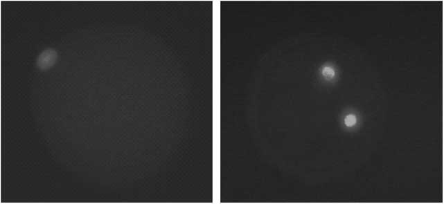
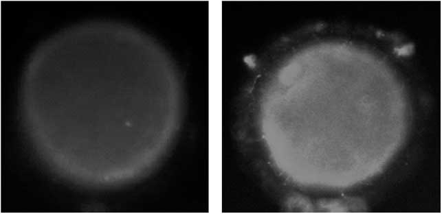
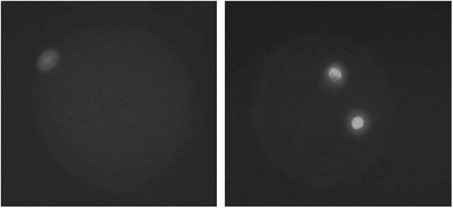
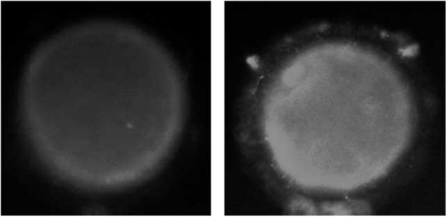
Expression of MAPK/ERK1-2 was determined before and after culture using immunocytochemistry protocol described earlier (Makarevich and Markkula, Reference Makarevich and Markkula2002) by using primary mouse monoclonal antiserum against both phosphorylated and non-phosphorylated MAPK/ERK1-2 (Santa Cruz, Santa Cruz, CA, USA) and secondary goat antiserum against mouse IgG labeled with FITC (Sevac, Prague, Czech Republic). Thereafter, oocytes were embedded in Vectashield mounting medium containing DAPI (Vector Laboratories, Inc., Burlingame, CA, USA). To verify the specificity of the immunocytochemical method, in each experiment, several randomly selected oocytes were treated with a secondary antibody omitting the primary antiserum used as a negative control. In this case, no substantial fluorescence of FITC was observed. Nuclear maturation (stage of meiosis) was determined in the same oocytes according to the morphological characteristics and positions of chromosomes stained by DAPI. The main read-out was the percentage of (a) matured oocytes and (b) oocytes containing MAPK. In each group, the percentage of oocytes containing visible MAPK/ERK1-2 (green fluorescence induced by FITC) and matured oocytes (at anaphase-metaphase II stage of meiosis, two groups of chromosomes marked with blue fluorescence induced by DAPI) was determined by fluorescent microscope (Leica Microsystems, Wetzlar, Germany). Illustrative photos of immature and mature oocytes (DAPI fluorescence) are shown in Figure 1. Oocytes containing low and high amounts of MAPK/ERK1-2 are shown in Figure 2.
Immature (diplotena-diakinesis I, left) and mature (telophase-metaphase II, right) oocytes. Staining with DAPI, magnification ×40. Intensive blue fluorescence of groups of chromosomes, only light signal in cytoplasm.
Oocyte with low (left) and high (right) expression of MAPK/ERK1-2. Staining with FITC, magnification ×40. Green fluorescence is localized mainly in the outer zone of cytoplasm along with the plasma membrane.
Statistics
Each experiment in the first series was conducted with 25 replications, and each experiment in the second series was repeated 19 times using different ovaries obtained at different times from different animals. In each experiment, all the groups/treatments were present (5 to 20 oocytes per group in each experiment). In the first series of experiments, each experimental group was characterized on the basis of analysis of 426 to 436 oocytes, and in the second series of experiments, each treatment was tested at 101 to 219 oocytes.
Significant differences between the treatments were determined using one-way ANOVA, followed by χ 2 and Student’s t-test to detect significant differences between the groups by using Sigma Plot 9.0 statistical software (Systat Software, GmbH, Erkrath, Germany). Differences from control at P < 0.05 were considered as significant.
Results
Fluorescent staining with DAPI demonstrated the presence of both immature (at meiotic stages diplotena-anaphase I) and mature (at telophase-metaphase II) oocytes in each experimental group (Figure 1). Fluorescent staining with FITC showed the presence of MAPK/ERK1-2 in oocytes. It was expressed as fine granules localized mainly in the outer zone of the cytoplasm along with the plasma membrane and in the nucleus of the oocyte (Figure 2). Expression of these markers of oocyte nuclear maturation and MAPK/ERK1-2 accumulation changed depending on the time of culture, addition of hormones, MAPK blocker PD98059 and the combination of these factors (Tables 1 and 2).
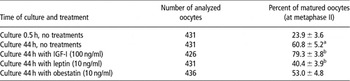
Effect of time of culture and of hormonal treatments on nuclear maturation of porcine oocytes

aEffect of time of culture: significant (P < 0.05) differences between oocytes cultured 0.5 and 44 h without treatments.
bEffect of hormonal treatments: significant (P < 0.05) differences between oocytes cultured 44 h with and without hormones.
Values are mean ± s.e.m.
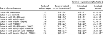
Effect of time of culture and of treatment with hormones, MAPK blocker PD98059 and their combination on nuclear maturation and expression of MAPK/ERK1-2 in porcine oocytes

aEffect of time of culture: significant (P < 0.05) differences between oocytes cultured 0.5 and 44 h without treatments.
bEffect of hormonal treatments: significant (P < 0.05) differences between corresponding groups of oocytes cultured 44 h with and without hormones.
cEffect of PD98059: significant (P < 0.05) differences between coresponding groups of oocytes cultured.
Values are mean ± s.e.m.
It was observed that the majority of oocytes isolated from the ovary before culture were immature and did not contain visible MAPK. On the other hand, some oocytes were already mature before culture, and the majority of these oocytes contained MAPK. Incubation of oocytes during 44 h resulted in a significant increase in proportion of matured oocytes and in the percentage of oocytes containing MAPK in both the matured and not matured groups (Tables 1 and 2).
The addition of IGF-I to the culture medium significantly increased the proportion of matured oocytes after culture; the addition of leptin, by contrast, significantly decreased the percentage of matured oocytes, while the addition of either ghrelin or obestatin did not substantially affect this parameter. The addition of hormones did not affect the expression of MAPK in either immature or mature oocytes (Tables 1 and 2).
The addition of MAPK/ERK1-2 blocker PD98059 alone dramatically decreased the percentage of oocytes undergoing maturation and containing MAPK/ERK1-2 after culture. No differences between immature and mature oocytes in response of MAPK/ERK1-2 to PD98059 treatment were found. When given together with hormones, PD98059 was able to reduce the stimulatory effect of IGF-I, to invert the inhibitory action of leptin to stimulatory and to induce the stimulatory action of ghrelin and obestatin on meiosis. Furthermore, in the presence of PD98059, IGF-I, ghrelin and obestatin, but not leptin increased accumulation of MAPK in either immature or mature oocytes (Table 2).
Discussion
Is MAPK involved in the control of oocyte nuclear maturation?
The nuclear maturation of porcine oocytes after isolation and culture, as well as the presence of MAPK/ERK1-2 in these oocytes, is in line with previous reports (Inoue et al., Reference Inoue, Naito, Aoki, Tayoda and Sato1995 and Reference Inoue, Naito, Nakayama and Sato1998; Motlik et al., Reference Motlik, Pavlok, Kubelka, Kalous and Kalab1998; Lee et al., Reference Lee, Miyano and Moor2000; Sirotkin et al., Reference Sirotkin, Dukesova, Makarevich, Kubek, Bulla and Hetenyi2000; Wehrend and Meinecke, Reference Wehrend and Meinecke2001; Ye et al., Reference Ye, Coleman, Hunter, Craigon, Campbell and Luck2007; Ebeling et al., Reference Ebeling, Schuon and Meinecke2007; Kimura et al., Reference Kimura, Hoshino, Totsukawa and Sato2007; Li et al., Reference Li, Liang, Xiong, Xu, Lin, Hou, Chen, Schatten and Sun2008). It was shown that a part of the oocytes were mature already before long-term culture. Maturation of these oocytes could be due to the natural disconnection between oocyte and surrounding follicular cells maintaining meiosis arrest before isolation (Kimura et al., Reference Kimura, Hoshino, Totsukawa and Sato2007; Li et al., Reference Li, Liang, Xiong, Xu, Lin, Hou, Chen, Schatten and Sun2008). Since isolation and culture induce the maturation of the majority of oocytes, the cells that matured before culture could represent a model to distinguish between the effects of culture and oocyte nuclear maturation on MAPK. It was shown that expression of MAPK/ERK1-2 in mature oocytes was higher before culture than in immature cells. It indicates that accumulation of MAPK/ERK1-2 is associated with nuclear maturation, but not with culture per se, and that MAPK/ERK1-2 can be involved in the promotion of porcine oocyte nuclear maturation. Requirement of MAPK/ERK1-2 for promotion of meiosis is confirmed by the inhibitory effect of MAPK blocker PD98059 on both MAPK/ERK1-2 accumulation and oocyte maturation after culture observed in our experiments. Our observations are in line with maturation-associated accumulation or activation of MAPK and with the ability of MAPK blocker to prevent meiosis in porcine oocytes observed by other authors (Inoue et al., Reference Inoue, Naito, Aoki, Tayoda and Sato1995 and Reference Inoue, Naito, Nakayama and Sato1998; Motlik et al., Reference Motlik, Pavlok, Kubelka, Kalous and Kalab1998; Lee et al., Reference Lee, Miyano and Moor2000; Sirotkin et al., Reference Sirotkin, Dukesova, Makarevich, Kubek, Bulla and Hetenyi2000; Wehrend and Meinecke, Reference Wehrend and Meinecke2001; Ye et al., Reference Ye, Coleman, Hunter, Craigon, Campbell and Luck2007; Ebeling et al., Reference Ebeling, Schuon and Meinecke2007; Kimura et al., Reference Kimura, Hoshino, Totsukawa and Sato2007; Li et al., Reference Li, Liang, Xiong, Xu, Lin, Hou, Chen, Schatten and Sun2008). All these data demonstrate the involvement of MAPK in promotion of porcine oocyte maturation. However, in our experiments, expression of MAPK in oocytes rose independent of their maturation in culture. Furthermore, oocytes cultured with IGF-I + MAPK blocker matured well despite a low level of MAPK. It suggests that a high level of intracellular MAPK during culture is not necessary for oocyte maturation, that is, that MAPK is probably not involved in the promotion of this process. In our experiments, we did not examine when the MAPK level started to increase during oocyte maturation, and when PD98059 started to influence this process. Nevertheless, the majority of previous publications (Inoue et al., Reference Inoue, Naito, Nakayama and Sato1998; Motlik et al., Reference Motlik, Pavlok, Kubelka, Kalous and Kalab1998; Lee et al., Reference Lee, Miyano and Moor2000; Sirotkin et al., Reference Sirotkin, Dukesova, Makarevich, Kubek, Bulla and Hetenyi2000; Wehrend and Meinecke, Reference Wehrend and Meinecke2001; Ye et al., Reference Ye, Coleman, Hunter, Craigon, Campbell and Luck2007; Ebeling et al., Reference Ebeling, Schuon and Meinecke2007; Kimura et al., Reference Kimura, Hoshino, Totsukawa and Sato2007; Li et al., Reference Li, Liang, Xiong, Xu, Lin, Hou, Chen, Schatten and Sun2008) have reported that this occurs before and during the start of meiosis reinitiation. It could be suggested that MAPK is necessary for reinitiation, but not for completion of the porcine oocyte nuclear maturation. Changes in MAPK/ERK1-2 at the end of oocyte maturation observed in our experiments could not be related to the start of maturation, which could be really promoted by MAPK.
Are metabolic hormones involved in the control of oocyte maturation?
In our experiments with hormonal treatments alone, the addition of IGF-I to culture medium increased, addition of leptin decreased, and ghrelin and obestatin did not alter the proportion of matured oocytes. It demonstrates the involvement of IGF-I in upregulation and of leptin in downregulation of porcine oocyte nuclear maturation. These observations confirm previous reports (Xia et al., Reference Xia, Tekpetey and Armstrong1994; Illera et al., Reference Illera, Lorenco, Illera and Petters1998; Sirotkin et al., Reference Sirotkin, Dukesova, Makarevich, Kubek, Bulla and Hetenyi2000) of stimulatory action of IGF-I on porcine oocyte nuclear maturation. However, they do not correspond to the previous reports of stimulatory action (Craig et al., Reference Craig, Zhu, Dyce, Wen and Li2005; Kun et al., Reference Kun, Shaohua, Yufang, Yankun, Hengxi, Xiuzhu, Yonghui, Yan, Yunping, Lei and Ning2007) or lack of effect (Jin et al., Reference Jin, Cui, Han and Kim2009; Suzuki et al., Reference Suzuki, Sasaki, Shimizu, Matsuzaki, Hashizume and Kuwayama2009) of leptin on porcine oocyte maturation. Furthermore, they do not confirm a previous report (Suzuki et al., Reference Suzuki, Sasaki, Shimizu, Matsuzaki, Hashizume and Kuwayama2009) of inhibitory action of ghrelin on this process. This is the first demonstration to show leptin can not only promote or influence oocyte maturation, but also it can inhibit oocyte maturation. Furthermore, this is the first evidence that ghrelin does not influence this process. The differences in leptin and ghrelin actions observed by different authors could be due to different states of oocytes used in experiments, including the state of their hormone receptors or post-receptor mediators of hormone action (protein kinases a.o.). This state and a corresponding variation in hormone action should be taken into account by the application of leptin and ghrelin for regulation of oocyte maturation in assisted reproduction and biotechnology. To our knowledge, this is the first examination of obestatin action on oocyte maturation, which demonstrated its lack of influence on this process.
Do metabolic hormones affect oocyte nuclear maturation via MAPK?
There exists evidence that MAPK may be a mediator of stimulatory action of EGF and gonadotropin action on porcine oocyte maturation. Both oocyte maturation and accumulation of MAPK were promoted by FSH, LH (Ebeling et al., Reference Ebeling, Schuon and Meinecke2007; Kimura et al., Reference Kimura, Hoshino, Totsukawa and Sato2007) and EGF (Li et al., Reference Li, Liang, Xiong, Xu, Lin, Hou, Chen, Schatten and Sun2008). MAPK microinjection promoted meiosis (Inoue et al., Reference Inoue, Naito, Nakayama and Sato1998). Furthermore, the MAPK blocker prevented EGF-induced porcine oocyte maturation (Li et al., Reference Li, Liang, Xiong, Xu, Lin, Hou, Chen, Schatten and Sun2008).
In our experiments, we used similar approaches (analysis of effect of hormones, MAPK blocker and their combinations on MAPK and oocyte maturation) to examine whether MAPK can mediate the effects of the metabolic hormones, IGF-I, leptin, ghrelin and obestatin, on this process. In our experiments, these metabolic hormones, when given alone, were able to affect oocyte maturation, but a high expression of MAPK occurred in either immature or mature oocytes, either treated or not treated with hormones.
Furthermore, in our experiments, MAPK blocker PD98059 was able to modify the effect of hormones on oocyte maturation and on MAPK expression, but it did not prevent the action of any hormone. It suggests the possible interference of hormones and MAPK-dependent intracellular mechanisms in oocytes. However, no influence of hormones on MAPK and lack of association between action of hormones and PD98059 on MAPK and meiosis suggest that MAPK is probably not a mediator of effect of IGF-I, leptin, ghrelin and obestatin on porcine oocyte nuclear maturation.
Taken together, the present results suggest the involvement of IGF-I, leptin and MAPK/ERK1-2, but not of ghrelin and obestatin, in porcine oocyte maturation. Although understanding fine interrelationships between these hormones and MAPK requires more detailed studies, our observations demonstrate the physiological importance and potential usefulness of some of these substances and their regulators for control of oocyte maturation in assisted reproduction and biotechnology.
Acknowledgments
The authors thank the administration of the Research Centre of Animal Production and Constantine the Philosopher University (Nitra, Slovak republic) for supporting this study, as well as K. Tothová and Ž. Kuklová for technical assistance.