Introduction
Patients with borderline personality disorder (BPD), who typically suffer from a pervasive instability in moods, interpersonal relationships, self-image, and behaviours, show a high prevalence of severe adverse life events and traumatic stress (Golier et al. Reference Golier, Yehuda, Bierer, Mitropoulou, New, Schmeidler, Silverman and Siever2003; McLean & Gallop, Reference McLean and Gallop2003; Pagano et al. Reference Pagano, Skodol, Stout, Shea, Yen, Grilo, Sanislow, Bender, McGlashan, Zanarini and Gunderson2004). Up until now several studies have investigated the psychophysiology of processing and neural representation of severe adverse life events in BPD. Functional abnormalities under resting-state conditions were repeatedly observed in the frontal cortex and in various subcortical areas (Goyer et al. Reference Goyer, Andreason, Semple, Clayton, King, Compton-Toth, Schulz and Cohen1994; De La Fuente et al. Reference De La Fuente, Goldman, Stanus, Vizuete, Morlan, Bobes and Mendlewicz1997; van Elst et al. Reference van Elst, Thiel, Hesslinger, Lieb, Bohus, Hennig and Ebert2001; Juengling et al. Reference Juengling, Schmahl, Hesslinger, Ebert, Bremner, Gostomzyk, Bohus and Lieb2003; Soloff et al. Reference Soloff, Meltzer, Becker, Greer, Kelly and Constantine2003). Some further studies indicated regional dysfunctions of the serotonergic system. In response to fenfluramine under positron-emission tomography (PET) conditions patients with BPD showed decreased glucose uptake in the right medial frontal and orbitofrontal cortex (OFC), in the left temporal and parietal lobe, and left caudatum (Soloff et al. Reference Soloff, Meltzer, Greer, Constantine and Kelly2000, Reference Soloff, Meltzer, Becker, Greer, Kelly and Constantine2003). In addition, α-[11C]methyl-l-tryptophan was found to be decreased in the medial OFC, the anterior cingulate cortex (ACC), temporal lobe and corpus striatum (Leyton et al. Reference Leyton, Okazawa, Diksic, Paris, Rosa, Mzengeza, Young, Blier and Benkelfat2001).
In a functional magnetic resonance imaging (fMRI) study by Herpertz et al. (Reference Herpertz, Dietrich, Wenning, Krings, Erberich, Willmes, Thron and Sass2001), BPD patients and healthy controls viewed emotional aversive and neutral pictures. In patients – but not in healthy subjects – aversive (versus neutral) pictures activated the amygdala as well as medial and inferolateral prefrontal areas (Herpertz et al. Reference Herpertz, Dietrich, Wenning, Krings, Erberich, Willmes, Thron and Sass2001). A PET study with sexually or physically abused subjects with and without BPD listened to individual scripts describing neutral and abusive events (Schmahl et al. Reference Schmahl, Vermetten, Elzinga and Bremner2004). The recall of abusive events was associated with an increased blood flow in different areas of the prefrontal cortex (right anterior cingulate, left orbitofrontal, right dorsolateral prefrontal cortex) and a decrease in the left dorsolateral prefrontal cortex in traumatized women without BPD but not in those with BPD (Schmahl et al. Reference Schmahl, Vermetten, Elzinga and Bremner2004). The authors concluded that these findings may reflect dysfunctions of these prefrontal areas in BPD. In an fMRI study, we compared the recall of traumatic events with negative but non-traumatic events in BPD patients with and without post-traumatic stress disorder (PTSD) (Driessen et al. Reference Driessen, Beblo, Mertens, Piefke, Rullkoetter, Silva-Saavedra, Reddemann, Rau, Markowitsch, Wulff, Lange and Woermann2004). In the subgroup without PTSD, activation of the OFC on both sides and of the Broca area predominated, while in the subgroup with additional PTSD activation was primarily observed in limbic areas, including the amygdala. In a recent fMRI study we compared the recall of unresolved adverse life events (ULE) and resolved negative life events (RLE) in patients with BPD and healthy volunteers (Beblo et al. Reference Beblo, Driessen, Mertens, Wingenfeld, Piefke, Rullkoetter, Silva-Saavedra, Mensebach, Reddemann, Rau, Markowitsch, Wulff, Lange, Berea, Ollech and Woermann2006a). Only in BPD patients did we find bilateral activation of the frontal cortex including parts of the insula and of the OFC, temporal activation including the amygdala, activation of the right occipital cortex, and of parts of the cerebellum. Patients but not controls reported higher levels of anxiety and helplessness during the ULE versus RLE memory condition.
In a very recent 12-week follow-up study under in-patient dialectic behavioural therapy, Schnell & Herpertz (Reference Schnell and Herpertz2006) investigated the response to standardized arousing images in six patients with BPD and six healthy controls. They found a decrease of blood oxygenation level-dependent (BOLD) responses in the right anterior cingulate, temporal and posterior cingulate cortices as well as in the left insula. In addition, they found an association between subjective arousal and activation of the left amygdala and hippocampus.
We here present the first fMRI follow-up study in BPD using individual and autobiographical stimulus material. We aimed to investigate changes of neural activation patterns in response to the recall of individual ULE compared with RLE over 1 year. We hypothesized that patients would show less activation of fronto-temporal and (para)limbic brain areas after 1 year, and that these changes would be associated with a decreased intensity of emotions during the recall and with clinical improvement at least of trauma-associated psychopathology after 1 year.
Method
Methods applied in this follow-up study are similar to those recently published by our work group (Beblo et al. Reference Beblo, Driessen, Mertens, Wingenfeld, Piefke, Rullkoetter, Silva-Saavedra, Mensebach, Reddemann, Rau, Markowitsch, Wulff, Lange, Berea, Ollech and Woermann2006a).
Subjects
Thirteen female right-handed BPD patients were included, who represent a subsample of our previous study. From those 20 subjects, four did not live any more in the region and three were not ready to take part in this follow-up study. All were treated for BPD as in-patients in the Department of Psychiatry and Psychotherapy Bethel (Ev. Hospital Bielefeld, Germany) at t1. We did not find differences between participating and non-participating patients in the present study with regard to psychopathology nor with regard to subjective evaluations of RLE and ULE (see below). The mean age of the present sample was 33.0 [standard deviation (s.d.) 8.8, range 19–47] years. Length of basic education was 11.1 (s.d.=1.6) years. Of the subjects, 76.9% (n=10) had passed a vocational education course and 23.1% had not (n=3). Of these three subjects, two were currently in education. None of them was married, but 38.5% (n=5) lived in a partnership. These characteristics did not change in the observation period. All patients met DSM-IV criteria of BPD which was considered to represent the main diagnosis as assessed and evaluated by their treating psychotherapists within the first week after admission. No participant was pregnant or had one of the following current or previous medical conditions, which were assessed by the medical history, by careful clinical examination, and by laboratory means: endocrine system disorders, malignant diseases, liver cirrhosis, neurological diseases, loss of consciousness (lifetime), or mental retardation. Further exclusion criteria were current infectious diseases, anorexia, schizophrenia, schizoaffective disorders, major depression disorder with psychotic symptoms, alcohol and/or drug abuse or dependence within 6 months prior to investigation at t1 or t2. Written informed consent was obtained from all subjects. Subjects received financial remuneration for their efforts (€75). The study was approved by the University of Muenster Ethics Committee. On average, 1 year after t1 (mean=358, s.d.=208, median 327 days) subjects again underwent most parts of the study programme at t1.
Clinical assessment
The clinical assessment was similar to that previously reported (Beblo et al. Reference Beblo, Driessen, Mertens, Wingenfeld, Piefke, Rullkoetter, Silva-Saavedra, Mensebach, Reddemann, Rau, Markowitsch, Wulff, Lange, Berea, Ollech and Woermann2006a). At t1 participants completed the full structured clinical interview for DSM-IV (SCID; First et al. Reference First, Spitzer, Gibbon, Williams and Benjamin1996; Wittchen et al. Reference Wittchen, Zaudig and Fydrich1997). Current psychopathology at both times was assessed by the symptom checklist (SCL-90-R; Franke, Reference Franke1995), the impact of event scale (Maercker & Schützwohl, Reference Maercker and Schützwohl1998), the Beck depression inventory (Beck et al. Reference Beck, Ward, Medelson, Mock and Erbaugh1961; Beck & Steer, Reference Beck and Steer1994), and the dissociative experiences scale (DES) (Bernstein & Putnam, Reference Bernstein and Putnam1986; Freyberger et al. Reference Freyberger, Spitzer and Stieglitz1999). In all subjects urinary drug screenings and a venous blood sample were obtained as part of the clinical routine. No pathological measures were found in any participant. To control for critical declarative memory dysfunctions the auditory verbal learning test was administered (Rey, Reference Rey1964).
Autobiographical interview
At t1, subjects underwent a semi-structured autobiographical research interview 1 week prior to fMRI investigation at t1 (Beblo et al. Reference Beblo, Driessen, Mertens, Wingenfeld, Piefke, Rullkoetter, Silva-Saavedra, Mensebach, Reddemann, Rau, Markowitsch, Wulff, Lange, Berea, Ollech and Woermann2006a). Together with the patients, two ULE and two RLE were selected for the fMRI session. ULE were defined as being of negative emotional valence, as being still a high burden from a subjective point of view, and as still evoking major emotional reactions. Participants reported not being able to successfully cope with them until the time of assessment (t1). By contrast, RLE were defined as being also negatively valenced but subjectively experienced as resolved or overcome and not leading to major emotional arousal during recall. Finally, subjects were asked to provide three key words for each of the four events which were subsequently used to trigger active recall during the fMRI measurement (cue-driven method). At t2, key words as well as the underlying events were again discussed until subjects and interviewers were certain that both were accurately remembered.
Stimulus presentation and design
At t1, anatomical MR scans were acquired a few days before the fMRI measurement to exclude brain damage and get the subjects used to the scanner. For fMRI acquisition we applied a box car design with two activation conditions (ULE and RLE) and a low level baseline condition (BC). Activation conditions were presented in a between-subjects randomly balanced order. The design consisted of 12 activation blocks, each anteceded and followed by a BC. Each block was introduced by one of the key words (cues) using the scanner's intercom. In response to the key words, subjects recalled the associated ULE or RLE. The beginning of the BC was indicated by the word ‘noise’ used as a cue to stop recall and focus on the sound of the MRI machine. Each activation condition and each BC lasted 30 s. Based on previous studies (Driessen et al. Reference Driessen, Beblo, Mertens, Piefke, Rullkoetter, Silva-Saavedra, Reddemann, Rau, Markowitsch, Wulff, Lange and Woermann2004; Beblo et al. Reference Beblo, Driessen, Mertens, Wingenfeld, Piefke, Rullkoetter, Silva-Saavedra, Mensebach, Reddemann, Rau, Markowitsch, Wulff, Lange, Berea, Ollech and Woermann2006a), this time interval was assumed to be optimum for the recall of each single autobiographical memory in the activation conditions as well as to ensure that the image contrast sufficiently returned to baseline in the BC. During each condition, 10 sets of 16 axial T2*-weighted MR slices were obtained.
Directly after the fMRI acquisitions at t1 and t2, subjects completed a four-point self-rating Lickert scale assessing the intensity of general (emotions) and specific affective qualities (anger, sadness, anxiety, helplessness, happiness) as well as sensoric qualities (visual, auditory, olfactory, tactile) during the recalls of ULE and RLE under fMRI.
MRI acquisition
MRI scanning was performed on a 1.5 T scanner (Siemens Magnetom Symphony, Erlangen, Germany) equipped with a standard head coil. On the day of fMRI scout images were obtained. Sagittal T1-weighted images were obtained in each subject scanning to position the axial T2*-weighted images along the anterior commisure–posterior commisure (AC-PC) line. For fMRI, 16 contiguous axial T2*-weighted images, slice thickness 7 mm, covering the whole brain were obtained using a standard echo-planer imaging (EPI) sequence [repetition time (TR)=3000 ms, echo time (TE)=50 ms, field of view (FOV) 192 mm, matrix 64×64]. Over a 12 min period 240 scans were acquired. For anatomical reference and to exclude gross brain pathology, a T1-weighted three-dimensional sequence [magnetization-prepared rapid gradient echo (MPRAGE), TR=11.1 ms, TE=4.3 ms, slice thickness 1.5 mm, FOV 201×230 mm, matrix 224×256] and an axial FLAIR data set (TR=9000 ms, TE=110 ms, TI=2500 ms, slice thickness 5 mm, FOV 201×230, matrix 220×256) were obtained in each subject.
Image and statistical analyses
fMRI data were analysed using spm99 (Wellcome Trust Centre for Neuroimaging, London, UK; www.fil.ion.ucl.ac.uk/spm/software/spm99) for all image preprocessing and voxel-based statistical analyses within the context of the general linear model. Image realignment was corrected for head movement using the spm99 default algorithm. This routine realigns a time series of images acquired from the same subject using a least-square approach and a six-parameter (rigid body) spatial transformation. The first image in the list specified by the user is used as a reference to which all subsequent scans are realigned. Spatial normalization reduced interindividual anatomical differences prior to test–retest analyses using default settings and the standard stereotactic space of spm99, the Montreal Neurological Institute (MNI, Montreal, PQ, Canada) brain. Spatial smoothing followed with a Gaussian kernel of 10 mm full-width at half maximum (FWHM) to increase both signal and anatomical conformity. Effects were computed at the random effects level (Friston, Reference Friston1998) to take into account within- and between-individual variability of changes of the BOLD contrast. On the second-level analysis paired-sample t tests against the null hypothesis of zero mean differences were estimated for each contrast using the appropriate option in SPM99.
Using random-effects statistical procedure on a voxel-by-voxel basis, contrasts Δt1/t2×ΔULE/RLE and Δt2/t1×ΔULE/RLE were analysed. For the results of the random-effects analysis, MNI coordinates of the major activations were transformed to the Talairach space using a correction procedure (http://www.neuro01.uni-muenster.de/t2t/t2ULEonv/conv3d.html) to obtain anatomical projections of maximum activation automatically, i.e. without any observer interaction.
Statistical analyses of clinical data were performed using SPSS version 14.0 (SPSS, Inc., Chicago, IL, USA). The paired t tests, analyses of variance and χ2 test were applied for the basic analyses of test–retest differences. Statistical analyses are two-tailed with α levels of significance of p<0.05.
Results
Psychopathology, trauma history and treatment
At t1 6.8 (s.d.=0.7, range 6–8) BPD symptoms were fulfilled by the patients with impulsivity, affective dysregulation/instability, instable relationships, and identity disturbances in 85% each (n=11), suicidal behaviours in 77% (n=10), feelings of emptiness in 69% (n=9), paranoid and/or dissociative symptoms in 60% (n=8) and self-mutilating behaviours in 46% (n=6). BPD was the main personality disorder in all patients, but some patients also fulfilled criteria of avoidant or depressive personality disorder (n=5), paranoid personality disorder (n=2), obsessive-compulsive, dependent, or negativistic personality disorder (n=1 each). Patients also fulfilled the criteria of one or two current Axis I disorders, i.e. panic disorder in 30.8% (n=4), major depression in 23.1% (n=3), bulimia (without current eating attacks) and obsessive-compulsive disorder in 15.4% (n=2), as well as dysthymia, agoraphobia, social phobia and specific phobia in 77% (n=1) each.
PTSD was the most frequent current Axis I disorder (53.8%, n=7), but even 84.6% (n=11) reported at least one trauma, which fulfilled criterion A1 according to DSM-IV PTSD (mean 2.0, s.d.=1.3, range 1–5). The mean age at the first trauma was 10.8 (s.d.=6.4) years. In addition, subjects reported extensive experiences of moderate to severe childhood trauma experiences with a childhood trauma questionnaire total raw score of 73.3 (s.d.=17.7) out of a maximum of 125 [subscales with 25 as maximum scores: emotional abuse, 18.8 (s.d.=4.3); physical abuse, 12.9 (s.d.=6.3); sexual abuse, 11.1 (s.d.=5.8); emotional neglect, 18.7 (s.d.=4.2); physical neglect, 11.9 (s.d.=3.6)].
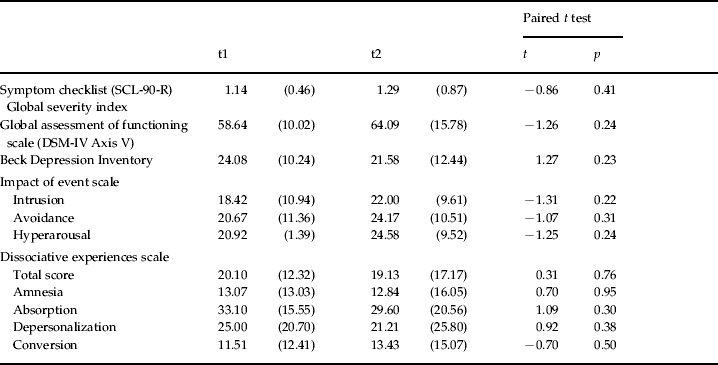
Patients showed moderate to severe general as well as specific depressive, post-traumatic, and dissociative psychopathology (Table 1). None of these assessments indicated significant changes from t1 to t2. The auditory verbal learning test (Rey, Reference Rey1964) revealed no deficits of declarative memory in the BPD subjects [mean total score 59.3 (s.d.=9.7)].
Psychopathology at t1 and t2

t1, Baseline; t2, 1 year after baseline; DSM, Diagnostic and Statistical Manual of Mental Disorders.
Values are given as mean (standard deviation).
On average, subjects had already been psychiatric in-patients 3.0 (s.d.=2.3) times prior to the index admission (t1). During the index period, all patients were treated by dialectic behavioural therapy. Between t1 and t2, 69.2% (n=9) underwent out-patient cognitive-behavioural or psychodynamic psychotherapy with one session every second week to three sessions per week in one case. In all cases psychotherapy focused on stabilizing patients, while trauma expositions were not applied in any case. In addition, 46% (n=6) of the subjects were readmitted to hospital for crisis intervention during the observation period. During index admission (t1) some patients received psychotropic medication due to depressive mood (selective serotonin reuptake inhibitors, n=4; tricyclics, n=2) or affective instability (neuroleptics, n=3; valproate acid, n=1; benzodiazepine, n=1).
Affective states and sensory qualities during recall
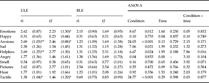
During ULE recall patients reported significantly higher levels of general emotions (p=0.01), anxiety (p<0.001), helplessness (p=0.02) and sadness (p=0.02) as well as more tactile experiences (p<0.001, Table 2) compared with RLE recall conditions. However, we did not observe any effects of time. Apart from helplessness (p<0.02) there were not any interaction effects (condition×time). Separate post-hoc analyses for t1 and t2 roughly yielded comparable results (Table 2).
Intensity of emotional and sensoric qualitiesFootnote a during recall of ULE and RLE under fMRI conditions

ULE, Unresolved negative life events; RLE, resolved negative life events; fMRI, functional magnetic resonance imaging; ANOVA, analysis of variance; t1, baseline; t2, 1 year after baseline.
Values are given as mean (standard deviation).
a 0=not at all, 1=low, 2=moderate, 3=intense, 4=extreme.
* p<0.05, ** p<0.01 (post-hoc paired t test comparing ULE versus RLE condition at t1 and t2, separately).
fMRI activation during recall of ULE and RLE at t1 and t2
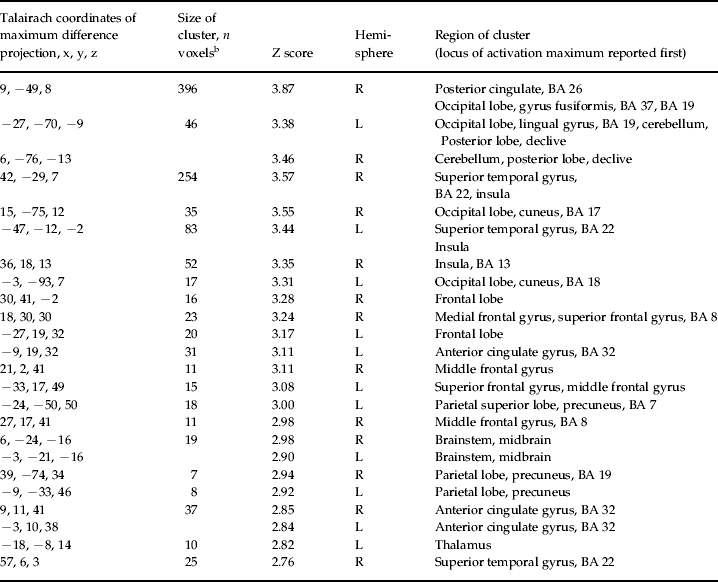
In the 2×2 factorial design, i.e. when contrasting t1 versus t2 (Δt1/t2×ΔULE/RLE, second-level analysis, p uncorrected=0.003), we found a bilateral right more than left cingulate and fronto-temporal activation pattern including extended activation of the right posterior cingulate cortex [PCC; Brodman area (BA) 31] and a minor cortical activation of ACC (BAs 24, 32) on both sides. In addition, we observed an extended right more than left activation of the superior temporal gyrus (BA 22) and a minor activation of the insula right more than left (Table 3, Fig. 1). Minor activation of the left superior and middle frontal gyrus, of the right medial frontal gyrus, and of the posterior lobe of the cerebellum on both hemispheres was also observed.
Increased activity of brain areas associated with the recall of unresolved compared with resolved negative life events at t1 compared with t2 (1 year later) in patients with borderline personality disorder (second-level analysis, p uncorrected=0.003). In this glassbrain presentation the right hemisphere is given on the right half (coronal projection) and on the lower half of the picture (transversal projection). SPM, Statistical Parametric Mapping.

Talairach coordinates of the maximum difference projection, cluster sizes and localization of the contrast Δt1/t2×Δ unresolved/resolved recall condition in BPD patientsFootnote a

BPD, Borderline personality disorder; BA, Brodmann area; L, left; R, right.
a Second-level analysis, p uncorrected=0.003, t=3.00.
b Clusters>five voxels are reported.
The opposite contrast, i.e. when contrasting t2 versus t1 (Δt2/t1×ΔULE/RLE), did not yield any significant activation.
Discussion
In this first 1-year fMRI follow-up study in patients with BPD we compared BOLD signal changes during the recall of highly adverse and subjectively ULE relative to RLE at two times. Contrasting t1 versus t2 we found a greater activation in the right more than the left PCC and ACC, the superior temporal gyrus (STG) and insula, left superior and middle frontal, the right medial frontal gyrus, and the cerebellum.
The major decrease of activation of the dorsal PCC from t1 to t2 in this study corresponds with previous cross-sectional observations of PCC activation when comparing victims with BPD and/or PTSD with healthy non-victims and the recall of trauma or ULE with RLE or neutral life events (Bremner et al. Reference Bremner, Narayan, Staib, Southwick, McGlashan and Charney1999; Driessen et al. Reference Driessen, Beblo, Mertens, Piefke, Rullkoetter, Silva-Saavedra, Reddemann, Rau, Markowitsch, Wulff, Lange and Woermann2004; Schmahl et al. Reference Schmahl, Vermetten, Elzinga and Bremner2004; Shin et al. Reference Shin, Wright, Cannistraro, Wedig, McMullin, Martis, Macklin, Lasko, Cavanagh, Krangel, Orr, Pitman, Whalen and Rauch2005, Reference Shin, Rauch and Pitman2006; Beblo et al. Reference Beblo, Driessen, Mertens, Wingenfeld, Piefke, Rullkoetter, Silva-Saavedra, Mensebach, Reddemann, Rau, Markowitsch, Wulff, Lange, Berea, Ollech and Woermann2006a). The PCC appears as a part of a neural network processing anxiety-related stimuli and even seems to be involved in fear conditioning (Doronbekov et al. Reference Doronbekov, Tokunaga, Ikejiri, Kazui, Hatta, Masaki, Ogino, Miyoshi, Oku, Nishikawa and Takeda2005). Indicating a more general function, the PCC was found to be involved in consciousness, in the recall of highly familiar autobiographical information as well as in visuospatial processing (Sugiura et al. Reference Sugiura, Shah, Zilles and Fink2005; Vogt & Laureys, Reference Vogt and Laureys2005; Vogt et al. Reference Vogt, Vogt and Laureys2006). This latter association is also supported by the additional activation of visual occipital cortex areas in the previous studies and a decline of activation of these areas in the present study.
We observed a substantial decline of activation of the STG right more than left as well as of the right insular cortex. The STG grey matter volume was reported to be greater in children and adolescents with PTSD than in controls (De Bellis et al. Reference De Bellis, Keshavan, Shifflett, Iyengar, Beers, Hall and Moritz2002). In adults with PTSD Lanius and co-workers found a relative hyperactivation of a neural network including the STG in response to traumatic scripts only in those subjects who showed dissociative states under stimulus conditions (Lanius et al. Reference Lanius, Bluhm, Lanius and Pain2006). They concluded this network to be associated with distorted body perceptions in dissociation. Our patients who reported a relative high degree of DES dissociative symptoms reported more tactile sensations during recall of ULE versus RLE at both times of investigation. This observation might be in line with such an assumption, but we did not assess dissociation under recall conditions. The right insular cortex was repeatedly found to play a role in anxiety-related internal stimuli in PTSD (Jatzko et al. Reference Jatzko, Schmitt, Kordon and Braus2005; Lanius et al. Reference Lanius, Bluhm, Lanius and Pain2006). It was assumed to be crucial for the access of visceral responses into awareness, and by this may represent a neural correlate for the evaluation of body equivalents of emotional states (Critchley, Reference Critchley2004).
The ACC is regarded as a central part of a supervisory attentional system which is activated in novel and difficult situations, error correction, and response inhibition (Gazzaniga, Reference Gazzaniga1998; Vollm et al. Reference Vollm, Richardson, Stirling, Elliott, Dolan, Chaudhry, Del Ben, McKie, Anderson and Deakin2004). In BPD cross-sectional neuroimaging studies under resting-state conditions indicated decreased as well as increased metabolism of the ACC (De La Fuente et al. Reference De La Fuente, Goldman, Stanus, Vizuete, Morlan, Bobes and Mendlewicz1997; Juengling et al. Reference Juengling, Schmahl, Hesslinger, Ebert, Bremner, Gostomzyk, Bohus and Lieb2003). Using fMRI we observed activation of the ACC when contrasting recalls of ULE versus RLE and comparing BPD patients with healthy subjects (Beblo et al. Reference Beblo, Driessen, Mertens, Wingenfeld, Piefke, Rullkoetter, Silva-Saavedra, Mensebach, Reddemann, Rau, Markowitsch, Wulff, Lange, Berea, Ollech and Woermann2006b). This finding is in agreement with the assumed function of the ACC to control emotion via the amygdala (Bush et al. Reference Bush, Luu and Posner2000). Activation of the ACC and other parts of the medial prefrontal cortex during the recall of adverse stimuli in PTSD was repeatedly found to be negatively associated with the activation of the amygdala (Shin et al. Reference Shin, Rauch and Pitman2006). This association may reflect an attempt to get control over an amygdala-mediated highly emotional state. Noteworthy, we here found a moderately extended decrease of ACC and frontal gyri activation over a 1-year period, but not any changes of activation of the amygdala. This latter finding coincides with subjects' reports of general emotional impact and anxiety during the recall of ULE being as high at t2 as 1 year before (see also below).
In sum, we observed a substantial right more than left hemispheric decrease of activation of brain areas, which can be considered to be involved in the processing of autobiographically relevant, traumatic or at least highly adverse and anxiety-related stimuli. Although the study by Schnell & Herpertz (Reference Schnell and Herpertz2006) in BPD is hard to compare with the present study (stable clinical conditions over 12 weeks, highly structured in-patient treatment, five times of measurements, standardized arousing pictures as stimuli), it is noteworthy that activation changes are more similar as could be assumed in both studies. Schnell & Herpertz, (Reference Schnell and Herpertz2006) also found a decreased activation of the anterior cingulate, insula, superior temporal gyrus, posterior cingulate, precuneus and cuneus, while activation changes of prefrontal areas were not observed.
Subjects in this study reported a higher emotional level, more intensive feelings of anxiety, sadness, and helplessness as well as more tactile sensations during the recall of ULE compared with RLE. Noteworthy, we did not find any effects of time or (apart from one single exception) interaction effects (condition×time) – in contrast to our hypothesis and in contrast to the changes of fMRI activation patterns from t1 to t2 [comparable observations were made by Schnell & Herpertz (Reference Schnell and Herpertz2006) with regard to arousal levels]. In addition, we did not find any effects of time regarding general, depressive, PTSD and dissociative pathology. This lack of clinical improvement despite ongoing treatment was surprising but may at least in part be due to the relatively short period of observation in BPD. In a recently published 10-year follow-up study, Zanarini et al. (Reference Zanarini, Frankenburg, Hennen, Reich and Silk2006) found that only 39% of treated patients with BPD showed a remission within the first 2 years. In addition, several characteristics of our sample at t1 (e.g. mean age >25 years, extensive trauma history, co-morbid PTSD in >50%, high psychopathological levels) were found to be negative predictors of early remission (Gunderson et al. Reference Gunderson, Weinberg, Daversa, Kueppenbender, Zanarini, Shea, Skodol, Sanislow, Yen, Morey, Grilo, McGlashan, Stout and Dyck2006; Zanarini et al. Reference Zanarini, Frankenburg, Hennen, Reich and Silk2006).
Hypothetically one might also conclude that although subjects had similar subjective feelings of distress in response to their ULE memories at t2 and t1 they needed less neural activation to cope on a comparable level at t2.
To interpret our findings limitations of this study have to be considered. First, our sample size is rather small and results must be confirmed in larger samples. Second, patients showed a variety of co-morbid axes I and/or II disorders, but BPD was regarded as the main diagnosis also in these patients. Symptoms of co-morbid disorders are typical in BPD and exclusion would have led to a sampling of a non-representative patient group. However, we cannot rule out that the results reported here may also be related to these co-morbid disorders rather than to BPD per se. Third, psychotherapy during the follow-up period in some patients as well as medication may have influenced our results but cannot explain within-group differences (ULE versus RLE). Fourth, we cannot completely exclude that habituation may play a role when neural activation decreases, although subjects reported comparable emotional and sensoric levels during the recalls in both conditions. An a priori test–retest reliability analysis over a short-term interval may have clarified this issue. Fifth, we only investigated within-group but not between-group differences by introducing a control group. Thus, we cannot confirm the validity of fMRI activation changes in BPD subjects. On the other hand, the lack of significant activation differences when comparing t2 and t1 contrasts with the assumption of merely artificial findings.
Conclusion
In this 1-year follow-up study we observed a significant decrease of a temporo-frontal neural activation pattern during the recall of ULE/trauma memories but this decrease was not associated with a decrease of highly emotional experiences during recall or with psychopathological improvement. If future research confirms our findings the question arises whether changes of complex neural activation patterns precede successful subjective coping with adverse life events and clinical improvement in BPD.
Acknowledgements
We would like to thank our subjects for their readiness to engage in this study. Parts of this study were supported by the DFG grant Dr 358/5-2 (Deutsche Forschungsgemeinschaft). In addition, we also thank the Society of Epilepsy Research Bethel for providing scan time.
Declaration of Interest
None.