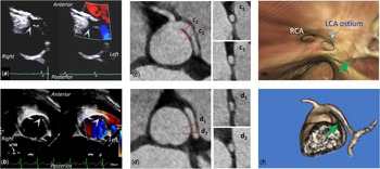
A 13-year-old, previously healthy, club-level, male athlete was referred for a cardiology evaluation of a murmur and found to have an anomalous aortic origin of a left coronary artery from the right aortic sinus with an interarterial course of the left coronary artery (Video S1, Fig 1a-b). An electrocardiography-gated CT angiography of the coronary arteries demonstrated left coronary artery origin was in close proximity to the right coronary artery ostium (Fig 1, aortic sinus 1b, level II)
Reference Krishnamurthy, Masand and Jadhav1
and interarterial course of the proximal left coronary artery (Video S2). The left coronary artery ostium appears round with no slit-like ostial stenosis on virtual angioscopy, and the proximal left coronary artery showed a round lumen on coronal oblique reconstructions (Fig 1c and 1e). The left coronary artery luminal shape then became elliptical for approximately 2 mm (Fig 1d1) before it again became round prior to its bifurcation (Fig 1d2). Based on the morphologic features, no intramural segment was reported on the CT angiography. A maximal exercise stress test including  -max was reassuring. A dobutamine stress cardiac magnetic resonance showed no inducible ischaemia or regional wall motion abnormalities.
Anomalous aortic origin of a left coronary artery from the right aortic sinus detected by echocardiography ( a , transthoracic; b , transesophageal). The ostium (arrowheads) is anterior to the intercoronary commissure (green arrow) and appears round without slit-like appearance ( c , e ). The left coronary artery appears round and robust in calibre except for a short segment of elliptical shape as the left coronary artery traverses the inter-coronary commissure ( c, d, f ).

The patient was presented at a multidisciplinary meeting of our Coronary Artery Anomalies Program, which is our usual practice. Typical high-risk morphological features of the anomalous aortic origin of the left coronary artery includes a slit-like ostium, acute-angled takeoff, intramural course, interatrial course, and high ostial location, which are commonly present together.
Reference Krishnamurthy, Masand and Jadhav1
The round shape of the left coronary artery ostium and proximal left coronary artery did not support an intramural course of the proximal left coronary artery. These features were similar to those reported in an adult who had only a brief intramural course of the left coronary artery as it traversed the inter-coronary commissure, in whom a patch ostioplasty with neo-ostium creation was performed.
Reference Giusti, Villano and Pozzi2
The patient’s risk of SCA was considered high due to interarterial course of the left coronary artery; therefore, surgery was recommended based on current practice guideline.
Reference Van Hare, Ackerman and Evangelista3
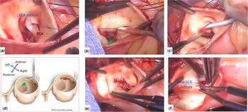
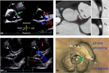
Intraoperative inspection revealed adjacent but separate right coronary artery and left coronary artery ostia with a round-appearing left coronary artery ostium from the right sinus, and a 10-mm long intramural course of the left coronary artery (Fig 2). Unroofing of the intramural left coronary artery was performed successfully which allowed the left coronary artery ostium to accept a 3-mm dilator easily with a direct takeoff from the left aortic sinus (Fig 3). The post-operative course was uneventful. A routine post-operative assessment following surgery was reassuring, including an echocardiogram (Video S3), coronary CT angiography (Video S4 & S5), exercise stress test with -max, and dobutamine stress cardiac magnetic resonance. The patient returned to competitive sport participation without any issues in the past 2.5 years.
Intraoperative inspection shows a round ostium of the left coronary artery ( a ), which is anterior to the intercoronary commissure (green arrow). A 3-mm probe was inserted into the left coronary artery and demonstrated a 10-mm intramural length (dotted line, b ), which provided guidance for unroofing procedure ( c-e ) bringing the new ostium to the left aortic sinus ( d-f ).

Post-operative echocardiographic and CT angiography images showing the new ostium (arrowheads, a–d ) of the left coronary artery from the left aortic sinus posterior to the intercoronary commissure (green arrows, a & d ) with patent flow by colour flow mapping ( a, b ).

This is an unusual case with a long intramural course of the left coronary artery despite a round ostium and round shape of the anomalous coronary artery. To our knowledge, this observation has not been reported and challenged our ability to detect intramural course of an anomalous coronary artery in the absence of elliptical luminal shape. In fact, such intramural left coronary artery cannot be ruled out even in the absence of some or all morphological characteristics typically visualised in anomalous aortic origin of the left coronary artery. It is possible that this separate entity poses a different SCA risk profile compared to the usual intramural anomalous aortic origin of the left coronary artery with slit-like ostium and abnormally elliptical shape, for which further large and longitudinal studies are needed.
Supplementary material
To view supplementary material for this article, please visit https://doi.org/10.1017/S104795112300001X
Acknowledgements
The authors appreciate the patient and his family for giving permission to use the medical and surgical information for this manuscript.
Financial support
This research received no specific grant from any funding agency, commercial, or not-for-profit sectors.
Conflicts of interest
None.