Introduction
Juvenile nasopharyngeal angiofibroma (JNA) originates around the sphenopalatine foramen area, and extends commonly in the lateral direction, through the sphenopalatine fossa and pterygomaxillary fissure, into the infratemporal fossa and cheek. During its course, it expands, and locally involves the adjoining bony walls to which it attaches firmly. It is not uncommon to find an hourglass-shaped constriction around the sphenopalatine foramina–sphenopalatine fossa–pterygomaxillary fissure region complex. Incomplete dissection of a tumour adherent to bone in this hourglass region complex is likely to result in residual disease.
On analysis of our past patient charts over many decades, we noted a high (18 per cent) recurrence rate.Reference Mishra and Mishra1 It is worth mentioning that our centre is known for having the highest incidence of JNA across the world.Reference Chew, Kerr and Groves2, Reference Mishra and Mishra3 The routine practice adopted at our facility for excision is the transpalatal approach (with modifications), except in certain advanced extensions. The majority of recurrences following transpalatal excisions occurred in cases with significant lateral extensions, further suggesting a residual disease in the pterygomaxillary fissure and infratemporal fossa.
Although endoscopic techniques (with pre-operative selective arterial embolisation) are undoubtedly superior, their availability in the Third World is limited. Hence, in order to reduce the chances of residual disease following open (transpalatal) surgery (which is practiced routinely worldwide), better instrumentation seems necessary to tackle these inaccessible areas. To date, the raspatory (Figure 1) is the only established instrument that can exert traction on the lateral extension of a JNA during a transpalatal approach. However, its engagement in the sphenopalatine fossa is not possible and the periosteal elevation of that region complex is out of reach.
Raspatory (left) and Mishra's SPF Dissector (right). Note the difference in lateral curvature and the width of the prongs.

Accordingly, a new instrument (Mishra's SPF Dissector) was designed. This novel device facilitates the removal of adhesions from the edges of the sphenopalatine foramen (and sphenopalatine fossa). It also exerts a deep traction on the most-lateral extension component. Our encouraging surgical results using this device with transpalatal excision have been reported previously.Reference Mishra, Mishra, Verma, Singh, Kumar and Tripathi4 This paper presents the evolution of the surgical design and establishes its novelty.
Materials and methods
When designing the instrument, the physical measurements of 20 adult cadaveric skulls and 10 computed tomography (CT) scans showing lateral extension of JNA were considered. These measurements included: the distances between the medial opening of the sphenopalatine foramen and the nasal septum and posterior boundary of nasopharynx; the distance between the hard palate and soft palate junction; and dimensions of the nasopharynx. The size and length of the actual dissecting end of the device was based upon the minimum size of sphenopalatine foramina, and the length of the sphenopalatine fossa–pterygomaxillary fissure–infratemporal fossa complex, as estimated on the CT scans. In addition, the angulation and length required to reach beyond the anatomical complex was calculated over several trials using malleable wire. Finally, the design of the handle and rigidity of the shaft were adapted from the already available raspatory. The resulting device was utilised for routine surgical practice from 2012 onwards wherever needed.
Using this device is quite simple, but some experience is needed for optimal results. Following a transpalatal incision, the tumour is exposed and its lateral extension through sphenopalatine foramina is palpated. The sphenopalatine fossa dissector is held firmly, like a raspatory, and introduced along this pathway laterally, towards the infratemporal fossa, and multiple circumferential movements are made around the tumour extension. This action separates tumour adhesions from surrounding bone or periosteum along this pathway. During the final stage of tumour delivery, the sphenopalatine fossa dissector is used as a lever to exert traction on the lateral most part that is already separated.
An extensive search of existing literature was conducted to identify existing patents for similar devices. Six related patents were found: (1) a raspatory for submucous conchotomy of the inferior turbinate; (2) a multifunctional periosteum raspatory; (3) a raspatory designed by Kosakovskyi and Semenov; (4) a flexible raspatory; (5) a facial bone contouring device that uses a hollowed rasp with non-plugging holes; and (6) surgical tools that facilitate the delivery of a neurostimulator into the pterygopalatine fossa.
Finally, in order to assess the efficacy of the new device, we compared the recurrence rates before and after 2012 at our institution. Specifically, we compared the previously published data for 3 years (2012 to 2015) in cases where the device was usedReference Mishra, Mishra, Verma, Singh, Kumar and Tripathi4 with our archival data of 400 cases operated on before 2012 without the device.Reference Mishra and Mishra1 It is worth mentioning again that the transpalatal approach is a routine practice at our facility and more than 90 per cent of JNA cases managed at our department undergo this technique.
Results
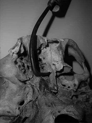
A comparison of the new device with the raspatory is shown in Figure 1. Its application around the sphenopalatine fossa is shown in Figure 2. The measurements used in the design of the device are shown in Figure 3.
Application of Mishra's SPF Dissector around the sphenopalatine foramen.

Design of Mishra's SPF Dissector. See text for details and measurements.

The new metal device consists of four parts: (1) the handle; (2) the first part of the shaft ‘X’ (5 cm length), which is straight in the axis of the handle; (3) the second part of the shaft ‘Y’ (6 cm), which is curved and angulated at a 45-degree angle ‘A’ with ‘X’; and (4) the third part ‘Z’, the main dissector shaft, which is sharply angulated at an angle of 75 degrees ‘B’ with ‘Y’. The length of part ‘Z’ is 2.5 cm; the main dissector shaft is flattened (Figure 3) to assist in periosteum elevation and dissection.
Regarding our institutional experience using this device, in a cohort of 63 JNA cases that primarily underwent transpalatal excision from 2012 to 2015, there was a recurrence in 3 cases and residual disease in 2 cases.Reference Mishra, Mishra, Verma, Singh, Kumar and Tripathi4 This is much lower than our archival recurrence rate of 17.59 per cent in cases that underwent similar transpalatal techniques without this device, from 1972 to 2014.Reference Mishra and Mishra3 Table I summarises the comparison details.
Comparative details of institutional experience using sphenopalatine fossa dissector

Our literature search of relevant patents shortlisted six related devices. These were compared with the sphenopalatine fossa dissector and the novelty of the current design was confirmed. Given the detailed specification and the substantial differences from the other devices, the instrument was patented as ‘Mishra's SPF Dissector’ (application number 201711009295, dated 17 March 2017).
Discussion
The advantage of the sphenopalatine fossa dissector lies in its direct contact with the deeper bony walls of sphenopalatine foramen and sphenopalatine fossa, where it actually peels off the tumour. In contrast, the raspatory is better suited for manipulating the tumour medial to the sphenopalatine fossa. Accordingly, the sphenopalatine fossa dissector assists in manipulating the tumour lateral to the sphenopalatine fossa. Hence, both instruments are complimentary to each other in terms of complete resection.
Moreover, even for a very lateral extension (e.g. in the infratemporal fossa; Figure 4), this instrument is more likely than other devices to exert a deep-seated traction and aid in its avulsion. The third part of the dissector can be made a bit longer to assist in cases with a more lateral extension. Alternatively, use of the dissector can be improved if the adjoining part of the hard palate is nibbled off and the sphenopalatine fossa visualised. There is the potential for injury to the maxillary artery during ‘blind’ use of this instrument, which is sometimes necessary, particularly when the hard palate is left intact. In such cases, bleeding increases momentarily, but can still be satisfactorily controlled with packing.
Coronal computed tomography scan showing a case with extreme lateral extension abutting the skull base. Such extension is only dealt with using Mishra's SPF Dissector. A longer prong may sometimes better facilitate the extreme extension.

Our design has certain limitations, especially when used in smaller children, as the design is based upon adult cadaveric dimensions. To partially overcome this, we incorporated CT parameters, but this again showed a selection bias for older adolescents. Moreover, the recurrence rates (Table I) do raise a few concerns. The majority of our 400 archival cases did not have the ‘luxury’ of endoscopic assistance (for removal of residual disease) or hypotensive anaesthesia (thereby limiting surgical time), resulting in increased recurrence.
The majority (90 per cent) of our archival cases underwent transpalatal excision (as routinely practised at our facility); however, some also underwent excision via an anterior approach or per via naturalis, making the comparison heterogeneous. Nevertheless, we found a definite advantage when this device was used as an addendum to the instruments already in use during transpalatal excision of JNA. However, it could be argued that the reduced recurrence is a result of the increased experience of surgeons, rather than the dissector itself. As the overall results reflect the work of the entire hierarchy of surgeons (from lecturers to senior professors) and the dissector was used universally (or, more correctly, it was made available for use if needed), it was not possible to segregate or present a heterogeneous comparison of surgeons using this device. However, it is quite likely that with experience of frequently using this device, the surgeon's preference for the device will increase. It is also worth mentioning that following filing of the patent, the new device was used by three surgeons at other state medical colleges, two of whom communicated their satisfaction, but only with small samples of patients. A well-planned trial should be undertaken for a more robust comparison, in order to establish the statistical advantage of this instrument in terms of residual-free resection.
In poor-resourced countries with increasing incidence of JNA, resection is primarily carried out via open surgery in which the transpalatal approach is very common. Hence, this instrument must be adapted routinely for dissecting the tumour from the sphenopalatine foramina–sphenopalatine fossa–pterygomaxillary fissure region complex.
Acknowledgements
The author would like to duly acknowledge Dr Navneet Chauhan (Department of Anatomy, King George's Medical University, Lucknow, India) for his permission to examine cadaveric skulls.
Competing interests
The instrument is patented as Mishra's SPF Dissector (application no. 201711009295, dated 17 March 2017). (The costs incurred while designing the instrument were solely contributed by the author himself.)