Introduction
Embryo cryopreservation is currently an integral part of embryo transfer of in vitro produced bovine embryos. However, the freezing and thawing processes decrease embryo viability, an effect attributed to physical and chemical damage induced during cryopreservation (Overstrom Reference Overstrom1996; Baguisi et al., Reference Baguisi, Lonergan, Overstrom and Boland2000). One cause of this damage could be the consequence of the toxic action of cryoprotectants that invade the cell during the freezing procedure.
Vitrification is a method in which not only cells but the entire solution is solidified without the crystallization of ice. In embryo cryopreservation, vitrification has advantages over slow freezing methods. For example, injuries related to ice crystallization are less likely to occur, and embryos can be cryopreserved in a shorter time period without a programmable freezer. Embryos can be cryopreserved by conventional vitrification using conventional straws or by ultra-rapid vitrification using minute tools such as electron microscopic grids, thin capillaries, minute loops or minute sticks, or as micro-drops (Kasai & Mukaida, Reference Kasai and Mukaida2004).
The minimum volume cooling (MVC) straw method is a type of ultra-rapid vitrification and a modification of the micro-drop method (Hamawaki et al., Reference Hamawaki, Kuwayama and Hamamno1999). Using MVC methods, we have reported that in vitro matured, vitrified bovine oocytes could develop normally to blastocysts (27.9%), and pregnancies followed embryo transfer (Kim et al., Reference Kim, Kim, Park, Lee, Weon, Nam, Lee, Park, Park, Yoon, Heo, Cho, Park, Chung and Lim2001). We also demonstrated that human embryonic stem cells can be successfully cryopreserved using MVC methods without losing their pluripotent characteristics (Kim et al., Reference Kim, Park, Yoon, Ghil, Lee, Lee, Tae, Kim, Lee, Chung, Park and Lim2004).
Apoptosis in embryonic cells has received increasing attention, primarily because of its potential role in the cellular response to suboptimal developmental conditions and stress (Betts & King, Reference Betts and King2001), and in eliminating defective cells by accumulating genetic damage (Metcalfe et al., Reference Metcalfe, Hunter, Bloor, Lieberman, Picton, Leese, Kimber, Brison, Ashkenazi and Dixit1999; Jurisicova et al., Reference Jurisicova, Latham, Casper, Casper and Varmuza1998). Increased incidence of cell death is an important indicator of an inadequate or suboptimal in vitro environment, and of embryo freezing damage. The TUNEL reaction is the most frequently used method to detect apoptotic cells, by labelling of extensive oligonucleosomal DNA fragmentation generated by endogenous DNase activity during the apoptotic process. However, the specificity of the TUNEL assay is relatively low, because nuclei of cells undergoing necrosis are also labelled.
Transcript analysis of apoptosis-related and/or stress-related genes is another useful method for examining the quality variation of embryos subjected to environmental changes (Gutierrez-Adan et al., Reference Gutierrez-Adan, Rizos, Fair, Moreira, Pintado, Dela, De La Fuente, Boland and Lonergan2004). Recently, examination of apoptosis (cell death) in frozen-thawed bovine oocytes and embryos using a Fluorescein-FragEL DNA fragmentation detection kit and a caspase-3 detection kit was reported (Hongshen et al., Reference Hongsheng, Monson, Parrish and Rutledge2003). However, there has been no information elucidating apoptosis-related gene expression patterns in frozen-thawed bovine blastocysts using real-time polymerase chain reaction.
In the present study we evaluated developmental competence and DNA fragmentation of frozen-thawed blastocysts using both conventional and MVC vitrification methods. We then evaluated quantitative differences in the expression of four selected genes ― caspase-3, Fas, survivin and Hsp 70 ― that are known to be related to apoptosis and stress responses in embryo development.
Materials and methods
Production of bovine in vitro matured/fertilized/cultured blastocysts
The culture procedures used to produce preimplantation embryos from follicular oocytes were as outlined by Park et al. (Reference Park, Kim, Kim, Park, Won, Yoon, Chung and Lim1998). Briefly, ovaries were obtained from a slaughterhouse, and cumulus―oocyte complexes (COCs) were aspirated from visible follicles (2–6 mm in diameter). The COCs were then washed with HEPES-buffered Tyrode's medium and cultured in maturation medium composed of TCM199 + 10% fetal bovine serum (FBS) supplemented with 0.2 mM sodium pyruvate, 1 μg/ml follicle stimulating hormone, 1 μg/ml estradiol-17β and 25 μg/ml gentamicin sulfate at 39 °C in a 5% CO2 incubator. After incubation for 22–24 h in in vitro maturation (IVM) medium, the COCs were fertilized using highly motile sperm recovered from frozen-thawed Hanwoo bull semen separated on a discontinuous Percoll column. Fertilization was assessed as the cleavage rate (≥2-cell) after 44 ± 2 h coincubation with the sperm. For in vitro culture, cleaved embryos were cultured in CR1aa medium supplemented with 3 μg/ml fatty-acid-free bovine serum albumin (BSA) and then transferred into CR1aa medium with 10% FBS added on day 4 after in vitro fertilization (IVF). Day 7 blastocysts produced in vitro after IVF were used in the study and were classified into early, expanded and early hatching stages, according to their developmental morphology, blastocoele expansion and zona thickness. We categorized the stages of blastocyst development as follows: early blastocyst, blastocoele comprises less than two-thirds of the whole embryonic cell; expanded blastocyst, blastocoele is greater than two-thirds of the whole embryonic cell with thinning of the zona pellucida evident; early hatching blastocyst, re-expanded from the shrunken state, caused by the osmotic difference between the blastocoele's fluid and culture medium entering via zona rupture.
Vitrification
Vitrification using the conventional straw method was modified slightly from the method described by Agca et al. (Reference Agca, Monson, Northey, Peschel, Schaefer and Rutledge1998). Solutions used for vitrification were prepared in Dulbecco's phosphate-buffered saline (D-PBS) supplemented with 10% FBS. Vitrification solution (40% EFS) was a mixture of 10.26% sucrose (S, Sigma), 18% Ficoll (F, Sigma) and ethylene glycol (EG, Sigma). Selected blastocysts for experiments were first exposed to 20% EG for 3 min, then exposed to 40% EFS. The blastocysts were then transferred into the vitrification solution (VS) portion of 0.25 ml French mini straws (IMV, L'Aigle, France). The open portions of the straws were sealed with powder and heat-sealed within 30–45 s after embryo loading. Straws were prepared for freezing as follows: 4 cm length was filled with 0.5 mol sucrose solution (prepared in D-PBS containing 10% FBS) followed by a 1.5 cm air bubble, 0.5 cm 40% EFS, 0.5 cm air bubble, 2 cm 40% EFS and 1.5 cm air bubble. The remaining portion of the straw was filled with 0.5 mol sucrose and then plunged into liquid nitrogen (LN2). An average of five embryos was loaded into the 2 cm VS portion of each straw.
The MVC straws were prepared by partially cutting off a 1 cm length of straw. For vitrification, blastocysts were pretreated in 10% EG for 5 min, then exposed to 30% EG for 30 s, and each blastocyst individually placed onto the inner wall of a prepared straw (IMV, L'Aigle, France) with a minimum volume (<2 μl) of VS. Then, straws were directly plunged into LN2. The mean number of blastocysts placed into one MVC straw was five.
Thawing
The conventional straws were thawed in air for 10 s and then in a 25 °C water bath for 20 s. The content of the straws was emptied into 0.8 ml of 0.3 mol S, and then recovered blastocysts were transferred into a fresh 0.8 ml of 0.3 mol S for 5 min. Finally, blastocysts were diluted in 0.8 ml of D-PBS for 5 min.
For thawing of the MVC straws, cryoprotectants were removed by a four-step procedure at 37 °C. MVC straws stored in LN2 were moved rapidly and placed into 1.0 mol S. Selected blastocysts were then serially transferred, 1 min for each solution, into 0.5 mol S, 0.25 mol S and 0.125 mol S. Finally, blastocysts were diluted in 0.8 ml of D-PBS for 5 min.
In vitro culture of thawed blastocysts, and assessment of survival
The frozen-thawed blastocysts were recovered, washed with CR1aa medium containing 10% FBS for 1 min, and then incubated in a co-culture drop for 48 h to determine in vitro survival. Cumulus cell drops were prepared with cumulus cells recovered from matured in vitro bovine blastocysts before IVF treatment. The post-thawing survival of embryos was assessed microscopically every 24 h. Blastocysts were morphologically judged as survivors if they were re-expanded within the first 24 h of culture, and were hatching or hatched out totally within 48 h of culture.
Terminal deoxynucleotidyl transferase-mediated dUTP nick-end labelling (TUNEL) assay
Blastocysts were washed three times in PBS (pH 7.4) containing polyvinylpyrrolidone (PVP, 1 mg/ml). This was followed by fixation in 3.7% paraformaldehyde in PBS for 1 h at room temperature (RT). After fixation, the embryos were washed in PBS/PVP and permeabilized by incubation in 0.5% Triton X-100 for 1 h at RT. The embryos were then washed twice in PBS/PVP and incubated with fluorescein-conjugated dUTP and the terminal deoxynucleotidyl transferase enzyme (Roche, USA) in the dark for 1 h at 37 °C. After being counterstained with 50 μg/ml RNase A in 40 μg/ml propidium iodide (PI) for 1 h at 37 °C to label all nuclei, embryos were washed in PBS/PVP, mounted with slight coverslip compression, and examined under a fluorescence microscope (Olympus).
mRNA extraction
The mRNA samples were prepared from pools of fresh blastocysts (n = 10) and frozen-thawed blastocysts (n = 10) using conventional and MVC straw freezing methods, for real-time reverse transcriptase polymerase chain reaction (RT-PCR) analyses using magnetic beads (Dynabeads mRNA purification kit; Dynal, Oslo, Norway) following the manufacturer's instructions. Briefly, embryos were resuspended in 100 μl lysis/binding buffer (100 mm Tris-HCl with pH 7.5, 500 mm LiCl, 10 mm EDTA with pH 8.0, 1% LiDS, 5 mM DTT), and vortexed at room temperature for 5 min to facilitate the lysis of the embryo and release of RNA. Fifty microlitres of oligo(dT)25 magnetic bead suspension was added to the samples, and they were incubated at room temperature for 5 min. The hybridized mRNA and oligo(dT) magnetic beads were washed twice with washing buffer A (10 mM Tris-HCl with pH 7.5, 0.15 M LiCl, 1 mm EDTA, 1% LiDS), and once with washing buffer B (10 mM Tris-HCl with pH 7.5, 0.15 M LiCl, 1 mm EDTA), respectively. Finally, mRNA samples were eluted in 15 μl double-distilled DEPC-treated water.
Real-time RT-PCR quantification
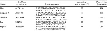
Single embryos were washed in Ca2+- and Mg2+-free PBS, snap-frozen in liquid nitrogen, and stored at 70 °C before analysis. The mRNA was extracted by using a Dynabeads mRNA Direct Kit, according to the manufacturer's instructions. In all experiments, histone H2a mRNA was used as an internal standard. First, standard cDNA synthesis was achieved by RT of the RNA by using the oligo (dT)12–18 primer and the Superscript reverse transcriptase enzyme (Invitrogen). The threshold cycle (Ct) value represents the cycle number at which sample fluorescence rises statistically above background. The reactions were conducted according to the protocol of the DyNAmo SYBR green qPCR kit, which contains modified Tbr DNA polymerase, SYBR Green, optimized PCR buffer, 5 mM MgCl, and a dNTP mix that includes dUTP (Finnzyme Oy, Espoo, Finland). The primer sequence, their approximate size and the GenBank numbers are listed in Table 2. The PCR protocol involved a denaturation programme (95 °C for 10 min) followed by an amplification and quantification programme repeated 40 times (94 °C for 10 s, 55 °C for 30 s, 72 °C for 30 s with a single fluorescence measurement), a melting curve programme (65–95 °C, with a heating rate of 0.28 °C/s and continuous fluorescence measurement) and, finally, a cooling step to 48 °C. Fluorescence data were acquired after the extension step during PCR reactions that contained SYBR Green. Thereafter, the PCR products were analysed by generating a melting curve. The melting curve of a product is sequence-specific, and can be used to distinguish between non-specific and specific PCR products. To use the mathematical model, it is necessary to determine the crossing points (CPs) for each transcript. The CP is defined as the point at which the fluorescence rises appreciably above the background fluorescence. The relative quantification of gene expression was analysed by the 2-ddCt method (Livak & Schmittgen, Reference Livak and Schmittgen2001). The sizes of the RT-PCR products were confirmed by gel electrophoresis on a standard 2% agarose gel stained with ethidium bromide and visualized by exposure to ultraviolet light.
Statistical analysis
The scores for in vitro survival and developmental rates of thawed blastocysts, apoptotic index, and the relative abundance of gene expression were subjected to analyses of variance using the general linear model (PROC-GLM) in the SAS software program (Anon., 1992). Where significant differences in the main effects were detected in each experimental parameter, treatment effects were compared by the least squares method. Differences of p < 0.05 were considered significant.
Results
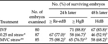
The in vitro survival of vitrified embryos, frozen according to the two different vitrification methods, was compared at 24 h post-thawing. The re-expansion rates of the C and MVC vitrified groups (77.0% and 88.2%, respectively) were significantly different (Table 1). The in vitro hatching rate of the C vitrified group (66.7%) was significantly lower than those of the non-frozen control and MVC groups (88.8% and 77.1%, respectively) at 48 h after thawing. Furthermore, the hatching rate of the non-frozen group (83.8%) was significantly higher than those of both the C and MVC vitrified groups (52.9% and 68.6%, respectively) (p < 0.05).
In vitro development of vitrified-thawed bovine embryos

Re-edB, expanded blastocyst; HgB, hatching blastocyst; Hd, hatched blastocyst.
* Conventional vitrification method.
** Modified vitrification method.
a-bp < 0.05.
Primer sequences and cycling conditions used in RT-PCR

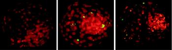
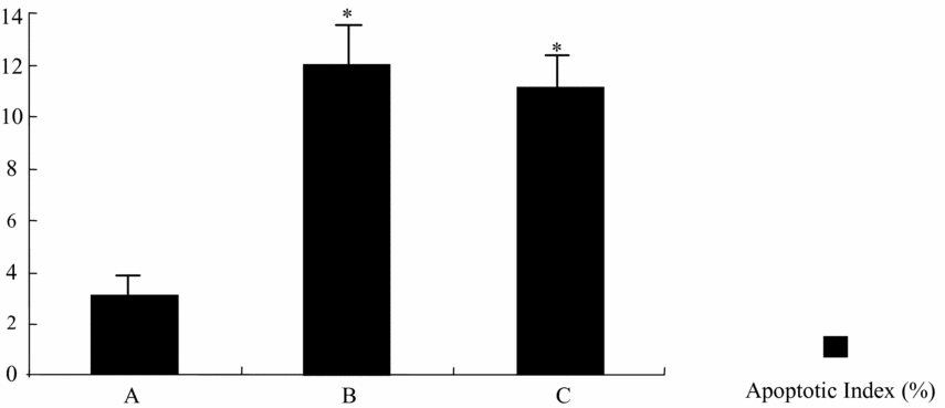
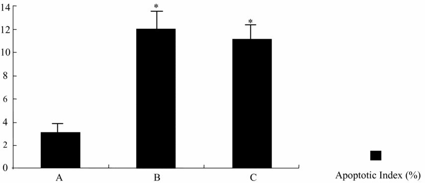
A total of 18 bovine blastocysts, non-frozen or frozen by one of the two vitrification methods, were semi-quantitatively analysed for the presence of DNA fragmentation using the TUNEL assay. Although dead cells were detected in almost all blastocysts, the number varied according to whether they had been frozen or not. Approximate values of the apoptotic index per blastocyst were measured using laser scanning confocal microscopic images (Fig. 1). The two vitrification methods resulted in blastocysts with a significantly higher apoptotic index (11.9% and 11.0%) than that of the non-frozen group (3.34%), regardless of the freezing method (Fig. 2). These results clearly indicate an increase in the apoptotic index of blastocysts that have been frozen-thawed.
Laser scanning confocal microscopic images of total nuclei and fragmented DNA in frozen-thawed bovine blastocysts (×400). (A) Non-frozen group. (B) Conventional vitrification group. (C) MVC vitrification group.

Apoptotic index in frozen-thawed bovine blastocysts (n = 18). (A) Non-frozen group. (B) Conventional vitrification group. (C) MVC vitrification group. Bars with different letters differ from each other statistically (p < 0.05).
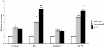
To investigate whether freezing and thawing affects the mRNA expression of apoptosis-related genes in bovine blastocysts, mRNA was prepared from single blastocysts that were either non-frozen or frozen-thawed, and each preparation was subjected to real-time RT-PCR analyses for survivin, Fas, caspase-3 and Hsp 70 gene transcripts. The relative abundance of survivin, Fas, caspase-3 and Hsp 70 gene expression increased in the frozen-thawed group in comparison with the fresh group (p < 0.01, Fig. 3). Also, the relative abundance of survivin and caspase-3 expression did not differ significantly between the two vitrification methods. However, the relative abundance of Fas and HSP expression significantly increased in the MVC group in comparison with the conventional vitrification group (p < 0.01, Fig. 3). These results suggest an increased expression of apoptosis-related genes when embryos are frozen-thawed, and this increase in the apoptotic index could be identified using the TUNEL assay.
Differential gene expressions in frozen-thawed bovine blastocysts. Bars with different letters differ from each other statistically (p < 0.05).

Discussion
In this study two vitrification methods, using either a conventional straw or minimum volume cooling (MVC; Hamawaki et al., Reference Hamawaki, Kuwayama and Hamamno1999) straw, were tested for their level of chilling damage to bovine blastocysts. The MVC method is a type of ultra-rapid vitrification, and was modified from minute tools protocols, such as the micro-drop technique. Kim et al. (Reference Kim, Kim, Park, Lee, Weon, Nam, Lee, Park, Park, Yoon, Heo, Cho, Park, Chung and Lim2001) reported that in vitro matured bovine oocytes vitrified by the MVC method could develop normally to blastocysts. In the present study, in vitro cultured bovine blastocysts vitrified by the MVC method showed higher survival rates compared with those vitrified by the conventional method.
The most frequently used methods for apoptosis detection in preimplantation embryos are based on the morphological observation of typical features of apoptosis, including condensation or fragmentation of nuclei (electron microscopy and fluorescence morphological staining), or on determination of the presence of specific DNA degradation products (TUNEL assay). It has been reported that the percentages of embryos containing at least one apoptotic cell are as high as 91–100% in bovine in vivo and in vitro produced blastocysts (Makarevich & Markkula, Reference Makarevich and Markkula2002; Matwee et al., Reference Matwee, Betts and King2000; Gjørret et al., Reference Gjørret, Knijn, Dieleman, Avery, Larsson and Maddox-Hyttel2003; Byrne et al., Reference Byrne, Southgate, Brison and Leese1999). In the present study, both vitrification methods showed a significantly higher apoptotic index (conventional group 11.9%, MVC group 11.0%) than the non-frozen group (3.0%). The apoptotic index (3%) of the non-frozen group, in this study, was lower than previously reported (4–9% in Makarevich & Markkula, Reference Makarevich and Markkula2002; Gjørret et al., Reference Gjørret, Knijn, Dieleman, Avery, Larsson and Maddox-Hyttel2003; Byrne et al., Reference Byrne, Southgate, Brison and Leese1999).
In our study we selected four apoptosis-related genes: caspase-3, Fas, survivin and Hsp 70. Caspase, a family of cysteine proteases, is important in mediating apoptosis and can be divided into two subgroups: ‘initiator’ and ‘effector’. The activation of caspase-3 that belongs to the caspase effector is the last step of the apoptotic cascade, leading to the cleavage of cellular substrates important for cell survival (Cryns & Yuan, Reference Cryns and Yuan1999). Caspase activity is regulated indirectly by the balance of different anti- and pro-apoptotic genes (Cohen, Reference Cohen1997; Cryns & Yuan, Reference Cryns and Yuan1999; Salveson & Dixit, Reference Salveson and Dixit1997).
Fas (48 kDa) is an integral cell membrane glycoprotein, similar to tumour necrosis factor-α receptor (TNFα-R). Apoptosis is induced in Fas-bearing cells when a Fas ligand binds to Fas. The expression of Fas and Fas L proteins in human gametes and embryos has been examined (Zaninovic et al., Reference Zaninovic, Rimarachin and Veeck1997; Kelkar et al., Reference Kelkar, Dharma and Nandedkar2003), and Kawamura et al. (Reference Kawamura, Fukuda, Kodama, Kumagai, Kumagai and Tanaka2001) reported that fragmented human embryos also expressed both Fas mRNAs, and suggested that the Fas–Fas L system may be involved in apoptosis regulation in early rat and human embryos. In this report, we observed that the transcription levels of both caspase-3 and Fas were lower in the vitrified blastocysts than in the non-frozen controls. No difference was observed between the two vitrification methods.
Survivin is a 16.5 kDa inhibitor of apoptotic protein (IAP) containing a single BIR (baculoviral IAP repeat) domain and an extended carboxyl-terminal helix (Ambrosini et al., Reference Ambrosini, Adida and Altieri1997). Survivin is known as a bifunctional protein that suppresses apoptosis and regulates cell division. Survivin acts as an inhibitor of apoptosis by directly binding caspases, although which caspases it can bind is still a controversial issue. Survivin has been shown to directly inhibit caspase-3 activity (Conway et al., Reference Conway, Pollefeyt, Cornelissen, DeBaere, Steiner-Mosonyi, Ong, Baens, Collen and Schuh2000; Shin et al., Reference Shin, Sung, Cho, Kim, Ha, Hwang, Chung, Jung and Oh2001), and conflicting data have been reported (Bnaks et al., Reference Bnaks, Plescia, Altieri, Chen, Rosenberg, Zhang and Ng2000). In this study, survivin expression increased with FAS and caspase-3 expression in the frozen-thawed groups. This increase in survivin mRNA expression may be expected because survivin acts along with FAS, and caspase-3, through the same pathways. This finding supported previous results that survivin enhances Fas ligand expression via up-regulation of specificity protein 1-mediated gene transcription in colon cancer cells (Asanuma et al., Reference Asanuma, Tsuji, Endoh, Yagihashi and Watanabe2004).
Heat shock factor (HSF)-1 is a protein responsible for the induction of expression of the heat shock protein (HSP) gene under both physiological and cell stress conditions. The HSP genes have numerous roles in cell function, including modulation of protein activity, regulation of protein degradation and transport across organelles, in addition to ensuring correct protein folding (Takayama et al., Reference Takayama, Reed and Homma2003). More recently, HSPs were implicated in the regulation of cell death, and Hsp 70 had been shown to prevent both caspase-dependent and caspase-independent cell death (Ravagnan et al., Reference Ravagnan, Gurbuxani, Susin, Maisse, Daugas, Zamzami, Mak, Jaattela, Penninger, Garrido and Kroemer2001). In this study, the expression level of Hsp 70 was increased in the groups frozen-thawed by both conventional and MVC methods.
In conclusion, our data indicate that the in vitro survival rate of vitrified-thawed embryos was higher in those cryopreserved by the MVC method than by the conventional method. However, both vitrification methods resulted in blastocysts with a significantly higher apoptotic index than that found in fresh blastocysts (non-frozen controls). The relative abundance of survivin, Fas, caspase-3 and Hsp 70 expression increased in frozen-thawed embryos in comparison with the non-frozen controls. These increased transcripts are indicative of a stress response and of poor quality of the preimplantation embryo. These results imply that cryopreservation caused damage that resulted in an increase in apoptosis- and injury-related gene transcription.
Acknowledgement
This work was sponsored by the Health 21 R&D Project, Ministry of Health & Welfare, Grant no. 01-PJ10-PG8-01EC01-0010, and the Korea Science and Engineering Foundation and National Research Laboratory Program of the Ministry of Science and Technology (to N.H.K.).