Congenital tracheal stenosis is a rare, life-threatening cause of acute upper airway obstruction, which may present with inspiratory stridor, respiratory distress, or difficult intubation.Reference Anton-Pacheco, Cano and Comas 1 It has been noted to occur slightly more frequently in children with trisomy 21 and may be associated with an increased incidence of cardiac anomalies, which often confers a poorer prognosis.Reference Wells, Landing, Shamszadeh, Thompson, Bove and Caron 2 – Reference Shapiro, Huang, Sangwan, Willner and Laks 3 Many surgical interventions have been reported for congenital tracheal stenosis, with slide tracheoplasty now being the preferred method for repair.Reference Manning, Rutter and Border 4 – Reference Anton-Pacheco, Comas and Luna 5
Case Report
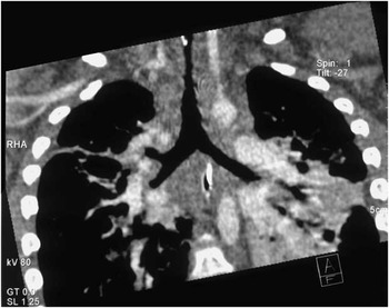
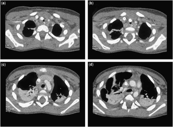
A 2-year-old boy with Down syndrome first presented to another institution with a history of stridor, fever, and coryzal symptoms. His symptoms were unresponsive to nebulised budesonide and adrenaline. He developed severe respiratory distress and carbon dioxide retention and required intubation and mechanical ventilation. His condition continued to deteriorate, and on day 2 of admission he was transferred to our unit and placed on venoarterial extracorporeal membrane oxygenation, ECMO. Direct laryngoscopy, bronchoscopy, and computed tomography showed a circumferential long segment tracheal stenosis in the mid trachea measuring 3 centimetres in length, with 1
 ${\scale50%{\vskip-6pt1}{\hskip-2.1pt\vskip-4.1pt\rot162\scale70%/}{\scale50%{\hskip-4.1pt\vskip0pt2}$
centimetres of normal proximal trachea and 2
 ${\scale50%{\vskip-6pt1}{\hskip-2.1pt\vskip-4.1pt\rot162\scale70%/}{\scale50%{\hskip-4.1pt\vskip0pt2}$
centimetres of normal distal trachea (Figs 1 and 2). The trachea was critically stenotic measuring 3 millimetres in diameter.
Computed tomogram image in coronal view demonstrating long segment critical tracheal stenosis.

Computed tomogram image in axial views demonstrating severe degree of tracheal stenosis with ECMO cannula in the superior vena cava and carotid artery. Note the size of the trachea relative to the nasogastric tube.

A median sternotomy and pericardiotomy was performed and the child was placed on cardiopulmonary bypass. Heparin was administered. The right atrium, inferior vena cava, and aorta were cannulated. He was decannulated from extracorporeal membrane oxygenation, and the right internal jugular vein and right carotid artery were repaired. A right atriotomy was performed and the left atrium was vented. The zone of apposition of the left atrioventricular valve was closed with 5/0 surgipro, and the primum atrial septal defect was closed with fresh autologous pericardium. The secundum atrial septal defect was closed primarily. The right atrium was closed in two layers; the heart was deaired and reperfused.
The trachea was dissected from the hyoid to the carina. A hyoid release procedure was performed by dividing the strap muscles superiorly, and the inferior pulmonary ligaments were divided to allow a tension-free anastomosis in the trachea. Tracheal rings were excised two centimetres and a slide tracheoplasty was performed with extended end-to-end anastomosis, with 6/0 Polydioxanone suture continuously posteriorly and interrupted anteriorly. The endotracheal tube was advanced beyond the anastomosis and Liga clips were placed as markers on either side of the anastomosis. He weaned easily off cardiopulmonary bypass; haemostasis was achieved and the surgical wounds were closed. A decision was made to excise 2 centimetres of trachea as well as to perform a slide tracheoplasty, as it was felt that this would optimise the final tracheal diameter. A standard technique of a posterior–superior incision and an inferior–anterior incision in the trachea was used to complete the slide tracheoplasty.
The post-operative course was complicated by an anterolateral myocardial infarction, which became apparent 24 hours after surgery with elevated cardiac enzymes, acute decrease in blood pressure, and ST segment changes on electrocardiogram. Transthoracic echocardiography demonstrated a right atrial thrombus. At 36 hours post-surgery, it became apparent that he had sustained a diffuse left-sided cerebral infarction causing right-sided hemiplegia. The myocardial and cerebral infarctions were presumed to be secondary to embolic events related to the right atrial thrombus, which developed during extracorporeal membrane oxygenation preoperatively, despite adequate anticoagulation. He was extubated 7 days post-operatively. He was transferred from the intensive care unit 2 weeks after surgery. He subsequently developed a clinically silent aspiration syndrome and required a percutaneous endoscopic gastrostomy tube insertion. His post-operative medications included diuretics and warfarin therapy. He was discharged home 2 months later. His right-sided hemiplegia improved with physiotherapy; his gastrostomy tube was removed at 3
 ${\scale50%{\vskip-6pt1}{\hskip-2.1pt\vskip-4.1pt\rot162\scale70%/}{\scale50%{\hskip-4.1pt\vskip0pt2}$
years of age and he is now doing well. He has no residual respiratory symptoms, and follow-up bronchoscopy showed no evidence of recurrent tracheal obstruction 6 months after surgery.
Discussion
Congenital long segment tracheal stenosis is a rare surgical emergency with significant morbidity and mortality. With the exception of neonates, children with coexisting cardiac anomalies have the worst prognosis and are at a high risk for significant morbidity and mortality.Reference Okamoto, Nishijima and Maruo 6 These patients require cardiopulmonary bypass, long periods of intensive care, artificial ventilation, and are susceptible to cardiac failure and other complications post-operatively.Reference Chiu and Kim 7 The optimum surgical management has evolved with slide tracheoplasty, showing the most promising results compared with patch tracheoplasty or end-to-end anastomosis.Reference Yong, d’Udekem, Robertson, Butt, Brizard and Konstantinov 8 However, slide tracheoplasty was previously used as a rescue therapy after failed patch tracheoplasty, but it is now the primary procedure of choice.Reference Anton-Pacheco, Comas and Luna 5 , Reference Yong, d’Udekem, Robertson, Butt, Brizard and Konstantinov 8 , Reference Elliott, Speggiorin and Vida 9
The simultaneous repair of cardiac anomalies has shown good results in a retrospective review of 27 patients who underwent simultaneous repair of congenital tracheal stenosis and cardiac anomalies.Reference Okamoto, Nishijima and Maruo 6 The majority of these simultaneous repairs involved simple intracardiac lesions such as secundum atrial septal defect, ventricular septal defect, and pulmonary artery stenosis; however, there were four deaths in children with longer durations of cardiopulmonary bypass and more complex cardiac defects, including critical pulmonary stenosis, tetralogy of Fallot, and cor triatriatum.Reference Okamoto, Nishijima and Maruo 6
Extracorporeal membrane oxygenation has been reported as a rescue therapy in a patient with tracheal dehiscence following slide tracheoplasty.Reference Raake, Johnson and Seger 10 This is the first reported case of a simultaneous slide tracheoplasty and repair of partial atrioventricular septal defect in a child with trisomy 21 stabilised with ECMO support before surgical intervention. The procedure was successful and to date no tracheal restenosis has occurred, which has been reported in up to 20% of patients. Patients may require reintervention including resection of a stenotic segment, balloon dilatation, or placement of a biodegradable stent.Reference Anton-Pacheco, Comas and Luna 5 However, satisfactory appearance of the trachea following slide tracheoplasty over the first year usually signifies good growth and the trachea remains stable over time.
This report highlights the role of extracorporeal membrane oxygenation in stabilising a child before successful slide tracheoplasty and repair of a partial atrioventricular septal defect. The lack of its availability in previous decades would invariably have resulted in a poor outcome.
Acknowledgement
We are grateful to Mr John Russell, Department of Ear, Nose, and Throat surgery for his assistance in the care of this patient. We are grateful to Mr Andrew Pendred for his assistance in preparation of the figures.
Conflicts of Interest
None.
Financial Support
This research received no specific grant from any funding agency, commercial or not-for-profit sectors.