

Pathogens on high-touch surfaces, on healthcare workers’ hands, and in the air can cause healthcare-associated infections (HAIs) directly and indirectly.Reference Otter, Yezli and French1,Reference Dancer2 Therefore, surface disinfection is an important element in reducing pathogen transmission to susceptible patients in hospitals.Reference Otter, Yezli, Perl, Barbut and French3,Reference Gebel, Exner and French4 The effectiveness of no-touch disinfection technology, such as ultraviolet (UV) light, in environmental disinfection and HAI reduction has been demonstrated in previous studies.Reference Stibich, Stachowiak and Tanner5–Reference Anderson, Moehring and Weber16
Xenon-based UV bulbs (Xe-UV) and mercury-based UV bulbs (Hg-UV) are 2 commonly used UV sources for most of the portable UV devices currently available. The Xe-based UV bulb produces broad-spectrum UV (200–320 nm) radiation in short pulses, killing organisms and spores.Reference Stibich, Stachowiak and Tanner5 The Hg-based UV bulb produces a germicidal range of UV (wavelength, 254 nm) light continuously.Reference Anderson, Gergen and Smathers17 Both forms of UV have previously demonstrated a 1–4 log reduction of various microbial organisms.Reference Nerandzic, Cadnum, Pultz and Donskey8,Reference Nerandzic, Thota and Sankar18
Because UV radiation is currently routinely used in disinfecting the hospital environment, examining the emergence of UV-resistant bacteria in the clinical environment is essential. Therefore, we sought to determine whether UV resistance develops after serial exposure to 2 widely used UV sources mimicking real-world irradiation dosing and time. To emulate the actual clinical environment, we performed 25 serial exposures to Xe-UV and Hg-UV sources at common doses and distances. We tested the effects on 3 common HAI-causing strains with multidrug resistance: methicillin-resistant Staphylococcus aureus (MRSA), carbapenemase-producing Klebsiella pneumoniae (KPC), and metallo-β-lactamase–producing Klebsiella pneumoniae (MBL).
Materials and methods
UV light source
This study was conducted in the Infectious Diseases Research Laboratory of the Central Texas Veterans’ Health Care System. Two separate UV sources were tested: a Hg-UV lamp (Puritech HNS 30W G13, G30T8, Osram Licht AG, Munich, Germany) used inside a laminar flow biosafety hood and a portable pulsed-xenon–based UV decontamination device (PPX-UV, Xenex Disinfection Services, San Antonio, TX).
Bacterial strains
We obtained 3 multidrug-resistant strains from the American Type Culture Collection (ATCC, Manassas, VA): methicillin-resistant Staphylococcus aureus (MRSA: ATCC 43300), carbapenemase-producing Klebsiella pneumoniae (KPC: ATCC 1705), and metallo-β-lactamase–producing Klebsiella pneumoniae (MBL: ATCC 2146).
UV exposure
Distance and time were optimized to mimic real-world dosages that the multidrug-resistant organisms (MDRO)s would receive in a healthcare setting. Other than a different UV light source, the experimental procedure and conditions were identical for both tests. The test strains were grown on blood agar plates (Remel, San Diego, CA) for MRSA and on Mueller-Hinton (MH) agar plates (Remel) for KPC and MBL for 18–24 hours in a 35°C non-CO2 incubator. Cells were harvested and suspended in sterile saline (B. Braun Medical, Irvine, CA), and 0.1 mL of the suspension (McFarland 0.5) was spread onto blood agar or Mueller-Hinton plates for UV exposure. The plates were then placed inside a laminar flow hood and exposed to Hg-UV at ˜90 cm (35 inches) for 10 minutes (93.6 mW/cm2). For Xe-UV exposure, the plates were placed perpendicular to the PX-UV lamps of the device and at distance of 122 cm (48 inches) for 5 minutes (38.1 mW/cm2). We avoided the edges of the plates because the UV exposure was not optimal around the edges of the plate. After the exposure, all plates were incubated at 35°C in a non-CO2 incubator for 18–24 hours in the dark. Saline sterility was tested by incubating 1.5 mL saline in trypticase soy broth (BD Biosciences, San Jose, NJ) for 18–24 hours at 35°C non-CO2 incubator. The initial number of colony-forming units (CFUs) in the bacterial suspension were measured by plating the suspension with serial dilution. After the incubation, CFUs of surviving organisms were measured and recorded. The log-kill rate was obtained by comparing the initial CFUs and the surviving CFUs for each cycle. After each cycle, MRSA was tested for coagulase, and KPC and MBL were tested for carbapenem resistance by plating onto MacConkey agar (Remel) and placing imipenem and meropenem disks (ROSCO diagnostica, Taastrup, Denmark) on the streak area. After each exposure, all of the surviving colonies were harvested and combined into a single suspension (McFarland 0.5) for the next cycle of UV irradiation. The sequence of survivor isolation, inoculation, UV exposure, and incubation was repeated 25 times.
Whole-genome sequence analysis
Bacterial strains from irradiation cycles 10, 20, and 25 and parental ATCC strains were subjected to whole-genome sequencing (WGS). From bacterial stock pooled after each irradiation cycle, 3 CFUs were randomly selected for sequencing. The DNA was extracted using the QIAamp DNA Micro Kit (Hilden, Germany). DNA libraries were prepared using the Nextera DNA Flex Library Prep Kit (Illumina, San Diego, CA). The whole-genome libraries were sequenced using next-generation sequencing on the Illumina NextSeq platform (Illumina). Whole-genome single nucleotide polymorphism (wgSNP) analysis was conducted using BioNumerics version 7.6 platform (Applied Maths NV, Sint-Martens-Latem, Belgium). Sequence variation was evaluated by comparing the sequences to the reference genome sequences (Staphylococcus aureus subsp aureus NCTC 8325, GenBank accession NC_007795.1 and Klebsiella pneumoniae subsp pneumoniae HS11286, GenBank accession NC_016845.1). De novo assembly was completed using the SPAdes version 3.7.1 assembler on the Bionumerics calculation engine (Applied Maths NV). Whole-genome multilocus sequence typing (wgMLST; assembly-free and assembly-based calls) was performed using the calculation engine on the Bionumerics version 7.6 platform. Any nucleotide changes of potential UV resistance genes of parental ATCC strains were analyzed by local alignment using the BLAST 2 sequences tool. After irradiation cycles 10, 20, and 25, WGS results were searched for nucleotide sequences of genes of corresponding parental strains.
Statistical analysis
Log reduction of bacteria by UV was calculated for each exposure cycle (24 for MRSA and 25 for KPC and MBL) for each organism using inoculum and surviving CFU count. Ordinary least-squares regression was used to assess a linear trend in each organism’s UV resistance to the Xe-UV or Hg-UV source over time. A type I error of α = 0.05 was assumed. All analyses were performed using SAS version 9.2 software (SAS Institute, Cary, NC).
Results
Effect of Xe-UV and Hg-UV exposure on MDROs following 25 cycles of serial exposure
For each cycle, the average number of organisms used for UV irradiation throughout the 25 cycles varied from 2.5 million to 7.8 million (Table 1). The average number of surviving organisms ranged between 11 and 32 CFU after exposure to Xe-UV and between 3 and 4 CFU after Hg-UV exposure. The average log reduction over 25 repetitions ranged from 5.37 to 5.89 for Xe-UV and from 5.73 to 6.31 for Hg-UV. The highest average log reduction by Xe-UV was for MRSA (5.89; SD, 0.46) followed by MBL (5.71; SD, 0.47) and KPC (5.37; SD, 0.45). The average log reduction by Hg-UV was greatest for MBL (6.39; SD, 0.40), followed by KPC (6.31; SD, 0.36) and MRSA (5.73; SD, 0.64).
Description of Inoculum and Survivor Counts With Log Reduction Across Time for Each UV Source

Note. KPC, carbapenemase-producing Klebsiella pneumoniae; MBL, metallo-β-lactamase–producing Klebsiella pneumoniae; MRSA, methicillin-resistant Staphylococcus aureus; Xe-UV, xenon-bulb–based ultraviolet light source; Hg-UV, mercury-bulb–based ultraviolet light source.
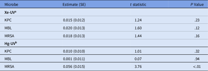
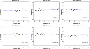
A trend in UV resistance did not appear to occur in MRSA, KPC, or MBL during the repeated exposure to Xe-UV (Table 2 and Fig. 1). Similarly, the log-kill rate did not vary considerably for KPC or MBL over time after Hg-UV exposure. However, although the initial reduction was lower, an upward trend in log-kill rate was observed for MRSA after Hg-UV exposure (P < .01). Following 18 sequentially irradiated organisms, log reduction was estimated to increase by 1. No phenotypic changes (coagulase positive for MRSA or carbapenem resistance for KPC and MBL) were observed, even with multiple UV irradiation cycles.
Ordinary Least-Squares Regression Results Assessing a Linear Trend in Microbe Resistance to Xenon- or Mercury-Bulb UV Exposure Over Time

Note. UV, ultraviolet light; SE, standard error; KPC, carbapenemase-producing Klebsiella pneumoniae; MBL, metallo-β-lactamase–producing Klebsiella pneumoniae; MRSA, methicillin-resistant Staphylococcus aureus; Xe-UV, xenon-bulb–based ultraviolet light source; Hg-UV, mercury-bulb–based ultraviolet light source.
Plot of log reduction over time of repeated UV exposure cycle for each organism exposed to each UV source and least-squares fitted regression line. Note. MRSA, methicillin-resistant Staphylococcus aureus; KPC; carbapenemase-producing Klebsiella pneumoniae; MBL, metallo-β-lactamase–producing Klebsiella pneumoniae; Xe-UV, xenon-bulb–based ultraviolet light source; Hg-UV, mercury-bulb–based ultraviolet light source.

WGS of MDROs after UV exposure
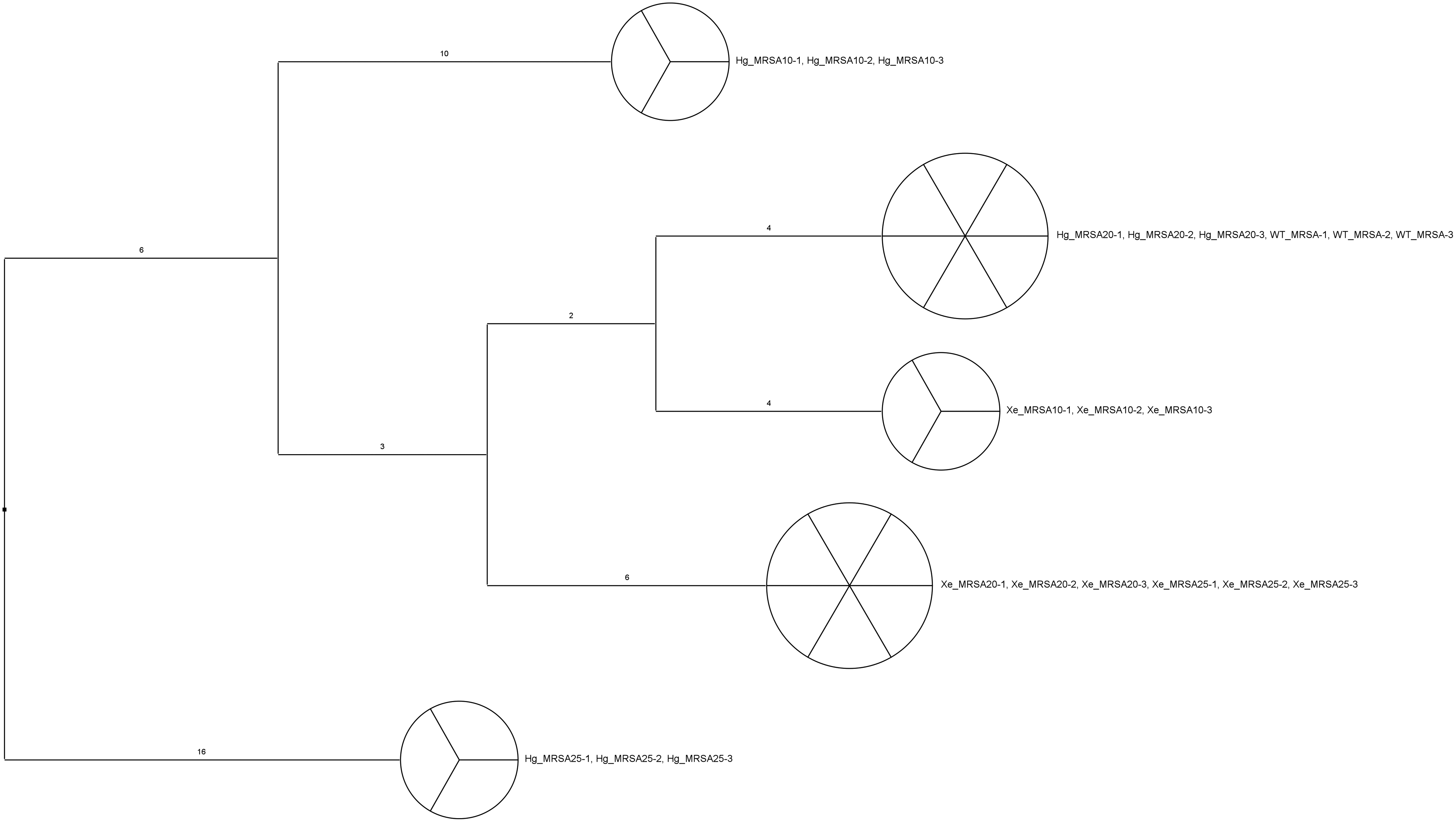
The SNP distance between MRSA CFUs after different UV irradiation cycles ranged between 4 and 6 bp for Xe-UV and between 4 and 16 bp for Hg-UV. No base changes among MRSA CFUs within each UV irradiation cycle were detected (Fig. 2). The SNP distances between MBLs CFUs of different UV irradiation cycles were <16 bp. The maximum SNP distance among different UV irradiation cycle KPCs was 33 bp, whereas the SNP distance between MBL and KPC was 4,856 bp, which was expected given their inherent genetic differences. The SNP distance between Klebsiella CFUs within same UV irradiation cycle was <4 bp (Fig. 3). The wgMLST analysis did not change the sequence type, even after multiple UV irradiation cycles for all 3 strains studied here. All MRSA CFUs were sequence type 39 (ST39). The sequence types of all MBL CFUs and KPC CFUs were ST51 and ST25, respectively. Potential UV resistance genes have been mapped in E. coli.Reference Alcantara-Diaz, Brena-Valle and Serment-Guerrero19 Homologous genes of radA, uvrA, dnaE, and polA in MBL, KPC, and MRSA have been examined. A single nucleotide change was found in MBL after irradiation cycle 25 (Fig. 4). This mutation is a silent mutation of Ile159 (ATC to ATT), and there was no significant codon usage difference between the 2 codons in K. pneumonia.Reference Nakamura, Gojobori and Ikemura20
Single-nucleotide polymorphism (SNP) differences in MRSA for wild-type isolates and isolates from cycles 10, 20, and 25.

Single-nucleotide polymorphism (SNP) differences for carbapenemase-producing Klebsiella pneumoniae (KPC) and metallo-β-lactamase–producing Klebsiella pneumoniae (MBL) for wild-type isolates and isolates from cycles 10, 20, and 25.

Nucleotides change found in polA gene of metallo-β-lactamase–producing Klebsiella pneumoniae (MBL) irradiation cycle 25.

Discussion
Ultraviolet disinfection can be used to supplement manual cleaning, and recently it has become an acceptable method of no-touch disinfection within healthcare facilities. However, the continuous and sometimes inadequate exposure of microorganisms to UV has the potential to induce bacterial evolution to become UV resistant, as has occurred in the development of antibiotic resistance due to subtherapeutic use, misuse, or overuse of antibiotics.
UV resistance can be developed either by natural or artificial UV exposure. UV-resistant bacteria have been found in extreme environments at high altitude.Reference Portero, Alonso-Reyes and Zannier24–Reference Rasuk, Ferrer, Kurth, Portero, Farias and Albarracin27 Additionally, previous studies have shown that Escherichia coli exposed to repeated UV exposure and growth cycles produced daughter strains that were more resistant to UV irradiation than the parent strains.Reference Alcantara-Diaz, Brena-Valle and Serment-Guerrero19,Reference Shibai, Takahashi and Ishizawa28,Reference Rames, Chaloupecky, Sojkova and Bencko29 Decreased efficacy of UV eradication have also been reported in S. aureus after repeated UV exposures.Reference Shoults and Ashbolt30 Although the development of UV-resistant strains of bacterial pathogens has not been reported in the hospital environment, increased and continuous exposure to UV can potentially induce bacterial resistance to UV.
This study was designed to test whether serial exposures of major HAI-causing MDROs to UV light would lead to new UV-resistant MDROs, which would cause additional challenges from an infection prevention standpoint. Moreover, 25 cycles of exposure was selected to mimic a realistic amount of UV disinfection that bacteria encounter in a hospital environment. The average patient stay in a CTVHCS hospital is ˜4 days. After each contact isolation discharge, environmental management services (EMS) staff manually clean the room and then deploy the Xe-UV device. Therefore, in a clinical setting, 25 exposures to Xe-UV, with an average of 4 days per exposure, would be equivalent to 100 days of patient care, mimicking a realistic survival time for these organisms.
As shown in Figure 1, no significant changes in log reduction occurred in correlation with the UV exposure cycle. One exception is the increase of the log reduction of MRSA when exposed to repeated Hg-UV exposure. We investigated the nature of potential DNA changes induced by multiple UV irradiation using SNP and wgMLST analyses. The SNP differences among the selected CFUs within an irradiation cycle ranged from 0 to 8 bp compared to 2 to 33 bp between different irradiation cycles. None of these SNP differences led to any meaningful sequence type changes in the wgMLST analysis (Figs. 2 and 3). The number of mutations introduced by UV exposure were within the predicted baseline mutation rate. Although there is little agreement for an appropriate SNP cutoff threshold, the number of SNPs separating any CFUs were below the standard SNP threshold used in published literature.Reference Hall, Holden and Srisomang31,Reference Miro, Rossen and Chlebowicz32 The scarcity of genetic changes observed in the potential UV resistance genes radA, uvrA, dnaE, and polA Reference Alcantara-Diaz, Brena-Valle and Serment-Guerrero19 showed that the possibility of the emergence of a UV-resistant strain by multiple UV irradiation cycles is very low.
Because UV irradiation increases mutations, UV irradiation can potentially induce UV resistance, as previously reported.Reference Alcantara-Diaz, Brena-Valle and Serment-Guerrero19,Reference Shibai, Takahashi and Ishizawa28–Reference Shoults and Ashbolt30 However, when the UV dose reaches high enough to kill the entire bacterial population, the emergence of a UV-resistant phenotype can be prevented. In the current experiment, very few colonies survived following administration of UV, likely due to random events such as cell shielding by aggregation. In the current study, higher UV doses (0.1–0.2 mW/cm2 vs 28.1–93.6 mW/cm2) were used, and higher UV log reduction (<4 log reduction vs 5.37–6.39 log reduction) was observed than in previous studies.Reference Alcantara-Diaz, Brena-Valle and Serment-Guerrero19,Reference Shibai, Takahashi and Ishizawa28–Reference Shoults and Ashbolt30 Consequently, our laboratory study indicates that UV disinfection is a safe and effective measure to employ in clinical settings without being concerned about the emergence of UV resistance. However, UV resistance might be observed at lower UV dose levels.
This study has several limitations. Although it was designed to closely resemble a real-world hospital environment with relevant doses and distances, the experiment was not performed in a clinical setting but rather in a laboratory setting using very few strains of each organisms. We did not have a mercury-based UV no-touch device but rather a mercury-bulb UV source in a laminar flow hood as a surrogate. We ensured that the dosage from the bulb mimicked a standard UV device dosage, and we believe that this factor did not alter the outcome, and in fact strengthens our argument for no resistance for either source.Reference Nakamura, Gojobori and Ikemura20
In conclusion, after 25 growth irradiation cycles from 2 separate types of UV exposure, MRSA, KPC, and MBL K. pneumoniae did not develop UV resistance. Our results indicate that there is very limited chance that HAI-causing bacteria could develop UV resistance with the proper use of UV disinfection devices. This outcome is significant because new UV disinfection devices are more frequently being utilized in healthcare facilities to supplement current disinfection protocols, especially during the current pandemic. These results should bring relief to healthcare providers and policy makers who were worried that the devices may become obsolete if organisms develop resistance to the UV devices.
Acknowledgments
The authors wish to express their gratitude to Dr. Thomas Huber for his invaluable effort and advice with this project. We thank environmental management services and infection prevention and control section of the Central Texas Veterans’ Health Care System.
Financial support
This work was supported by a merit review grant (no. IIR 12–347) from the Department Veterans Affairs to J.Z. The laboratory activity was supported by a grant from Xenex Healthcare Services. This work was also supported by the Central Texas Veterans Health Care System (Temple, TX), with additional support from Baylor Scott & White Health through the Center for Applied Health Research (Temple, TX).
Conflicts of interest
The authors declare that they have no competing interests.