Introduction
Nanotechnology-based medical techniques have been under development for decades, and molecular imaging and targeted drug delivery using nanoparticles (NPs) have made tremendous progress in theranostics. Theranostics, a portmanteau of therapeutics and diagnostics, is the combination of therapeutic and diagnostic agents within a single platform, Reference Janib, Moses and MacKay1 and theranostic NPs are expected to realize the promise of personalized medicine. NPs have unique physical, chemical, and biomedical properties due to their small size, large surface area, and pharmacokinetic characteristics. Reference Janib, Moses and MacKay1,Reference Shchukin and Sukhorukov2 Current theranostic NPs utilize a variety of targeting motifs and imaging labels, and they have been playing more significant roles in early-stage diagnosis and effective therapy.
Theranostic NPs have, in particular, been extensively studied to challenge the limitations of conventional cancer treatment. Reference Yu, Park and Jon3,Reference Ahmed, Fessi and Elaissari4 Chemotherapy, the most common type of conventional cancer treatment, often shows poor prognostic outcomes in spite of the effective anticancer activity of the drugs. This is mainly attributed to the systemic toxicity of anticancer drugs, due to the non-specific distribution of the drugs in normal tissues. Most drugs also alter the biological metabolism of normal cells, thus there is an urgent need for targeted drug delivery for reducing the toxicity of anticancer drugs in normal tissues. In this point of view, drugs formulated within NPs have been regarded as a good tumor-targeting strategy. Since the late 1980s, selective accumulation of macromolecular drugs in tumors, the so-called enhanced permeability and retention (EPR) effect, has attracted the interest of many researchers. Reference Matsumura and Maeda5 Based on size-related properties, NPs can easily penetrate the coarse structure of new blood vessels in tumors, and the poor lymphatic drainage of solid tumors leads to retention of the NPs in tumors. Reference Yhee, Son, Son, Joo, Kwon, Bae, Mrsny and Park6,Reference Yuan, Dellian, Fukumura, Leunig, Berk, Torchilin and Jain7 However, EPR effects differ in various tumors because the characteristics in tumor vascularization and molecular markers of cancer cells may depend on the type and stage of tumors. Reference Yu, Park and Jon3,Reference Bae and Park8,Reference Bae9 Therefore, diverse attempts, including surface modification of NPs, have been made to achieve more accurately targeted drug delivery.
To design smart NPs for cancer theranostics, a variety of physiochemical properties, including size, surface charge, and deformability, should be carefully considered. Theranostic NPs have been developed using various organic and inorganic materials, including lipids, synthetic and natural polymers, proteins, metals, and other carbon structures. In particular, organic materials are of major interest in the synthesis and preparation of NPs. In addition to the size-dependent properties, organic NPs provide other advantageous properties. Organic NPs are usually flexible and easy to modify, and highly stable in biological fluids. Reference Nayak and Lyon10,Reference Sundar, Kundu and Kundu11 Moreover, certain organic NPs show special characteristics, such as biodegradability and affinity to specific molecular biomarkers for cancer or other diseases. Reference Feracci, Gutierrez, Hempel, Gil, de la Fuente and Grazu12–Reference Choi, Chung, Min, Yoon, Kim, Park, Kwon and Jeong14
In this article, we review the application of diverse organic NPs. The article focuses on recent developments in organic NPs, which are grouped into four major kinds of base materials: lipids (cationic lipid), polysaccharides, peptides/proteins, and synthetic polymers. Modification of organic NPs with targeting moieties, use of contrast agents and nanoprobes, and theranostic applications are discussed in various aspects of cancer research.
Lipid-based NPs
Based on the advantages of low toxicity and easy cellular uptake, lipid-based nanocarriers play a major role in cancer therapy. Due to the EPR effect and enhanced half-life in blood, lipids and liposomes, which are lipid bilayer-based vesicles, have been considered to be popular base materials for theranostic NPs from the early days of nano-biotechnology. Reference Peer, Karp, Hong, Farokhzad, Margalit and Langer15,Reference Estella-Hermoso de Mendoza, Campanero, Mollinedo and Blanco-Prieto16 Lipid nanosuspensions, the first concept of lipid-based nano-formulation, were used in clinics for nutrition supply in the 1950s. Reference Altmayer, Grundmann, Ziehmer and Larsen17 Other studies followed, such as oil-in-water emulsions to ameliorate the pain of Diazepam-Lipuro injections. Reference Muller, Mader and Gohla18 Diazepam is a typical drug for the treatment of anxiety, seizures, and muscle spasms, but the intravenous injection of diazepam would be painful and damaging to veins. The nano-formulation of an oil-in-water emulsion could reduce pangency (pungency) of diazepam and prevent the pain of injection. However, the first generation of these lipid products suffered from limitations such as low drug solubility and instability. To date, extensive research has been done to overcome such obstacles.
In general, liposomal and lipid-based nanocarriers have been considered as drug carriers because they show high drug loading capacity for both hydrophilic and hydrophobic drugs. Reference Yang, Lu, Cai, Zhu, Liang and Yang19,Reference Lim, Lee and Kim20 For instance, lipid micelles, aggregates of surfactant molecules dispersed in a liquid colloid, consist of phospholipid bilayers and an aqueous core, and large amounts of hydrophobic drugs could be encapsulated between the two components. Reference Rawat, Singh, Saraf and Saraf21,Reference Onyuksel, Sejourne, Suzuki and Rubinstein22 The outer lipid shell is attributed to simple and easy cellular uptake of NPs, and it also protects the loaded therapeutics against enzymatic degradation before reaching the target site. In addition to these advantages, lipid-based materials are biocompatible. Commonly used lipid-based materials for NP formulation such as cholesterol, phosphatidylcholine, and 1,2-distearoyl-sn-glycero-3-phosphoethanolamine-N-methoxy-poly(ethylene glycol) 2000 (DSPE-PEG2000) are all approved by the US Food and Drug Administration (FDA). Reference Koo, Rubinstein and Onyuksel23,Reference Barenholz24
Lipid-based nanocarriers include liposomes, lipid micelles, solid lipid NPs, nanosuspensions, and nanoemulsions. In particular, liposomes have been studied as cancer theranostic NPs for more than 30 years. Reference Malam, Loizidou and Seifalian25 Liposomes are colloidal self-closed vesicles that form a hydrophobic phase between lipid bilayers and the inner core. To prepare their single bilayers, lipid films are hydrated following sonication or extrusion. Reference Sharma, Mayhew, Bolcsak, Cavanaugh, Harmon, Janoff and Bernacki26 Liposomal NPs may be classified as multilamellar- (500 to 5,000 nm in diameter), small unilamellar- (around 100 nm in diameter), and large unilamellar- (200 to 800 nm in diameter) liposomes by the number of layers. Reference Torchilin27 Liposomal NPs are biocompatible vesicles that can be loaded with either hydrophilic or hydrophobic drugs in their multimeric bilayers. Practically, many hydrophobic chemo-drugs for cancer, such as paclitaxel and doxorubicin, have been loaded in liposomal NP formulations, and they are already in the clinical pipeline or marketed. Reference Constantinides, Chaubal and Shorr28,Reference Samad, Sultana and Aqil29 In addition, liposomal NPs have been investigated for protein and peptide drug delivery. Considering the hydrophilic side chains of proteins/peptides, protein/peptide drugs could be entrapped by the inner core of liposomes for delivery to tumors without loss of therapeutic bioactivity. Reference Martins, Sarmento, Ferreira and Souto30,Reference Torchilin, Omel’yanenko, Klibanov, Mikhailov, Gol’danskii and Smirnov31
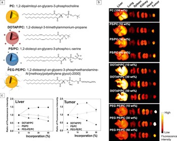
The in vivo biodistribution and tumor targeting ability of liposomes differ depending on the charge and surface modification. Lee et al. comparatively analyzed various liposome formulations with different lipid compositions. Reference Lee, Lee, Park, Ryu, Na, Koo, Lee, Jeon, Kwon, Kim and Jeong32 Different types of liposomes based on a neutral lipid, a cationic lipid, an anionic lipid, or a poly-(ethylene glycol)-modified (PEGylated) lipid were fabricated ( Figure 1 a, PC, DOTAP/PC, PS/PC, and PEG-PS/PC, respectively). The PEGylated lipid-based liposomes demonstrated superior tumor accumulation even though they were all similar in particle size (Figure 1b). In contrast, the cationic DOTAP/PC (1,2-dioleoyl-3-trimethylammonium-propane/1,2-dipalmitoyl-sn-glycero-3-phosphocholine) liposomes highly accumulated in the liver and resulted in the lowest tumor targeting ability (Figure 1c).
Tumor targeting ability of various modified lipid nanoparticles (NPs). (a) Illustration and chemical structures of the liposomes with different lipid compositions (PC, neutral liposme; DOTAP/PC, cationic liposome; PS/PC, anionic liposome; PEG-PE/PC, PEGylated liposome). (b) In vivo distribution of the liposomes in tumor-bearing mice models. High fluorescence intensity means greater accumulation of NPs, and the PEGylated liposome shows the highest tumor accumulation. (c) Relative ratio of near infrared fluorescence intensity in the liver and tumor to that of the unmodified liposome-treated group. PEGylated liposomes selectively accumulate in the tumor. Reprinted with permission from Reference 32. © 2012 Wiley.

Solid lipid NPs are a new colloidal delivery system, composed of a solid lipid core matrix, which is solid at room temperature and body temperature. They are useful for carrying the lipophilic drugs, such as doxorubicin, because the lipid core is able to solubilize lipophilic drugs. Reference Zara, Cavalli, Fundaro, Bargoni, Caputo and Gasco33 Solid lipid NPs have remarkable advantages, such as increased stability in biological fluids, the possibility of mass production, and low toxicity compared to other liposomes and polymeric nanocarriers. Reference Estella-Hermoso de Mendoza, Campanero, Mollinedo and Blanco-Prieto16 In addition, solid lipid NPs can not only be injected but also orally administered. For example, methotrexate (MTX), an anti-cancer drug, could be orally administered using solid lipid NPs, and MTX-bearing solid lipid NPs increased MTX concentration in intestinal lymphatic regions and showed enhanced drug bioavailability. Reference Paliwal, Rai, Vaidya, Khatri, Goyal, Mishra, Mehta and Vyas34 To make up for the low drug encapsulating capacity of solid lipid NPs, modified solid lipid NPs, lipid drug conjugates, and nanostructured lipid carriers have been extensively studied. Reference Yang, Lu, Cai, Zhu, Liang and Yang19,Reference Lim, Lee and Kim20,Reference Podlogar, Gasperlin, Tomsic, Jamnik and Rogac35 These studies have contributed to the wider application of lipid-based NPs.
Cationic lipid nanocarriers, such as lipofectamine, are able to bind to negatively charged genes to form lipid-therapeutic gene complexes, Reference Martin, Sainlos, Aissaoui, Oudrhiri, Hauchecorne, Vigneron, Lehn and Lehn36,Reference Kostarelos and Miller37 and a cationic lipid-based emulsion (CLE) has been applied for in vivo gene delivery. Reference Kwon, Nam, Nam, Park, Lee, Kim, Kwon, Kim, Kang, Park and Jeong38 The CLE/DNA complex showed prolonged blood half-life with effective cellular delivery of DNA and low cytotoxicity, which leads to effective gene delivery in vivo. Although lipid-DNA complexes were evaluated in a non-tumoral animal model, the results suggested that CLEs have various advantages as a gene carrier, such as ease of manufacturing, low immunogenicity, and wide range of target gene delivery. However, the insufficient effect of gene delivery due to low transfection efficacy and colloidal instability may serve as a hurdle for clinical applications. Reference Scheule, St. George, Bagley, Marshall, Kaplan, Akita, Wang, Lee, Harris, Jiang, Yew, Smith and Cheng39,Reference Felgner, Kumar, Sridhar, Wheeler, Tsai, Border, Ramsey, Martin and Felgner40
Over the past decades, numerous lipid-based drug delivery systems have been developed for cancer theranosis, simultaneous cancer therapy and diagnosis. Lipid-based materials can be utilized with diverse anticancer drugs, and they are relatively safe. The combination of nanotechnology and lipid-based colloidal systems plays important roles in tumor imaging and therapy. In particular, liposomal and lipid-based NPs are a promising approach for delivery of peptides/protein drugs and therapeutic genes via different routes of administration. Despite many remaining caveats of using lipid-based NPs such as low transfection efficacy and disease-targeting ability, liposomal NPs are anticipated to be good candidates for future drug delivery systems. As indicated by the successful marketing of commercial anti-cancer drug formulation Doxil (PEG-liposomal doxorubicin), more of the anticancer drugs will be formulated in lipid-based NPs for clinical applications in the future.
Polysaccharide-based NPs
Polymers are the most prevalent materials for building NP structures. With the earliest report of the use of a polymer for cancer therapy dating back to 1979, the emergence of polymeric nanomedicine was boosted by the increasing need for novel drug delivery carriers with better features, such as controlled release of anti-cancer agents, easily modifiable chemical and physical properties, biocompatibility and biodegradability, and more. Reference Peer, Karp, Hong, Farokhzad, Margalit and Langer15,Reference Gupta, Agrawal and Vyas41,Reference Couvreur, Kante, Roland and Speiser42 A variety of polymers have been utilized to develop theranostic NPs, and they can be subdivided into two major categories of natural and synthetic polymers. Although synthetic polymers show low batch to batch variation and have easily predictable mechanical properties, natural polymers are also important for their own advantages. Extracted from living organisms, natural polymers have the benefit of biocompatibility and intrinsic properties related to biodegradation. In addition, certain natural polymers possess biological properties, such as targeting moieties and mucoadhesion, where the material can attach to the surface of mucosa. Reference Dang and Leong43,Reference Yhee, Koo, Lee, Choi, Kwon and Kim44
Natural polymers include polysaccharides and proteins, but polysaccharides are the major natural polymeric materials for nanoparticle fabrication. Polysaccharides have various functional groups in their molecular chains that contribute to easy chemical modification for fabrication of NP structures. In addition, hydrophilic groups such as hydroxyl, carboxyl, and amino groups are notable for their bio-adhesive properties and ability to form non-covalent bonds with surface glycoproteins of mucosa cells. Reference Liu, Jiao, Wang, Zhou and Zhang45
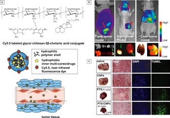
Among polysaccharide-based NPs, chitosan particles have been popular from the early days of theranostic NPs. Chitosan, deacetylated chitin, is a natural polymer that consists of β-(1, 4)-2-amido-d-glucose linked via (1–4) glycosidic bonds. A variety of chemically modified chitosan derivatives, including carboxymethyl chitosan, trimethyl chitosan, PEGylated chitosan, or glycol chitosan, have been widely utilized for cancer theranosis. Reference Sahu, Mallick, Santra, Maiti, Ghosh and Pramanik46–Reference Mao, Roy, Troung-Le, Janes, Lin, Wang, August and Leong49 In practice, water-soluble glycol chitosan (GC) and hydrophobic 5β-cholanic acid conjugates self-assembled to form tumor-homing chitosan-based NPs (CNPs) ( Figure 2 a). Reference Park, Kim, Nam, Lee, Nam, Kim, Park, Kim, Choi, Kim and Kwon50,Reference Kwon, Park, Chung, Kwon, Jeong and Kim51 Different molecular weights of GC and varying degrees of hydrophobic substitution can significantly influence the distributions of resulting CNPs in vivo. Reference Park, Kim, Nam, Lee, Nam, Kim, Park, Kim, Choi, Kim and Kwon50,Reference Na, Lee, Lee, Koo, Min, Jeong, Yuk, Kim and Kwon52 In addition, the physicochemical properties and pharmacokinetic alterations depend on the stability and deformability of NPs. Reference Na, Koo, Lee, Min, Park, Yoo, Lee, Park, Kwon, Jeong and Kim53 Consequently, the base material and properties of assembled NPs should be carefully considered, even if the researcher selected from among the chitosan derivatives. The optimized CNP (250 kDa GC, with 23 wt% 5β-cholanic acid) showed excellent tumor-targeting property, mainly due to the enhanced blood circulation and EPR effect. The CNPs typically labeled with Cy5.5, a near infrared fluorescence dye, can be traced in the body using whole body fluorescence scanners. The intravenous injection of Cy5.5-labeled CNPs enabled superb diagnostic imaging of various tumors, including flank, brain, and liver tumor models in mice (Figure 2b). Reference Na, Koo, Lee, Min, Park, Yoo, Lee, Park, Kwon, Jeong and Kim53 Concurrently, water-insoluble anticancer chemo-drugs, such as paclitaxel (PTX), docetaxel, and camptothecin (CPT), also can be encapsulated into the Cy5.5 labeled CNPs for targeted cancer therapy. Reference Kim, Kim, Park, Kim, Park, Nam, Lee, Park, Park, Kim, Choi, Kim, Park and Kwon48,Reference Min, Park, Kim, Bae, Lee, Jo, Park, Kim, Jeong, Kim and Kwon54,Reference Hwang, Kim, Kwon and Kim55 The drug-loaded CNPs delivered chemo-drugs to tumors with high specificity, leading to an effective therapeutic effect (Figure 2c). As a result, the theranostic CNPs are certainly one of the most promising systems for theranostic NPs for cancer treatment.
Tumor-homing chitosan-based nanoparticles (CNPs). (a) Schematic diagram of CNPs for cancer theranosis. CNPs can penetrate the coarse structure of newly forming blood vessels in the tumor. (b) Diagnostic tumor images using Cy5.5 (a near infrared fluorescence dye)-CNPs in various tumor models, including flank, brain, and liver. Tumor regions can be discriminated (arrows: tumor site, color bars indicate the fluorescence intensity). (c) Comparative therapeutic effect of paclitaxel loaded CNPs in tumor-bearing mice (original magnification ×100 in microscopic images). Reprinted with permission from Reference 48. © 2010 Elsevier, and from Reference 53. © 2011 Elsevier. Note: H&E, hematoxylin and eosin stain; DAPI, 4’,6-diamidino-2-phenylindole; TUNEL, terminal deoxynucleotidyl transferase dUTP nick end labeling; PTX, paclitaxel.

Hyaluronic acid (HA), a negatively charged copolymer of d-glucuronic acid and N-acetyl-d-glucosamine, is also a type of extensively investigated polysaccharide for preparing NPs. Reference Choi, Chung, Min, Yoon, Kim, Park, Kwon and Jeong14,Reference Choi, Min, Na, Choi, Kim, Park, Kwon and Jeong56 Since various cancer cells overexpress the HA receptor (CD44), HA has been utilized as a cancer-targeting macromolecule because of the affinity of HA to CD44 in cancer cells. Reference Yu, Park and Jon3,Reference Platt and Szoka57,Reference Lee, Lee and Park58 More recently, PEGylated HA NPs and self-assembled HA NPs consisting of amphiphilic HA-5β-cholanic acid conjugates were synthesized and characterized for cancer theranosis. Reference Choi, Chung, Min, Yoon, Kim, Park, Kwon and Jeong14,Reference Choi, Min, Na, Choi, Kim, Park, Kwon and Jeong56,Reference Choi, Yoon, Kim, Bae, Park, Kang, Kim, Kwon, Choi, Jeong, Kim and Park59–Reference Min, Son, Lee, Koo, Yoon, Na, Choi, Park, Lee, Han, Park, Kim, Jeong, Rhee, Kim, Kwon and Kim61 The PEGylated HA NPs showed efficient drug delivery in tumors. They could release the loaded anticancer drugs in the presence of Hyaluronidase-1, an enzyme known to be implicated in cancer cell proliferation and neovascularization in the tumor. The Hyaluronidase-1 specific drug release enables tumor cell-specific drug delivery. Reference Choi, Yoon, Kim, Bae, Park, Kang, Kim, Kwon, Choi, Jeong, Kim and Park59 The hydrophobic photo-sensitizer, chlorin e6 (Ce6), could be also loaded in PEGylated HA NPs, and tumor-targeted delivery of Ce6 allows simultaneous photodynamic imaging and therapy by fluorescence and singlet oxygen upon laser irradiation. Reference Yoon, Koo, Choi, Lee, Kim, Kwon, Leary, Park, Yuk, Park and Choi60
HA-based NPs were used in ultrasound imaging applications as well as in optical imaging. Perfluoropentane, a hydrophobic molecule with a boiling point of 27°C, is in its gas form at body temperature (37°C). Based on this property, it can be used as an ultrasound contrast agent. Perfluoropentane was encapsulated in HA NPs to discriminate a tumor from normal liver by the tumor’s distinct low echogenicity, and the HA NPs showed exceptionally long echo persistence.61As base materials for building up NPs, other polysaccharides include alginate, heparin, cellulose, and dextran. Reference Choi, Jang, Joung, Kwon and Park62–Reference Zahoor, Sharma and Khuller65 Alginate is a water-soluble biopolymer that can be gelled in cation-positive conditions. Based on this unique property, alginate has been widely used in the fabrication of hydrogel and inhalable NPs. Reference Zahoor, Sharma and Khuller65,Reference Damge, Reis and Maincent66 Heparin, a variably sulfated repeating disaccharide unit, is also a common base material for building up NPs to facilitate efficient delivery of protein/peptide drugs to tumors. Reference Byrne, Betancourt and Brannon-Peppas68 Heparin-binding domains in various proteins allow for high binding affinity of protein with heparin. Reference Na, Kim, Park, Kim, Woo, Kwon, Chung and Park67 High affinity of dextran derivatives toward iron oxide facilitated the development of dextran NPs as a magnetic resonance imaging nanoprobe. Iron oxide carrying dextran NPs have drawn attention for clinical cancer imaging using MRI, Reference Harisinghani, Barentsz, Hahn, Deserno, Tabatabaei, van de Kaa, de la Rosette and Weissleder69,Reference Park, von Maltzahn, Zhang, Schwartz, Ruoslahti, Bhatia and Sailor70 and these organic–inorganic hybrid NPs will be further discussed in the subsequent “Other NPs” section.
Peptide/protein-based NPs
Proteins and peptides are attractive base materials for building up theranostic NPs. Due to the abundance of primary amine groups in the structure, proteins and peptides generally show high binding affinity with various negatively charged drugs and/or optical probes. In addition, reticuloendothelial system (RES) clearance, an immune reaction that clears foreign materials in the liver, is low against the peptide or protein NPs. The aqueous steric barrier of the protein carrier results in less clearance, and it contributes to the advanced pharmacokinetic properties of the NPs. Reference Elzoghby, Samy and Elgindy71
Drug conjugates and complexes are typical structures of peptide/protein-based NPs. They can be simply prepared through diverse physical and covalent interactions between the drug and carrier. In fact, tumor imaging using protein-dye conjugates has been studied since the early 1990s. Becker et al. utilized transferrin (Tf)-indotricarbocyanine (ITCC) and human serum albumin-ITCC conjugates for optical imaging of tumors; both of the protein-dye conjugates accumulated in tumors for EPR effects, showing high tumor-to-tissue contrast. Reference Becker, Riefke, Ebert, Sukowski, Rinneberg, Semmler and Licha72
Abraxane also is a good example of a protein-drug nanocomplex. Abraxane is an albumin-bound paclitaxel NP, which was approved by the FDA in 2005 and is now in clinical use. Reference Hawkins, Soon-Shiong and Desai73 It can be produced in a high-pressure homogenizer, resulting in 130 to 150 nm-sized NPs. Reference Janib, Moses and MacKay1,Reference Elzoghby, Samy and Elgindy71 Based on the particle size and the characteristics of the albumin carrier, Abraxane demonstrated longer blood circulation and lower off-targeting to achieve an improved therapeutic effect compared to naked paclitaxel. Reference Hawkins, Soon-Shiong and Desai73,Reference Miele, Spinelli, Miele, Tomao and Tomao74 More recently, Tf-modified stealth NPs encapsulating PEG-CPT conjugates were synthesized for selective delivery of CPT to tumor tissues. Reference Hong, Zhu, Jiang, Tang, Sun, Fang, Shi and Pei75 The Tf-based NPs also have similar advantages of prolonged blood circulation (up to 72 h) and increased EPR effect by the steric stabilization of PEG-CPT conjugates. Additionally, improved intracellular delivery based on Tf receptor-ligand mediated endocytosis, internalization of material into the cytoplasm, is expected, since proliferating cancer cells commonly overexpress Tf receptors on their plasma membrane. Reference Gatter, Brown, Trowbridge, Woolston and Mason76
In recent years, proteins have also been considered as nanoscale carriers for gene delivery. In particular, small interfering RNA (siRNA) molecules have attracted much attention, because they can induce selective gene silencing. However, they require effective carrier systems to compensate for low stability and poor cellular uptake. Therapeutic genes, including siRNA, consist of oligonucleotides, and they are negatively charged molecules that show insufficient binding affinity to neutral proteins. Therefore, chemical modification of carrier proteins is often required to form NP structures with genes. Thiol-modified or PEG-modified proteins generally show better binding affinity for therapeutic genes. In particular, thiol group-introduced proteins can be folded at disulfide bonds within the protein structures. As a result, folding of tertiary or quaternary protein structures contributed to the formation of compact NPs for gene delivery. Reference Kommareddy and Amiji77–Reference Son, Song, Lee, Min, Kim, Yhee, Huh, Kwon, Jeong, Byun, Kim and Kim80
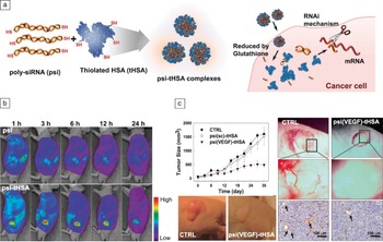
Recently, thiolated protein carriers have been extensively developed using structurally modified genes, including siRNA polymers. Reference Lee, Son, Yhee, Choi, Kwon, Kim and Kim81 The siRNA polymer (poly-siRNA), which represents self-polymerization of 5’-end thiol-modified siRNA molecules, exhibited improved physiochemical properties with higher molecular weight and functional chemical groups, and they could be contrastively incorporated in thiolated protein carriers, including thiolated albumin, thiolated gelatin, and thiolated Tf. Reference Lee, Yhee, Kim, Kwon and Kim78–Reference Son, Song, Lee, Min, Kim, Yhee, Huh, Kwon, Jeong, Byun, Kim and Kim80 Son et al. utilized human serum albumin (HSA), the most abundant protein in serum, as a protein carrier for siRNA. Reference Son, Song, Lee, Min, Kim, Yhee, Huh, Kwon, Jeong, Byun, Kim and Kim80 To provide sufficient binding affinity between HSA and siRNA, thiol groups were introduced into both of the molecules. The resulting poly-siRNA and thiolated HSA (tHSA) formed condensed NPs (denoted as psi-tHSA NPs, 169.3 ± 10.2 nm) by chemical cross-linking and self-cross-linking. The natural glutathione in animal cells reduces the disulfide bonds of the NPs structure. Consequently, the psi-tHSA NPs demonstrated efficient gene silencing after cellular uptake, which was mainly dependent on the reducibility in the cytoplasm as other glutathione-responsive nano-vehicles ( Figure 3 a). Reference Cheng, Feng, Meng, Deng, Feijen and Zhong82 Cy5.5 labeled psi-tHSA NPs showed time-dependent kinetics of the NPs, indicating delivery of NPs in a tumor-targeted manner (Figure 3b). More importantly, vascular endothelial growth factor (VEGF) targeting siRNA molecules, which suppress the neovascularization in the tumor, were delivered using psi-tHSA NPs to successfully inhibit tumor growth by suppression of angiogenic vessel formation (Figure 3c). This result suggests that the application of thiolated protein carriers for gene delivery can be expanded in in vivo therapy with virtually any therapeutic genes, as the physicochemical properties of poly-siRNA would be practically the same regardless of the sequential differences according to the target messenger RNA.
Tumor imaging and therapy using poly-siRNA-thiolated human serum albumin nanoparticles (psi-tHSA NPs). (a) Self-cross-linking psi-tHSA NPs and gene silencing process after cellular uptake (RNAi, RNA interference; mRNA, messenger RNA; VEGF, vascular endothelial growth factor; psi, naked poly-siRNA; CTRL, control). (b) Tumor imaging using tumor targeted delivered psi-tHSA NPs in whole body fluorescence scanning. The psi-tHSA NPs selectively accumulate in the tumor region. (c) In vivo therapeutic effects of the psi(VEGF)-tHSA NPs. Tumor growth curve and blood vessel formation in the tumor of the psi(VEGF)-tHSA injected mice show that the psi(VEGF)-tHSA can reduce blood vessel formation in the tumor to delay tumor growth (black arrows = tumoral microvessels). The psi(sc)-tHSA, the tHSA NPs including scrambled siRNA, could not delay the tumor growth. Reprinted with permission from Reference 80. © 2013 Elsevier.

Synthetic polymer-based NPs
In the last several decades, there also have been tremendous advancements in the use of synthetic polymer-based NPs. Compared to natural polymers, synthetic polymers offer advantages in many ways. They are generally easier to handle and synthesize, and they also show low variation between batches and low chance of microbial contamination. In addition, synthetic polymers usually exhibit sufficient surface area and functioning groups, which can be used to conjugate bioactive agents for targeting and/or contrast agents for imaging. Some examples of synthetic polymers for nano-formulations include polylactic acid (PLA), poly(lactic-co-glycolic) acid (PLGA), poly(ethylene glycol) (PEG), and polyethyleneimine (PEI). With these synthetic polymers, several types of carriers have been designed including polymer micelles, polymersomes, polymeric conjugates, and polymeric NPs. In this section, cancer theranostic NPs formulated with most commonly used synthetic polymers will be discussed.
PEG is one of the most widely used synthetic polymers. Since the first approval of PEG for clinical use in the 1990s, it is the gold standard for polymers as drug carriers. Reference Knop, Hoogenboom, Fischer and Schubert83 In fact, most current FDA approved polymer-based drug delivery systems are PEG-functionalized products, and other synthetic polymers have not yet been approved. Reference Knop, Hoogenboom, Fischer and Schubert83,Reference Ledet84 PEG has been widely used because of its hydrophilicity and solubility, as well as reduced interactions with the immune system and blood components, hence the stealth effect. Reference Knop, Hoogenboom, Fischer and Schubert83 Other advantageous features of PEG include low PDI, low hydrodynamic volume, high solubility in organic solvents, and more. Reference Veronese and Pasut85,Reference Zacchigna, Cateni, Drioli and Bonora86 Although there were earlier reports of using PEGylated proteins to enhance drug circulation time, Yokoyama et al. first demonstrated the use of PEG block copolymers for drug delivery. Reference Yokoyama, Miyauchi, Yamada, Okano, Sakurai, Kataoka and Inoue87 Subsequently, an extensive number of reports of PEGylated entities followed, such as on PEG-drug conjugates, copolymers, and PEGylated micelles. Reference Knop, Hoogenboom, Fischer and Schubert83,Reference Yoo and Park88,Reference Pasut and Veronese89 It is also important to note that PEG is not a synthetic polymer that is used alone, rather it is used as a di- or multi-block copolymer, since it is easy to synthesize it as a block copolymer. Reference Chan, Valencia, Zhang, Langer and Farokhzad90
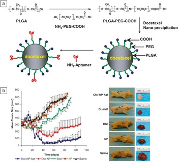
Similar to PEG, PLGA has a long history of use in biomedical applications, from biodegradable sutures to micelles. PLGA, a FDA-approved copolymer of lactic acid and glycolic acid, exhibits excellent biocompatibility, mechanical properties, and complete biodegradability. Reference Dinarvand, Sepehri, Manoochehri, Rouhani and Atyabi91 Since PLGA is a copolymer, its mechanical strength, capacity to undergo hydrolysis, and biodegradation rate are directly influenced by the type and molar ratio of the individual monomer components in the copolymer chain. Reference Dinarvand, Sepehri, Manoochehri, Rouhani and Atyabi91,Reference Jain92 Glycolic acid is more hydrophilic than lactic acid, thus glycolic acid-rich PLGA tends to be more hydrophilic and subsequently degrades more slowly. Reference Park93 Because both of the components, lactic acid and glycolic acid, are endogenous and are easily metabolized in biological fluids, minimal systemic toxicity is expected in drug delivery applications. Reference Kumari, Yadav and Yadav94 In practice, PLGA has been utilized in various forms, such as NPs and micelles. Many anticancer drugs, including paclitaxel and docetaxel, have been incorporated into PLGA NPs, but they are somewhat limited for effective tumor-targeting, due to RES clearance or opsonization. Reference Owens and Peppas95 To address such limitations, surface modification of PLGA NPs with a hydrophilic entity, such as PEG and other aptamers, has been a popular method in drug delivery. Omid et al. developed docetaxel loaded PLGA-PEG NPs, and they successfully reduced tumor volumes compared to the free docetaxel treated group ( Figure 4 ). Reference Farokhzad, Cheng, Teply, Sherifi, Jon, Kantoff, Richie and Langer96 In particular, aptamer-conjugated NPs significantly reduced cytotoxicity and anti-tumor efficacy of NPs. Other reports have discussed PLGA copolymers and anticancer drug-loaded PLGA-b-mPEG, which have been approved for clinical applications. Reference Chan, Valencia, Zhang, Langer and Farokhzad90,Reference Locatelli and Franchini97
(a) Schematic representation of the PLGA-PEG-COOH copolymer based nanoparticles (NPs) for delivery of docetaxel (Dtxl). Surface modification of NPs with an aptamer can enhance the target-specific delivery of NPs (PLGA, poly(lactic-co-glycolic) acid; PEG, poly(ethylene glycol); Apt, aptamer). (b) Growth curve of the tumors treated with Dtxl loaded NPs in tumor xenograft nude mice. Aptamer introduced PLGA-PEG copolymer NPs show the best therapeutic effects. (Single intratumoral injection of saline [black]; PEGylated PLGA NP without drug [NP, brown]; free Dtxl [Dtxl, green]; Dtxl-encapsulated NPs [Dtxl-NP, red]; Dtxl-encapsulated NP-Apt bioconjugates [Dtxl-NP-Apt, blue], 40 mg/kg Dtxl in each Dtxl formulation, * means that p < 0.05 by ANOVA test; data points for the Dtxl-NP-Apt group that were statistically significant compared with all other groups by statistical [ANOVA] test at 95% confidence interval. Arrows: tumor sites). Reprinted with permission from Reference 96. © 2006 National Academy of Sciences. Note: m, n, and o, number of repeating units.

PEI has been exceptionally useful in the field of non-viral gene delivery. Traditional gene delivery methods had involved adenoviral or liposomal vectors before 1995, but safety concerns over using viral vectors raised interest in non-viral vectors. Boussif et al. first reported that PEI is also a versatile vector for gene delivery. Reference Boussif, Lezoualc’h, Zanta, Mergny, Scherman, Demeneix and Behr98,Reference Godbey, Wu and Mikos99 The backbone of PEI contains repeating units of two carbons and a nitrogen, and the branched form of PEI (bPEI) exhibits high potential to be protonated due to the primary, secondary, and tertiary amines. This unique property contributes to the ability to carry a large amount of gene in a compact and condensed form, as nucleic acids are negatively charged with their phosphate backbone. Reference Boussif, Lezoualc’h, Zanta, Mergny, Scherman, Demeneix and Behr98 In addition, PEI is easy to modify and is safer and also more convenient to use compared to viral vectors, because viral vector modifications are not simple and may cause serious complications related to infectious diseases.
Research in gene delivery with PEI has seen significant progress to date, as bPEI, 25 kDa, in particular, is considered to be the gold standard of polycationic gene delivery vectors. Reference Mahato, Kumar and Sharma100 The use of PEI has expanded as PEI-peptide conjugates, and modified PEI with various natural or synthetic polymers, have been widely developed. For instance, PEGylated PEI with a targeting ligand, Reference Ogris, Walker, Blessing, Kircheis, Wolschek and Wagner101 poly(ethylene oxide)–poly(propylene oxide)–poly(ethylene oxide) (PEO–PPO–PEO) tri-block (denoted as Pluronics)-PEI copolymer, Reference Nguyen, Lemieux, Vinogradov, Gebhart, Guerin, Paradis, Bronich, Alakhov and Kabanov102 and chitosan-PEI copolymer, Reference Zhao, Li, Pan, Liu, Lv, Leung and Lu103 have been studied with or without imaging agents and anticancer drugs. With the modified PEI NPs, the weakness of conventional PEI NPs could be addressed in terms of relatively low toxicity and biocompatibility.
Other than the synthetic polymers introduced previously, continuing efforts are under way to improve physiochemical and biological properties of polymer-based NPs, especially for in vivo applications. Recently, Pluronics, poly(glutamic acid) (PGA), and poly(N-(2-hydroxypropyl)methacrylamide) (PHPMA) have been used as alternatives to the conventional formulation using PEG. Reference Li and Wallace104,Reference Duncan105 These new polymers could reduce the side effects of PEG, such as incomplete biodegradability and instability under mechanical stress. Reference Knop, Hoogenboom, Fischer and Schubert83,Reference Herold, Keil and Bruns106,Reference Zhang and Granick107 The use of poly(ε-caprolactone), poly(ε-l-lysine), or poly(methyl methacrylate) represents other good examples of synthetic polymer-based NPs. Reference Gupta, Agrawal and Vyas41,Reference Chawla and Amiji108–Reference Xie, Bagby, Cohen and Forrest110 Physiochemical properties of each base polymer provide useful characteristics to the NPs, such as pH sensitivity, stimuli-responsiveness, and good lymphatic targeting. Moreover, the versatility of these synthetic polymers is arguably one of the most important strengths, as they can be easily modified to work as copolymers, targeting ligands, and/or imaging agents. Based on these controlled and unique properties, synthetic polymers are anticipated to see continued development for cancer theranostic NPs.
Other NPs
Many organic materials have been extensively studied as base materials for theranostic NPs, as seen from the previous descriptions. In spite of the advantageous characteristics of these respective materials, no magic formula for perfect tumor-targeting has been developed with a single material. Although remarkable progress has been achieved based on nanotechnology, more advanced nanocarriers are needed in order to respond to increasing unmet demands. Consequently, various strategies have been employed for further application for theranosis, for instance, hybrid NPs. Hybrid NPs include inorganic-organic hybrid NPs and organic-organic hybrid NPs.
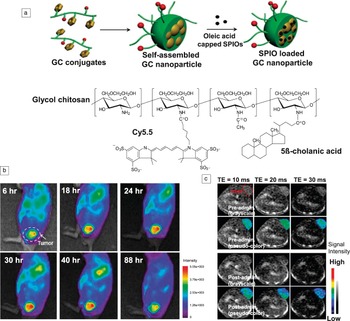
Inorganic materials, such as gold, silica, iron oxide, and other metals are also important for theranostic NPs. In particular, non-optical imaging modules, including computed tomography, magnetic resonance (MR), and positron emission tomography, require inorganic materials as imaging contrast agents. To improve tumor targeting and reduce cytotoxicity, the surface of these inorganic particles is usually modified with organic materials. For instance, Key et al. encapsulated superparamagnetic iron oxide (SPIO) NPs in GC NPs for optical-MR dual imaging. Reference Key, Cooper, Kim, Dhawan, Knapp, Kim, Park, Choi, Kwon, Park and Leary111 As we described for polysaccharide-based NPs, GC-5β-cholanic acid-based CNPs showed highly selective accumulation in tumors. Loaded SPIOs in the CNP greatly improved MR contrast tumor imaging ( Figure 5 ), and the organic part of the NPs played a successful role in tumor-targeting ability. Dual or multi-modal imaging offers more opportunities for early diagnosis, even for tumors in deep tissues. As with other inorganic-organic hybrid NPs, PEGylated or transferrin grafted gold NPs have been developed for cancer theranosis. Reference Li, Wang, Liu, Zhang, Guo, Liu and Tang112,Reference Jain, Hirst and O’Sullivan113 Gold is useful for carrying negatively charged therapeutic genes, and it also produces heat after irradiation. Tumor thermal therapy using gold NPs has been one of the intensively studied topics in the 2000s. Reference Visaria, Griffin, Williams, Ebbini, Paciotti, Song and Bischof114 However, gold NPs alone show low targeting ability with poor stability in blood. Modified gold NPs, including PEG-Tf grafted gold NPs, showed improved cellular uptake and targeted delivery to tumors with comparatively low toxicity. The combined use of base materials for building up NPs can be a complementary method to overcome the limitations of traditional NPs.
(a) Schematic diagram of superparamagnetic iron oxide (SPIO)-loaded glycol chitosan (GC) nanoparticles (NPs) for magnetic resonance imaging (MRI) and chemical structures of GC-5β-cholanic acid conjugates. (b) In vivo near infrared fluorescence images of Cy5.5-GC NPs injected tumor-bearing mice provide anatomical information of tumors up to 88 h. (c) The SPIO loaded GC NPs provide in vivo MR images using 3T MRI and an eight-channel head coil. (Red circle: tumor site, TE: echo time). Reprinted with permission from Reference 111. © 2012 Elsevier.

Organic-organic hybrid NPs are also available. For example, a peptide-based optical probe can be conjugated to a polysaccharide-based NP. Ryu et al. designed a cathepsin B-sensitive nanoprobe (CB probe) using the peptide sequence, for monitoring of cathepsin B activity. Reference Ryu, Kim, Koo, Yhee, Lee, Na, Youn, Choi, Kwon, Kim and Kim115 Cathepsin B is closely related to tumor invasion and metastasis, and the probe alone indeed showed cathepsin B-specific near infrared fluorescence (NIRF) signals in vitro and in vivo. Reference Ryu, Kim, Koo, Yhee, Lee, Na, Youn, Choi, Kwon, Kim and Kim115 To achieve tumor-targeted delivery and longer blood circulation of the probe, the CB probes were conjugated to CNPs, which demonstrated amplified NIRF intensity with prolonged activity in vivo ( Figure 6 ). The CNP-CB probe could provide valuable biological information on enzyme activity in tumors with enhanced imaging contrast.
Cathepsin B (CB) probes and chitosan-based nanoparticle (CNP)-CB probe. (a) Structural designs of the CB probe. CB probes recover fluorescence signals in the presence of CB. BHQ3 is a dark quencher that has no native fluorescence but absorbs fluorescence in the range of 620 to 730 nm (FRET, fluorescence resonance energy transfer; gly, glycine; Arg, arginine; Lys, lysine). (b) Near infrared fluorescence (NIRF) imaging of the excised tumor and visceral organs after CB probe and CNP-CB probe injection in the tumor-bearing mice. CB probes were chemically conjugated to CNPs to achieve the tumor-targeted delivery of the probes. (c) NIRF signal intensity at each tumor and visceral organs. CNP-CB probe indicates high fluorescence signals in tumors, suggesting cathepsin B activity. Reprinted with permission from Reference 115. © 2011 The Royal Society of Chemistry. Note: Cy5.5, near infrared fluorescence dye.

Conclusions
Deliberate efforts to develop effective theranostic nanoparticles (NPs) have made great strides in cancer treatment. Among cancer-theranostic NPs, organic NPs have shown a variety of advantages for theranostics purposes. In summary, diverse organic NPs have their own strengths, depending on the physiochemical and biological properties of the base materials. Lipid-based NPs have high drug loading capacity for both hydrophilic and hydrophobic drugs, and the outer lipid shell enables easy cellular uptake of NPs. Polymers are versatile due to their easily modifiable properties. Natural polymer-based NPs are fundamentally biocompatible and have intrinsic properties related to biodegradation. Polysaccharide and protein-based NPs have been studied since the early days of nanomedicine, and the properties, mechanisms for tumor accumulation, and theranostic applications of each NP formulation were excogitated and well-described over long periods of time. Particularly, unique biological properties, including mucoadhesion, possession of tumor-specific ligands, or rapid degradation by tumor-rich enzymes, would be further utilized for designing rational NPs for cancer theranosis. Synthetic polymers are chemically refined ones that show distinct pros and cons with low variation. Consequently, synthetic polymer-based NPs are relatively easy to design with predictable properties. Finally, mutual complementary use of base materials by combination is a common choice for development of NPs. Organic-inorganic hybrid and organic-organic hybrid NPs indicate interesting characteristics, and they may provide multi-modal tumor imaging or monitoring of cancer-related enzyme activities.
Recent progress in the application of cancer theranostic NPs has focused on the safe and efficient delivery of contrast agents and anticancer drugs. In this review, practical performance of organic NPs and the most frequently used base materials were introduced. Further research will offer clinical use of organic NPs for cancer theranostics in the near future, and the significant advances in theranostic NPs may provide better opportunities for the early diagnosis of and therapy for cancer. We expect that smarter and advanced theranostic NPs will be developed for more effective cancer treatment and diagnosis.
Acknowledgments
This work was supported by the Global Research Laboratory Project (NRF-2013K1A1A2032346) and the GiRC Project (2012K1A1A2A01055811) of MSIP, and the Intramural Research Program (Global RNAi Carrier Initiative) of KIST.