Introduction
A congenital cholesteatoma is an epidermoid cyst behind an intact tympanic membrane, which arises from congenital remnants of keratinising squamous epithelium in the temporal bone. Congenital cholesteatoma may develop in various temporal bone sites, including the petrous apex, cerebellopontine angle, middle-ear cavity and mastoid process. The mastoid process is the least frequent site of occurrence, with only a small number of cases having been reported worldwide.Reference Derlacki and Clemis1–Reference Warren, Bennett, Wiggins, Salzman, Blevins and Shelton8 Moreover, congenital cholesteatoma originating in the mastoid is more difficult to diagnose, as the clinical presentation is variable.Reference Cureoglu, Osma, Oktay, Nazaroglu, Meric and Topcu5
We report herein an unusual case of congenital cholesteatoma of mastoid origin manifesting as acute mastoiditis, and we provide a brief overview of the literature. At surgery, the cholesteatoma was found to be restricted to the mastoid process. This was confirmed by pathological and immunohistochemical analysis (the latter using involucrin, a specific marker for keratin).
Case report
A 65-year-old man presented to the ENT department of Iwaki Kyoritsu General Hospital with a two-week history of left-sided, post-auricular swelling and pain. He had no history of acute otitis media, previous ear surgery or local trauma. The patient was receiving medical treatment for diabetes. He had noted a gradually progressive, bilateral hearing loss over the previous 10 years, and in addition had recognised a slight worsening in left-side hearing over approximately the past year.
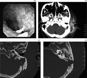
On physical examination, bulging of the posterior wall of the external auditory canal was observed. However, there was no apparent perforation or retraction of the tympanic membrane, including Schrapnell's membrane (Figure 1a).
(a) Otoscopic view of the left ear showing a bulging posterior wall of the external auditory canal. However, there was no apparent perforation or retraction of the tympanic membrane, including Schrapnell's membrane. (b) Pre-operative, axial, enhanced computed tomography (CT) scan showing mastoiditis and post-auricular abscess. (c) & (d) Pre-operative, axial, high-resolution CT scans showing an expansile, lytic lesion eroding the bony plate of the posterior fossa and the bony plate covering the sigmoid sinus. Although soft-density shadows are apparent in the attic, no enlargement was found in the aditus ad antrum at operation.

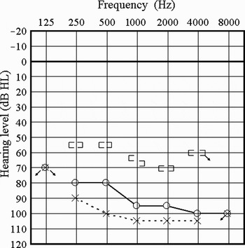
Audiometric evaluation showed a mixed-type hearing loss bilaterally (Figure 2). Averaged air conduction thresholds on the left and right sides were 103.8 and 90 dB, respectively.
Pre-operative audiogram.

No symptoms of balance disorder or nystagmus were observed.
Computed tomography (CT) with gadolinium enhancement showed an abscess in the post-auricular region. Enhancement was observed in areas corresponding to the sigmoid sinus, and there were no signs of sigmoid sinus thrombosis (Figure 1b).
High-resolution CT of the temporal bone revealed an expansile, lytic lesion in the left mastoid eroding the bony plate of the posterior fossa and the bony plate covering the sigmoid sinus (Figure 1c and 1d). Mastoid pneumatisation was fairly well developed bilaterally. The inner ear was normal. Although soft tissue density was apparent in the middle ear, no enlargement or destructive findings were observed in the aditus ad antrum, attic or ossicles (Figure 1d).
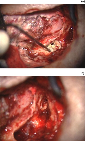
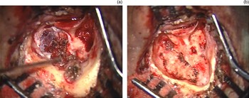
Based on the CT findings, acute otitis media with coalescent mastoiditis was suspected. An emergency mastoidectomy was thus conducted. A post-auricular abscess connecting to the mastoid was drained. The peripheral part of the mastoid was found to be filled with cholesteatoma, which had destroyed the bony plate of the posterior cranial fossa along with the bony plate over the sigmoid sinus. However, the cholesteatoma was limited to the mastoid region, with only hypertrophic mucosa, granulation tissue and a bloody effusion observed anterior to the aditus ad antrum (Figure 3a and 3b). The posterior wall of the external auditory canal was found to be intact.
Findings at initial surgery. (a) The lower part of the mastoid was found to be filled with cholesteatoma (indicated by the suction tube). (b) The cholesteatoma matrix did not show any connection to the aditus ad antrum or attic.

Pathological analysis of the surgical specimen revealed that the mastoid contents comprised keratinous material surrounded by bacterial colonies, consistent with infection. Conversely, specimens from the aditus ad antrum comprised granulation tissue infiltrated with lymphoid and plasma cells (data not shown).
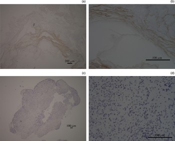
In order to analyse for the existence of squamous epithelium, immunohistochemical staining with involucrin was performed on paraffin-embedded sections of mastoid content, using the avidin-biotin-peroxidase complex immunoperoxidase technique, as previously described.Reference Suzuki, Ohtani, Aikawa, Sato, Sakuma and Ogawa9 Involucrin positivity was seen in areas corresponding to keratinous material (Figure 4a and 4b). Conversely, a specimen from the aditus ad antrum yielded negative results for involucrin staining (Figure 4c and 4d). These findings confirmed that the cholesteatoma was limited to the mastoid area, and a diagnosis of congenital cholesteatoma of the mastoid region was established.
Photomicrographs showing involucrin staining. (a) Mastoid specimen showing involucrin positivity, corresponding to areas of keratinocytes. (b) Higher magnification view of same section. (c) Aditus ad antrum specimen showing infiltration of plasma cells and lymphocytes, with no involucrin positivity. (d) Higher magnification view of same section. Magnification as shown.

About two months after the first operation, a second surgical procedure was conducted to extirpate the cholesteatoma matrix. The mastoid was found to be filled with cholesteatoma, which had almost destroyed the tegmen mastoideum, the bony plate of the posterior fossa, and the bony plate covering the sigmoid sinus (Figure 5a). The middle ear, aditus ad antrum and attic were unaffected by the cholesteatoma. Granulation tissue surrounding the cholesteatoma matrix was adhesive to both the posterior fossa dura and the sigmoid sinus, and was gently separated then completely removed via an intact canal procedure (Figure 5b). Pathological and immunohistochemical analysis of surgical specimens showed the same characteristics seen for specimens from the first procedure (data not shown).
Findings at second surgical procedure. (a) The cholesteatoma matrix was seen to connect with the dura and sigmoid sinus of the posterior fossa. (b) Surgical appearance after total resection of cholesteatoma.

Discussion
The clinical presentation and imaging characteristics of congenital cholesteatoma are variable, nonspecific and determined by location.Reference Luntz, Telischi, Bowen, Rees and Balkany3 Quite rarely, cholesteatoma arises from the mastoid, presenting with symptoms including neck pain, post-auricular swelling, pain and dizziness.Reference Lee, Hong and Jung7
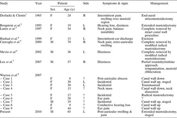
Until a recent report of nine cases by Warren et al., only seven cases of congenital cholesteatoma of the mastoid had previously been reported (Table I).Reference Derlacki and Clemis1–Reference Warren, Bennett, Wiggins, Salzman, Blevins and Shelton8 Four of Warren and colleagues' nine cases were identified incidentally from a database of neuroradiology cases.
Reported cases of congenital cholesteatoma of the mastoid region

Y = years; F = female; M = male; R = right; L = left; ? = unknown; canal wall down = canal wall down mastoidectomy; canal wall up = canal wall up mastoidectomy; transmastoid = transmastoid approach
In cases of congenital cholesteatoma of the mastoid region, post-auricular pain or abscess is not always initially present; only three of the 16 previously reported cases showed such symptoms and signs.
We report herein an unusual case of congenital cholesteatoma of the mastoid region, manifesting as acute mastoiditis and diagnosed after emergency mastoid surgery.
Mevio et al. stated that the particular site and rarity of congenital cholesteatoma of the mastoid region results in difficulty differentiating it from other possible tumours, such as paraganglioma, endolymphatic sac tumour and meningioma.Reference Mevio, Gorini, Sbrocca, Artesi, Lenzi and Lecce6 Because the present case required emergency surgical drainage, due to the periauricular abscess identified on physical examination and CT, pre-operative magnetic resonance imaging was not conducted.
Another reason for diagnostic difficulty was the fact that cholesteatoma of mastoid origin cannot be verified surgically if it occupies the entire middle-ear cleft including the mesotympanum.Reference Luntz, Telischi, Bowen, Rees and Balkany3, Reference Warren, Bennett, Wiggins, Salzman, Blevins and Shelton8
Although our patient's cholesteatoma was limited to the mastoid, we had to bear in mind the fact that it may have developed from implantation of epithelium into the middle ear due to either trauma or previous tympanic membrane perforation.Reference House and Sheehy10, Reference Friedberg and Congenital11 For instance, Suzuki et al. reported a case of acquired cholesteatoma without any connection to the tympanic membrane, which might have been misdiagnosed as congenital cholesteatoma.Reference Suzuki, Ohtani, Aikawa, Sato, Sakuma and Ogawa9 In their analysis, based on computer-aided, three-dimensional reconstruction, the cholesteatoma was observed not to be connected with the tympanic membrane. However, the tympanic membrane had been deformed as a result of a healed perforation, and showed retraction of Schrapnell's membrane. Immunohistochemical analysis using involucrin produced positive results for granulation tissue in the attic, but no epithelial tissue was observed on haematoxylin and eosin staining. Based on these findings, Suzuki et al. concluded that cholesteatoma matrix had originally been present in the tympanic membrane region but had resolved, and they thus defined their case as acquired cholesteatoma.Reference Suzuki, Ohtani, Aikawa, Sato, Sakuma and Ogawa9 In our patient, surgical and pathological findings indicated that the cholesteatoma matrix was limited to the mastoid region and did not connect to the aditus ad antrum or attic. Moreover, no enlargement was found in the aditus ad antrum on CT or intra-operatively. To confirm these findings, we conducted immunohistochemical studies, using the same procedures as Suzuki et al. Reference Suzuki, Ohtani, Aikawa, Sato, Sakuma and Ogawa9 Mastoid material showed only positive results for involucrin, establishing a diagnosis of cholesteatoma of mastoid origin.
Derlacki and Clemis defined congenital cholesteatoma as an entity fulfilling the following criteria: (1) development behind an intact tympanic membrane in the absence of a history of otitis media; and (2) absence of any demonstrable connection with the external auditory canal.Reference Derlacki and Clemis1 Regarding the first point, our case showed poorly developed mastoid air cell pneumatisation (differing from previous reports of mastoid congenital cholesteatoma such as that of Luntz et al.).Reference Luntz, Telischi, Bowen, Rees and Balkany3 This would suggest a history of otitis media, although no such history was elicited. However, previous history of otitis media was not strictly included in the definition of congenital cholesteatoma, as more than 70 per cent of children under two years of age suffer from otitis media.Reference Levenson, Parisier, Chute, Wenig and Juarbe12 The key to diagnosing congenital cholesteatoma is the absence of previous local trauma or infection and the presence of an intact drum, both of which were seen in the present case.Reference Rashad, Hawthorne, Kumar and Welsh4
Another notable point in the current case was the patient's presentation relatively late in adulthood. This is uncharacteristic for congenital cholesteatoma, which often presents in childhood with a white mass behind an intact tympanic membrane or with hearing loss, due either to ossicular chain destruction caused by cholesteatoma growth or to associated congenital ossicular anomaly.Reference Suetake, Kobayashi and Takasaka13 In the 17 reported cases of congenital cholesteatoma in the mastoid region (including the current case), the mean age of presentation was 31.5 years, with a female:male ratio of 2:1.
• Congenital cholesteatoma may develop in various temporal bone sites, including the petrous apex, cerebellopontine angle, middle-ear cavity and mastoid process
• Occurrence in the mastoid process is the least frequent; only a small number of such cases have been reported worldwide
• This paper describes a case of congenital cholesteatoma presenting as acute mastoiditis in an adult
Our patient's audiogram showed mixed-type hearing loss bilaterally, with almost symmetrical bone conduction thresholds. While the middle ear of the unaffected side was apparently normal, based on tympanic membrane examination and CT findings, an air–bone gap of approximately 25 dB existed nevertheless. No ossicular erosion or destruction was found during surgery, and no anatomical continuity was evident between the mastoid cholesteatoma and the attic. The patient's air conduction threshold asymmetry (approximately 14 dB) could thus have been attributable to an effusion or granulation tissue in the tympanum and attic.
Warren et al. identified conductive hearing loss as the second most frequent presentation in their nine cases of mastoid-origin cholesteatoma.Reference Warren, Bennett, Wiggins, Salzman, Blevins and Shelton8 These nine patients were divided into mastoid-only cholesteatoma and mastoid and middle-ear cholesteatoma groups. The former group showed a higher incidence of dural exposure, facial nerve exposure and auditory canal erosion, presumably because disease restricted to the mastoid may be clinically silent as the lesion does not impinge upon any critical neurovascular structures. Four of the nine patients fell into the latter group, and almost all showed ossicular erosion at surgery. From his surgical and pathological findings, our patient would have been included in Warren and colleagues' mastoid-only group, and his dural and sigmoid sinus destruction was consistent with these authors' findings.Reference Warren, Bennett, Wiggins, Salzman, Blevins and Shelton8
As stated above, cholesteatoma of mastoid origin is not able to be correctly diagnosed if it occupies the entire middle ear.Reference Luntz, Telischi, Bowen, Rees and Balkany3, Reference Warren, Bennett, Wiggins, Salzman, Blevins and Shelton8
A small number of publications have reported double cholesteatomas of the middle ear. Huang reported double intratemporal congenital cholesteatomas located in the mastoid and petrous pyramid.Reference Huang14 Moriyama and Honda reported three cases with independent cholesteatoma located behind an attic (acquired) cholesteatoma.Reference Moriyama and Honda15 Sinkawa et al. reported several similar cases demonstrating the presence of a highly extensive cholesteatoma along with a small lesion located among the tegmental cells of the mastoid region.Reference Sinkawa, Hozawa, Usami, Ichijo, Saitoh and Tominaga16 Based on these reports, cases of congenital cholesteatoma of the mastoid region may not be as rare as previously reported. With the ongoing development of diagnostic modalities, it is possible to identify such cases with increasing accuracy.
Acknowledgments
We thank Drs Shigeyuki Asano (Department of Pathology, Iwaki Kyoristu General Hospital), Iwao Ohtani and Hando Hakozaki (Fukushima Rousai Hospital) for their helpful advice.