Introduction
Dizziness is one of the most common presenting symptoms, affecting approximately 20–30 per cent of the general population.Reference Neuhauser, Von Brevern, Radtke, Lezius, Feldmann and Ziese1–Reference Furman and Cass6 Dizziness of vestibular cause has a lifetime prevalence of 7.4 per cent, while its annual prevalence suggests that nearly 5 per cent of the adult population are affected every year.Reference Neuhauser, Von Brevern, Radtke, Lezius, Feldmann and Ziese1–Reference Furman and Cass6 This has implications not only for an affected individual's quality of life but also for their professional activities, with 40 per cent of affected patients reporting interruptions in their daily activities and increased sick leave, and nearly 20 per cent avoiding leaving their home. Interestingly, females seem to be affected more frequently than males. In 88 per cent of all cases, vertigo appears to be recurrent.Reference Neuhauser, Von Brevern, Radtke, Lezius, Feldmann and Ziese1–Reference Kontorinis, Tyagi and Crowther7 Such numbers make accurate diagnosis and efficient management of the dizzy patient a necessity.
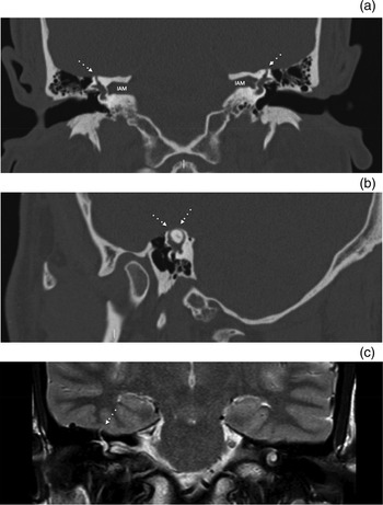
Benign paroxysmal positional vertigo, labyrinthitis and vestibular neuronitis, and Ménière's disease, are the most common peripheral causes of dizziness, followed by uncommon causes such as vestibular schwannomas and vestibular manifestations of systemic diseases.Reference Baloh5–Reference Kontorinis, Tyagi and Crowther7 In 1998, Minor et al. described for the first time superior semicircular canal dehiscence, which is a defect in the bone over the arcuate eminence of the superior semicircular canal (Figure 1).Reference Minor, Solomon, Zinreich and Zee8 This condition presents primarily with sound- and pressure-induced vertigo, adding to the differential diagnosis of the dizzy patient. Sound- and pressure-induced vertigo is a less well-known symptom, but is relevant to the differential diagnosis of peripheral dizziness. In addition to sound- and/or pressure-induced vertigo, patients with superior semicircular canal dehiscence can present with oscillopsia and nystagmus, alongside other vestibular or audiological manifestations such as autophony and conductive hearing loss.Reference Minor, Solomon, Zinreich and Zee8
Coronal high-resolution computed tomography scans at the level of the internal auditory meatus (IAM) in a patient with bilateral superior semicircular canal dehiscence (dotted arrows) (a), and reconstruction on an oblique sagittal plane showing the extent of the dehiscence (dotted arrows) (b). Although less utilised for superior semicircular canal dehiscence diagnostics, coronal T2-weighted magnetic resonance imaging of the IAM shows protrusion of the right superior semicircular canal deep in the middle fossa and temporal lobe (dotted arrow) in a patient with superior semicircular canal dehiscence (c).

The reported incidence of superior semicircular canal dehiscence based on cadaveric studies is 0.3 per cent, with an additional incidence of 1.3 per cent for thin temporal bone (0.1 mm or less) covering the roof of the superior semicircular canal.Reference Carey, Minor and Nager9,Reference Williamson, Vrabec, Coker and Sandlin10 However, the radiological prevalence of superior semicircular canal dehiscence is 9 per cent.Reference Williamson, Vrabec, Coker and Sandlin10 The significant difference between the radiological and the cadaveric confirmation of dehiscence may be due to a low specificity of imaging in the diagnosis of superior semicircular canal dehiscence and possible over-diagnosis. However, considering the imaging and clinical presentation, superior semicircular canal dehiscence is much more common than indicated in cadaveric studies.
Although superior semicircular canal dehiscence is believed to be a disease of adults, a few studies have shown high incidence in the paediatric population.Reference Schutt, Neubauer, Samy, Pensak, Kuhn and Herschovitch11–Reference Sugihara, Babu, Kitsko, Haupert and Thottam13 Despite these references, the reported mean age of superior semicircular canal dehiscence patients is 51 years.Reference Zhou, Gopen and Poe14 With respect to gender, there does not appear to be a sex predominance. A few studies have reported a slightly increased incidence in males without, however, identifying any significant difference.Reference Sugihara, Babu, Kitsko, Haupert and Thottam13,Reference Jackson, Allen, Morell, Carpenter, Givens and Kakade15,Reference Saxby, Gowdy, Fandiño, Chadha, Kozak and Sargent16
Overall, as with most newly diagnosed conditions, the presence of superior semicircular canal dehiscence can be missed, resulting in patients with long-standing dizziness and disequilibrium that affects their day-to-day activities, including professional, individual and social aspects. Zhou et al. named superior semicircular canal dehiscence a great otological mimicker, highlighting the diagnostic challenges of the condition as well as the necessity for awareness of the disease's existence.Reference Zhou, Gopen and Poe14 Additionally, given the associated auditory symptoms, mainly autophony, low-frequency conductive hearing loss and tinnitus, superior semicircular canal dehiscence has been misdiagnosed, and many times mismanaged as patulous Eustachian tube (autophony) or even otosclerosis (conductive hearing loss with normal tympanic membranes and tympanometry) over the years.Reference Sugihara, Babu, Kitsko, Haupert and Thottam13–Reference Saxby, Gowdy, Fandiño, Chadha, Kozak and Sargent16 Indeed, with symptoms varying from pressure-induced vertigo to autophony and tinnitus, there is a long list of otological and neurotological conditions that can be confusing for otorhinolaryngologists. The present review focuses on the symptoms, underlying pathophysiology, origin and cause of the condition, diagnosis, and available treatment options.
Search strategy
Data for this review were identified by searches of Medline, Embase and Epistemonikos databases, using the search terms ‘superior semicircular canal dehiscence’ and ‘superior semicircular canal’. All papers were written in English or German language, and published from 1998 until October 2019. The references of relevant articles were also searched. We excluded case reports, conference abstracts, and papers focusing on animals and fetal development. The current literature review focused on the incidence and symptoms of superior semicircular canal dehiscence, and the diagnostic techniques and treatment options.
Presenting symptoms and pathophysiology
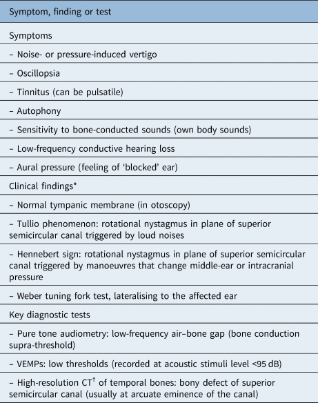
Superior semicircular canal dehiscence typically presents with: vertigo; oscillopsia (the illusion of a moving visual world, with an inability to stabilise visualised objects, which appear to constantly oscillate); and nystagmus, with a rotational component on the plane of the superior semicircular canal triggered by loud noises (Tullio phenomenon), or/and by manoeuvres that change the middle-ear or intracranial pressure (Hennebert sign).Reference Minor, Solomon, Zinreich and Zee8–Reference Martin, Chahine, Veyret, Richard, Prades and Pouget23 The patients may additionally describe a feeling of ‘blocked’ ear or aural pressure, and some drop in their hearing (Table 1). Most patients are able to identify certain triggers of their symptoms, which are usually related to either loud noises or pressure changes.Reference Minor, Solomon, Zinreich and Zee8–Reference Martin, Chahine, Veyret, Richard, Prades and Pouget23
Key presenting symptoms, clinical findings and diagnostic tests

*In an otherwise healthy individual. †Cone beam or digital volume tomography can better identify the bony dehiscence, given the reduced partial volume effect. VEMPs = vestibular-evoked myogenic potentials; CT = computed tomography
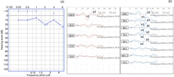
Many patients also report a hypersensitivity to sounds, being able to hear their own sounds very loudly. Typically, the patient with superior semicircular canal dehiscence will report autophony, but also the ability to hear movements of their eyes or even their hair very loudly; the ability of patients with superior semicircular canal dehiscence to hear their eyeballs moving is an auditory ‘hallmark’ of the disease, mostly because of the oddness of the symptom rather than its scientific background or clinical significance.Reference Minor, Solomon, Zinreich and Zee8–Reference Martin, Chahine, Veyret, Richard, Prades and Pouget23 This hypersensitivity audiologically corresponds to a negative bone conduction threshold in the low frequencies: a supra-threshold (Figure 2a). Essentially, the inner ear is too sensitive to bone-conducted sounds, a condition called conductive hyperacusis. This phenomenon is also responsible for the low-frequency air–bone gap (conductive hearing loss). Following this hypersensitivity, Weber tuning fork testing typically lateralises to the affected ear, and, interestingly, it can be heard even if the tuning forks are placed far from the head (Table 1).Reference Minor, Solomon, Zinreich and Zee8–Reference Martin, Chahine, Veyret, Richard, Prades and Pouget23
(a) Pure tone audiogram of the left (affected) ear of a patient with superior semicircular canal dehiscence, showing a bone conduction supra-threshold (thin line marked with ‘>’) up to −10 dB and an air–bone gap (gap between the bone conduction ‘>’ and the air conduction ‘×’). (b) Cervical vestibular-evoked myogenic potentials of the same patient, showing a low (abnormal) threshold in the left (L) ear (n1 and p1 waveforms) up to 65 dB, while in the right (R) ear, healthy ear cervical vestibular-evoked myogenic potentials are recorded at 100 dB.

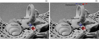
The reported symptoms are related to the ‘third window’ effect, which, although not completely understood, provides a rational explanation of the patient's symptoms.Reference Minor, Solomon, Zinreich and Zee8,Reference Nadgir, Ozonoff, Devaiah, Halderman and Sakai12,Reference Sohmer, Freeman and Perez24,Reference Hiroven, Carey, Liang and Minor25 Instead of having two windows on the osseous cochlea, namely the round and the oval windows, patients with superior semicircular canal dehiscence have an additional, third one at the level of the superior semicircular canal (Figure 3). This is believed to increase the inner-ear immittance and affect the endolymphatic homeostasis, resulting in hypersensitivity of the inner ear and generating a vestibular response to lower sound level stimuli than anticipated.Reference Minor, Solomon, Zinreich and Zee8,Reference Nadgir, Ozonoff, Devaiah, Halderman and Sakai12,Reference Sohmer, Freeman and Perez24,Reference Hiroven, Carey, Liang and Minor25
Model of an inner and middle ear embedded in the temporal bone, with the upper part removed to enable visualisation of the middle- and inner-ear structures of an intact inner ear, showing: (a & b) the direction of travel of the acoustic energy through the oval window (OW, thick red arrows) and the round window (RW, thin blue arrows). This balance is being disturbed following the bony dehiscence of the superior semicircular canal (b), with abnormal energy transmission (dashed arrows) through the ‘third window’. PSC = posterior semicircular canal; SSC = superior semicircular canal; LSC = lateral semicircular canal

The third window allows bone-conducted sounds to reach the inner ear through the labyrinth rather than through the oval window, disseminating the acoustic energy and resulting in cochlear hypersensitivity (Figure 3). The presence of a third window lowers the impedance of the cochleovestibular system, causing lower resistance for pressure and, essentially, sound transmission.Reference Minor, Solomon, Zinreich and Zee8,Reference Nadgir, Ozonoff, Devaiah, Halderman and Sakai12,Reference Sohmer, Freeman and Perez24–Reference Rosowski, Songer, Nakajima, Brinsko and Merchant27 Recent studies in a chinchilla model by Hiroven et al. have shown such mechanisms of pressure sensitivity following a third window.Reference Hiroven, Carey, Liang and Minor25 The effect of the third window on inner-ear pressure changes as well as the ease of transmission of intracranial pressure changes through the ‘window’ can explain both the vertiginous and auditory symptoms related to superior semicircular canal dehiscence, particularly autophony and hyperacousis.Reference Sohmer, Freeman and Perez24–Reference Rosowski, Songer, Nakajima, Brinsko and Merchant27
The theory and concept of the third window is not new; several middle-ear surgeons were using this concept to treat conductive hearing loss in patients with otosclerosis until 1955, prior to the introduction of stapedectomy and stapes replacement prostheses.Reference Lenarz and Boenninghaus28 Otosclerosis is a progressive fixation of the stapes due to abnormal bone metabolism around the footplate and osseous inner-ear capsule, causing primarily significant conductive hearing loss.Reference Eshraghi and Telischi29,Reference Nazarian, McElveen and Eshraghi30 Middle-ear surgeons used to create a small bony opening, a third window, on the osseous cochlea or vestibule, to improve patients’ conductive hearing loss. As the third window causes a milder conductive hearing loss in the low frequencies, the surgeons used this technique to bypass the footplate fixation, which was preventing the sound conduction. Thus, the surgeons aimed for a better hearing threshold via the transmission of sounds through the third window rather than the fixed stapes.Reference Lenarz and Boenninghaus28
It is this third window effect that explains the symptoms related to superior semicircular canal dehiscence. However, it still leaves a few questions unanswered. In particular, it is unclear why some patients will experience more severe symptoms than others, and why some patients will be completely asymptomatic, with superior semicircular canal dehiscence being an incidental imaging finding.
To date, there is no known factor affecting vestibular symptom severity. The extent of the bony defect has not been associated with dizziness or oscillopsia severity. However, the size and location of the bony defect have been linked to the presence, character and severity of the auditory symptoms.Reference Chien, Janky, Minor and Carey31–Reference Niesten, Stieger, Lee, Merchant, Grolman and Rosowski35 It seems that most patients with a very small defect do not experience severe auditory symptoms. In contrast, patients with a wide dehiscence describe more severe autophony and have a larger air–bone gap in the low frequencies. Additionally, these patients, as well as patients with a dehiscence near the superior petrosal sinus, report more troublesome tinnitus, which can be pulsatile in nature. This happens because of transmission of the heartbeat through the middle fossa dura vessels and/or the superior petrosal sinus.Reference Niesten, Hamberg, Silverman, Lou, McCall and Windsor34,Reference Niesten, Stieger, Lee, Merchant, Grolman and Rosowski35
Despite the impact of the size and location of the defect on the auditory symptoms, it still remains unclear why some individuals are asymptomatic, at least at the time of the radiological diagnosis. The presence of a very thin bony layer that is too thin to be visualised on computed tomography (CT), or even cone beam CT, because of technical and resolution limitations, is a possible explanation. Despite radiological and electrophysiological confirmation of superior semicircular canal dehiscence, in some cases there might still be a very thin area of bone overlying the superior semicircular canal, preventing the clinical appearances of superior semicircular canal dehiscence. Nonetheless, symptomatic patients with a very thin bony cover, reflecting ‘near-superior semicircular canal dehiscence’, have been reported.Reference Ward, Wenzel, Ritzl, Gutierrez-Hernandez, Della Santina and Minor36,Reference Lookabaugh, Kelly, Carter, Niesten, McKenna and Curtinet37
An alternative theory is that not every patient with superior semicircular canal dehiscence will develop symptoms because of the lack of a ‘second event’.Reference Minor19,Reference Chilvers and McKay-Davies22,Reference Whyte Orozco, Martinez, Cisneros, Obon, Gracia-Tello and Angel Crovetto38 This theory is based on the concept that many individuals can have superior semicircular canal dehiscence but will not develop any symptoms unless there is a second event that initiates them. In the literature, these second events (possible mechanisms of symptom initiation in a patient with existing superior semicircular canal dehiscence) have included: head trauma; bone metabolism abnormalities, for example osteoporosis; increased middle-ear or intracranial pressure; recent flight; or rapid changes of pressure. Given the communication of the inner-ear space with the cerebrospinal fluid,Reference Pisano, Niesten, Merchant and Nakajima26,Reference Rosowski, Songer, Nakajima, Brinsko and Merchant27 even changes in the intracranial pressure can affect patients’ symptoms, acting as a ‘trigger’ for symptom initiation (the second event).
Additionally, some patients might not experience any significant symptoms, as a result of a ‘non-functional’ superior semicircular canal either, because of a previous infection (labyrinthitis causing failure, fibrosis or obliteration of the canal) or any other event that has resulted in superior semicircular canal failure. In such cases, the patients can be asymptomatic despite the radiological evidence of superior semicircular canal dehiscence.
In summary, the otherwise healthy symptomatic patient with superior semicircular canal dehiscence will typically present with pressure- or sound-generated dizziness and nystagmus, oscillopsia, autophony, and conductive hyperacusis, and will have normal otoscopic findings, and low-frequency conductive hearing loss with a bone conduction supra-threshold, while Weber's tuning fork test will lateralise towards the affected side (Table 1).
Origin
Given the available evidence, there is an ongoing debate as to whether superior semicircular canal dehiscence is a congenital, developmental or an acquired condition. There are good numbers of studies in favour of both theories, indicative of the scientific controversies and possibly the co-existence of both; the precise mechanism by which the ‘third window’ is created is unclear. Based on the current evidence, explained below, there are patients with superior semicircular canal dehiscence of a congenital origin, but also patients with a pre-existing thinning of the bony overlay of the superior semicircular canal, which gradually was either absorbed or disrupted, leading to the presence of superior semicircular canal dehiscence and its related symptoms. Careful analysis of the available studies indicates that no one theory can explain all cases; thus, it appears that superior semicircular canal dehiscence could be the result of the combination of an acquired factor and an existing congenital or developmental background (often referred to as a ‘second event’ theory).
Takahashi et al. compared macroscopically the middle cranial fossa of adult and fetal cadavers, including imaging findings.Reference Takahashi, Tsunoda, Shirakura and Kitamura39 They showed that the superior semicircular canals protrude into the cranium in fetal life, while in adulthood they are embedded in bone. This observation raised the hypothesis that this protrusion into the cranium could lead to adhesions between the dura and the superior semicircular canal, and increase the likelihood of a superior semicircular canal dehiscence in later life. Additionally, the bilateral presentation of superior semicircular canal dehiscence in some cases, or even the contralateral thinning of the bony cover of the superior semicircular canal reported by many authors, supports a congenital background of superior semicircular canal dehiscence. Furthermore, studies showing multiple tegmen defects and/or ear malformations in patients with superior semicircular canal dehiscence are in favour of such an origin.Reference Suryanarayanan and Lesser40–Reference Oh, Vivas, Hudgins and Mattox43
A recent case–control study showed better-developed (pneumatised) temporal bones and lower-lying middle fossa dura anteriorly in patients with superior semicircular canal dehiscence, raising the possibility of a developmental cause of superior semicircular canal dehiscence.Reference Tikka and Kontorinis44 Furthermore, the description of superior semicircular canal dehiscence in paediatric patients, aged even younger than one year, and the decreased prevalence of thin bony cover of the superior semicircular canal with age, also support a congenital background.Reference Jackson, Allen, Morell, Carpenter, Givens and Kakade15,Reference Saxby, Gowdy, Fandiño, Chadha, Kozak and Sargent16,Reference Meiklejohn, Corrales, Boldt, Sharon, Yeom and Carey45,Reference Lagman, Ong, Chung, Elhajjmoussa, Fong and Wang46 Overall, certain aspects of temporal bone anatomy have been sporadically associated with superior semicircular canal dehiscence. However, it is difficult to determine whether such anatomical differences, like the thin overlying bone, have a causal effect or not.
On the other hand, the prevalence of superior semicircular canal dehiscence has been found to be higher in older patients and in those with osteoporosis, suggesting an acquired rather than a congenital origin.Reference Nadgir, Ozonoff, Devaiah, Halderman and Sakai12,Reference Crovetto, Whyte, Rodriguez, Lecumberri, Martinez and Fernandez47–Reference Mahulu, Fan, Ding, Quaye, Mambo and Mafuru50 For instance, an assessment of 306 temporal bone CT scans showed increased radiological prevalence in older patients, with nearly 8 per cent of the scans showing superior semicircular canal dehiscence and nearly 45 per cent showing thinning of the bony cover of the superior semicircular canal.Reference Nadgir, Ozonoff, Devaiah, Halderman and Sakai12 Two years later, using similar settings, a study involving temporal bone imaging of 312 patients showed slight osteopenia of the superior semicircular canal roof associated with ageing.Reference Crovetto, Whyte, Rodriguez, Lecumberri, Martinez and Fernandez47 Additionally, a recent study investigating bone metabolism markers concluded that such markers could be important in the clinical assessment and management of patients with superior semicircular canal dehiscence.Reference Nguyen, Lagman, Sheppard, Duong, Ong and Poon48
Despite the abovementioned evidence, and the possibility of superior semicircular canal dehiscence being an acquired condition, the otic capsule undergoes little remodelling once its development is completed, mostly because of factors related to genetics of the inner ear.Reference Sørensen, Frisch and Bretlau51,Reference Thys and Van Camp52 Thus, the direct impact of bony changes of the temporal bone on the superior semicircular canal itself is debatable.
In addition to age-related changes of the temporal bone and bone metabolism, body mass index (BMI) and the presence of increased intracranial pressure have been examined as factors contributing to the pathophysiology of superior semicircular canal dehiscence. Although Schutt et al. showed a possible causal correlation between increased BMI, obstructive sleep apnoea and superior semicircular canal dehiscence,Reference Schutt, Neubauer, Samy, Pensak, Kuhn and Herschovitch11 and El Hadi et al. showed a possible association between increased intracranial pressure and superior semicircular canal dehiscence,Reference El Hadi, Sorrentino, Calmels, Fraysse, Deguine and Marx53 the causal impact of increased intracranial pressure is controversial. Indeed, Kuo et al. failed to identify an association between increased intracranial pressure and the presence of superior semicircular canal dehiscence.Reference Kuo, Bagwell, Mongelluzzo, Schutt, Malhotra and Khokhar54
If one considers the controversial available evidence indicating a congenital or developmental origin for some cases, and an acquired origin for others, the second event theory seems to offer the most rational explanation for the origin of superior semicircular canal dehiscence. In particular, a congenital or developmental background (first event) may well be present in most cases; however, it will be an additional factor such as a head trauma, a rapid increase in middle-ear or intracranial pressure, or age-related factors that will result in the superior semicircular canal dehiscence.Reference Minor19,Reference Chilvers and McKay-Davies22,Reference Whyte Orozco, Martinez, Cisneros, Obon, Gracia-Tello and Angel Crovetto38,Reference Tikka and Kontorinis44,Reference Brandolini, Modugno and Pirodda55 Essentially, the second event causes disruption of the thin overlying bone, leading to the bony defect. Although a second event cannot always be identified, it is assumed that this is mostly related to the retrospective character of many of the available studies, and the gaps in the clinical information and the medical history.
A gradual progressive thinning of bone occurring over time in predisposed individuals currently offers the most logical explanation for the origin of superior semicircular canal dehiscence.
Key diagnostic batteries
Although the suspicion of superior semicircular canal dehiscence is based on purely clinical grounds and detailed medical history, it is the imaging studies, with specific focus on the high-resolution CT of the temporal bones (Figure 1), and the audiovestibular assessment, mainly vestibular-evoked myogenic potential testing, that will confirm the diagnosis (Figure 2 and Table 1).
Additional tests such as pure tone audiometry, globally used as the first-line method of hearing assessment, can show typical but not pathognomonic findings, namely a low-frequency supra-threshold (indicative of hypersensitivity to sounds) and a low-frequency air–bone gap (conductive hearing loss in the low frequencies) (Figure 2).Reference Minor, Solomon, Zinreich and Zee8–Reference Chilvers and McKay-Davies22
While other tests have been sporadically described, the combination of clinical suspicion, low-frequency conductive hearing loss, the radiological presence of superior semicircular canal dehiscence and the registration of vestibular-evoked myogenic potential responses in lower than normal hearing thresholds are considered pathognomonic for superior semicircular canal dehiscence (Table 1).
Imaging
High-resolution CT of the temporal bones can visualise the bony defect on a reconstruction in the coronal plane (Figure 1). Typically, thin sections are adequate to identify the dehiscence, and describe not only its extent but also its relation to neighbouring structures and the overall anatomy of the temporal bone, which could be of surgical significance should surgical intervention be considered.Reference Minor, Solomon, Zinreich and Zee8–Reference Williamson, Vrabec, Coker and Sandlin10,Reference Minor18–Reference Martin, Chahine, Veyret, Richard, Prades and Pouget23,Reference Yuen, Boeddinghaus, Eikelboom and Atlas32–Reference Niesten, Hamberg, Silverman, Lou, McCall and Windsor34,Reference Tikka and Kontorinis44 Given the technical limitations of the CT scanners provided, with thin sections of 0.5–0.625 mm in most places, as well as the resolution limitations of the monitors used to interpret and report the scans, very thin areas of overlying bone (near-dehiscence) can be misdiagnosed as superior semicircular canal dehiscence. Although this limitation could to a certain extent explain the high radiological incidence of superior semicircular canal dehiscence, symptomatic patients with radiologically evident ‘near-dehiscence’ have been well described in the current literature.Reference Ward, Wenzel, Ritzl, Gutierrez-Hernandez, Della Santina and Minor36,Reference Lookabaugh, Kelly, Carter, Niesten, McKenna and Curtinet37 A positive CT scan for superior semicircular canal dehiscence cannot diagnose superior semicircular canal dehiscence without clinical and electrophysiological correlation.
It is worth mentioning that superior semicircular canal dehiscence is not a purely radiological diagnosis; imaging along with electrophysiology confirms the clinical suspicion. Several studies have reported overestimating the presence of a bony defect over the superior semicircular canal by high-resolution CT scans.Reference Crovetto, Whyte, Rodriguez, Lecumberri, Martinez and Elexpuru56–Reference Mondina, Bonnard, Barreau, Darrouzet and Franco-Vidal59 Thus, careful interpretation of the clinical findings and correlation with the clinical presentation are crucial. In cases where there is clinical suspicion of superior semicircular canal dehiscence, high-resolution CT of the temporal bone is indicated.
Studies have reported on various CT techniques and reconstructions, namely flat panel CT and oblique reformatted images.Reference Branstetter, Harrigal, Escott and Hirsch60,Reference Tunkel, Carey and Pearl61 Although flat panel CT and intra-operative correlation showed more accurate measurements of the dehiscence, it appears that high-resolution CT scan with reconstruction in the coronal plane offers adequate quality to confirm the radiological diagnosis in most cases.Reference Branstetter, Harrigal, Escott and Hirsch60–Reference Belden, Weg, Minor and Zinreich63 Additional reformatting could be used in equivocal cases to challenge the limitations of the available CT scanners.
Interestingly, despite the known limitations of magnetic resonance imaging (MRI) in assessing bony structures, recent studies have assessed the efficacy of MRI in identifying superior semicircular canal dehiscence, showing high specificity and sensitivity (Figure 1).Reference Browaeys, Larson, Wong and Patel64,Reference Beyazal Çeliker, Özgür, Çeliker, Beyazal, Turan and Terzi65 Although this finding is not against the utilisation of CT in suspected superior semicircular canal dehiscence cases, it does highlight the focus on evolving MRI techniques and the developments in temporal bone imaging. Future studies involving MRI might show to what extent the use of this imaging modality will be favoured.
With respect to imaging, it is also worth mentioning that, following technological advances, cone beam CT, also known as digital volume tomography, has been utilised to provide radiological confirmation of superior semicircular canal dehiscence.Reference Bremke, Luers, Anagiotos, Gostian, Dorn and Kabbasch66 Being less prone to the partial volume effect, and offering high resolution with a voxel size as low as 0.08 mm3, the flexibility of reconstruction on multiple planes, and lower exposure to radiation, cone beam CT is a very appealing alternative to CT.Reference Bremke, Luers, Anagiotos, Gostian, Dorn and Kabbasch66–Reference Bremke, Luers, Stenner, Bovenschulte, Dorn and Unkel68 However, mostly for economic reasons, such equipment is not available in all units; accurate reporting also requires experienced, cone beam CT-accustomed radiologists.Reference Bremke, Luers, Anagiotos, Gostian, Dorn and Kabbasch66–Reference Bremke, Luers, Stenner, Bovenschulte, Dorn and Unkel68 On these grounds, to date, high-resolution CT remains the standard in imaging superior semicircular canal dehiscence.
Electrophysiology
Vestibular-evoked myogenic potential testing has high sensitivity and specificity for the diagnosis of superior semicircular canal dehiscence.Reference Rosowski, Songer, Nakajima, Brinsko and Merchant27,Reference Bremke, Luers, Stenner, Bovenschulte, Dorn and Unkel68–Reference Streubel, Cremer, Carey, Weg and Minor72 Thus, this testing is carried out routinely in patients with clinical suspicion and/or radiological confirmation of the condition. The recording of vestibular-evoked myogenic potentials is generally based on the knowledge that loud sounds can, in addition to cochlear hair cells, stimulate the vestibule (because of the presence of residual hair cells). Reduced vestibular-evoked myogenic potential thresholds and enlarged amplitudes are recorded in patients with superior semicircular canal dehiscence because of the ‘faster’ and ‘more effective’ transmission of the acoustic energy to the inner ear following the lower impedance of the vestibular system.Reference Rosowski, Songer, Nakajima, Brinsko and Merchant27,Reference Zuniga, Janky, Nguyen, Welgampola and Carey69–Reference Streubel, Cremer, Carey, Weg and Minor72
There are two main types of vestibular-evoked myogenic potentials, cervical and ocular; they both reflect electromyographic signals. Cervical vestibular-evoked myogenic potentials are recorded through electrodes placed on the forehead, neck and ipsilateral sternocleidomastoid muscle by projecting loud sounds (mostly clicks) and detecting the myogenic activity. Ocular vestibular-evoked myogenic potentials reflect extraocular electromyographic activity (following exposure to sounds) through electrodes placed beneath the contralateral eye.Reference Chien, Ravicz, Rosowski and Merchant73–Reference Manzari, Tedesco, Burgess and Curthoys77 Although the precise origin of cervical and ocular vestibular-evoked myogenic potentials is not completely understood, it is believed that cervical vestibular-evoked myogenic potentials originate from the saccule, while ocular vestibular-evoked myogenic potentials originate mostly from the utricule.Reference Chien, Ravicz, Rosowski and Merchant73–Reference Manzari, Tedesco, Burgess and Curthoys77 Typically, patients with superior semicircular canal dehiscence will have reduced vestibular-evoked myogenic potential thresholds (increased inner-ear sensitivity), which in a healthy individual are recorded at a level of not lower than 95–100 dB (Figure 2).
Although cervical vestibular-evoked myogenic potential testing has been more widely used, there has been an increasing scientific interest in ocular vestibular-evoked myogenic potential testing, suggesting that the latter is better for diagnosing superior semicircular canal dehiscence. Recent studies have demonstrated 90 per cent sensitivity and specificity of ocular vestibular-evoked myogenic potential testing in diagnosing superior semicircular canal dehiscence.Reference Bremke, Luers, Stenner, Bovenschulte, Dorn and Unkel68,Reference Zuniga, Davalos-Bichara, Schubert, Carey and Janky78,Reference Thabet79 These studies strengthen the position of ocular vestibular-evoked myogenic potential testing in the key diagnostic batteries for superior semicircular canal dehiscence, but further research is required.
The diagnostic techniques for both ocular and cervical vestibular-evoked myogenic potential testing are currently evolving. Recently, Noij et al. optimised the technique of cervical vestibular-evoked myogenic potential testing for superior semicircular canal dehiscence using different stimuli (2 kHz bursts instead of clicks), demonstrating 100 per cent specificity and 96 per cent sensitivity.Reference Noij, Herrmann, Guinan and Rauch80
To date, cervical vestibular-evoked myogenic potential testing remains more widely used, not only because of the experience of audiology teams with this technique, but also because cervical vestibular-evoked myogenic potentials were initially used in describing the electrophysiology in patients with superior semicircular canal dehiscence.Reference Brantberg, Bergenius and Tribukait81
Treatment options
Based on the increasing evidence on dealing with superior semicircular canal dehiscence, surgical interventions are currently being considered as a sensible therapeutic approach for the symptomatic patient. The decision-making follows detailed consultation, careful assessment of symptom severity, patients’ realistic expectations, desired outcomes and related risks. Overall, surgical intervention in a limited variety of approaches and methods has shown promising outcomes in dealing with the vestibular symptoms and oscillopsia, while the impact on the auditory symptoms, although they are mostly improved, remains unpredictable.Reference Minor19,Reference Ziylan, Kinaci, Beynon and Kunst20,Reference Vlastarakos, Proikas, Tavoulari, Kikidis, Maragoudakis and Nikolopoulos82–Reference Nguyen, Lagman, Sheppard, Romiyo, Duong and Prashant86 There is increasing evidence that surgery, in the sense of either resurfacing the bony defect of the superior semicircular canal or obliterating the superior semicircular canal, can achieve good results in the symptomatic patient with superior semicircular canal dehiscence, while the risks for associated complications, mainly irreversible severe sensorineural hearing loss and long-term imbalance, are relatively low in experienced centres.Reference Minor19,Reference Ziylan, Kinaci, Beynon and Kunst20,Reference Vlastarakos, Proikas, Tavoulari, Kikidis, Maragoudakis and Nikolopoulos82–Reference Nguyen, Lagman, Sheppard, Romiyo, Duong and Prashant86 However, the available evidence, mostly a result of the limited timeframe since the initial description of the condition and the issues with diagnosing the disease, is limited to small cohorts and retrospective case series.
In contrast, superior semicircular canal dehiscence patients who are asymptomatic or have much milder symptoms are managed conservatively, undergoing a thorough consultation and receiving generic vestibular rehabilitation advice. To date, there is no direct evidence on the efficacy of the conservative management of superior semicircular canal dehiscence, but such management is reserved for patients with milder clinical presentation, who generally do not require any active treatment.
With respect to resurfacing the bony defect, this can be achieved through either a middle fossa (intracranial) approach or a transmastoid (extracranial, through the mastoid) approach. The risks have overall been described as being slightly higher for the middle fossa approach, mainly because of the additional potential risks associated with intracranial operations, such as seizures or stroke.Reference Minor19,Reference Ziylan, Kinaci, Beynon and Kunst20,Reference Vlastarakos, Proikas, Tavoulari, Kikidis, Maragoudakis and Nikolopoulos82–Reference Nguyen, Lagman, Sheppard, Romiyo, Duong and Prashant86 However, in most available studies, there seems to be no statistically significant difference in the complication rates involving irreversible sensorineural hearing loss or permanent balance problems.Reference Minor19,Reference Ziylan, Kinaci, Beynon and Kunst20,Reference Vlastarakos, Proikas, Tavoulari, Kikidis, Maragoudakis and Nikolopoulos82–Reference Nguyen, Lagman, Sheppard, Romiyo, Duong and Prashant86
Transmastoid obliteration of the superior semicircular canal has been gradually gaining popularity. It involves identification of the superior semicircular canal through mastoidectomy, gentle opening of the canal and its obliteration (elective cancellation or destruction of the superior semicircular canal). This has been shown to have great vestibular outcomes without higher complication rates compared with resurfacing techniques; additionally, it is an extracranial procedure.Reference Minor19,Reference Ziylan, Kinaci, Beynon and Kunst20,Reference Vlastarakos, Proikas, Tavoulari, Kikidis, Maragoudakis and Nikolopoulos82–Reference Nguyen, Lagman, Sheppard, Romiyo, Duong and Prashant86
Both interventions described here, middle fossa or transmastoid resurfacing and transmastoid obliteration of the superior semicircular canal, can achieve resolution of the vestibular symptoms, with a potential, but not definite, positive impact on the auditory symptoms. Thus, they are considered well-established surgical techniques. The even more recently introduced obliteration of the round window (which aims to block the natural second window, the round window),Reference Nikkar-Esfahani and Banerjee87 despite the initial enthusiasm associated with its technical simplicity (it can be performed as a day-case procedure), has not shown adequate control of the patients’ symptoms.Reference Succar, Manickam, Wing, Walter, Greene and Azeredo88,Reference Ahmed, Rajagopal and Lloyd89 Ahmed et al., in 2019, reviewed the available evidence on round window obliteration; they concluded that the evidence is scant and that this procedure is unlikely to replace the better-established techniques of superior semicircular canal resurfacing or obliterating.Reference Ahmed, Rajagopal and Lloyd89
An early review paper published in 2009 on the efficacy and complications of surgical approaches showed only studies with short-term follow up, and an absence of standardised assessment and documentation methods. Still, it concluded that surgery was a valid therapeutic option for symptomatic patients with superior semicircular canal dehiscence, as the overall reported outcomes were promising.Reference Vlastarakos, Proikas, Tavoulari, Kikidis, Maragoudakis and Nikolopoulos82 Gradually, more studies focusing on surgical outcomes were published, shedding more light on approaches and results.
More recent reviews have shown no superiority of any of the approaches. In particular, Gioacchini et al., in 2016, identified no significant differences among the different modalities; a year later, Ziylan et al. came to the same conclusion.Reference Ziylan, Kinaci, Beynon and Kunst20,Reference Gioacchini, Alicandri-Ciufelli, Kaleci, Scarpa, Cassandro and Re85 Additionally, they showed lower complication and revision rates, and a shorter hospital stay, with the transmastoid approach, indicating this as a method of choice in the severely affected superior semicircular canal dehiscence patient. Interestingly, a systematic review published in 2018, conducted by Nguyen et al., which included 24 retrospective studies (230 patients), showed that resurfacing through the middle fossa approach can achieve higher odds of resolving auditory symptoms compared with the transmastoid approach; however, it did not include factors beyond symptom resolution and associated hearing loss (in-patient stay or revision rates).Reference Nguyen, Lagman, Sheppard, Romiyo, Duong and Prashant86 The authors also commented on the better exposure that the middle fossa approach can offer compared with the transmastoid approach; this factor could reduce the risk of deafness as a complication of the surgery.Reference Nguyen, Lagman, Sheppard, Romiyo, Duong and Prashant86
Conclusion
As it has been just over 20 years since the first description of superior semicircular canal dehiscence, the available evidence is based on retrospective cohorts. The main presenting symptoms, namely sound- or pressure-induced vertigo, oscillopsia, autophony, conductive hyperacusis, and tinnitus, should trigger additional investigations. The radiological presence of superior semicircular canal dehiscence on CT or cone beam CT, and electrophysiological confirmation attained via vestibular-evoked myogenic potential testing, will confirm the diagnosis of superior semicircular canal dehiscence in the presence of the listed symptoms.
Given the current level of evidence, the best surgical approach is controversial and mostly dependent on the surgeon's experience. However, surgery has been recognised as a sensible treatment for the symptomatic patient with superior semicircular canal dehiscence, which primarily aims to improve vestibular symptoms. Data and knowledge from a longer period are essential to overcome the existing controversies and propose universal treatment methods.
Acknowledgement
The authors would like to greatly thank the Library Network, Queen Elizabeth University Hospital, University of Glasgow, Scotland, for their help with the comprehensive review of the literature on superior semicircular canal dehiscence.
Competing interests
None declared