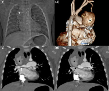
A 10-month-old infant with a history of tetralogy of Fallot, palliated with a modified Blalock–Taussig shunt at 5 months of age, presented to the emergency room with cyanosis and respiratory distress. Her oxygen saturation at room air was 72%, while the rest of her vital signs were normal. Chest X-rays revealed a large right paratracheal mass, and a transthoracic echocardiogram indicated stenosis and hypoplasia of the right pulmonary artery. A cardiac catheterization was performed to clarify the diagnosis.
The aortogram showed occlusion of the shunt’s proximal segment and the angiography pointed to severe stenosis of the right pulmonary artery at the level of the shunt anastomosis, with contrast entering into the right mediastinum. However, catheter-directed angiography was limited by its 2D nature, and the impossibility of concomitantly evaluate the systemic and pulmonary vascular systems.
Therefore, a CT angiography was done, and it showed a 27-mm pseudoaneurysm arising from the stenotic portion of the right pulmonary artery ((Fig 1).
Surgery was decided. Interestingly, an unexpected, huge collection arising from a pseudoaneurysm of the right pulmonary artery was noticed and drained. Ignoring at that moment the nature of this collection, a complete surgical repair of the tetralogy of Fallot was performed. After microbiological cultures of the collection tested positive for Salmonella, complicated endocarditis was considered and intensive antimicrobial treatment was established.
This case illustrates the utility of CT angiography in the assessment and surgical planning in patients with complex CHDs, as well as its superiority in producing images with a higher temporal and spatial resolution compared to those produced in catheter-directed angiography.Reference Haramati, Glickstein and Issenberg1,Reference Dillman and Hernandez2
Right pulmonary artery (RPA) pseudo-aneurysm, stenosis and occluded right BT shunt. The chest X Ray shows a right mediastinal mass in a patient with history of previous right BT shunt (White asterisk on a). CTA of the chest (posterior view of a 3D rendering on b and coronal-reformatted images on c and d) shows the big pseudo-aneurysm (black asterisk on b to d) as well as the RPA stenosis (White arrows on b and c). A communication with the RPA is also depicted (Black arrow on d).

Acknowledgments
None.
Financial support
This research received no specific grant from any funding agency, commercial or not-for-profit sectors.
Conflict of interest
None.
Ethical standards
The research protocol was approved by the Research Committee of the Fundación Cardioinfantil.