Introduction
Schistosomiasis affects over 200 million people with more than 250,000 deaths per year. In 2017, 220.8 million needed schistosomiasis prophylaxes, with only 102.8 million receiving treatment (Bajracharya et al., Reference Bajracharya, Pandit and Bhandari2020). Most infections occur in rural and low-income areas of sub-Saharan Africa (Adenowo et al., Reference Adenowo, Oyinloye, Ogunyinka and Kappo2015). Depending on the region, different Schistosoma species can infect humans. Schistosoma mansoni and Schistosoma haematobium are endemic in Africa and the Middle East, accounting for 85% of global cases (Barakat et al., Reference Barakat, El Morshedy, Farghaly, McDowell and Rafati2014). Both S. mansoni and S. haematobium are found in Egypt (Adenowo et al., Reference Adenowo, Oyinloye, Ogunyinka and Kappo2015).
The World Health Organization recommends praziquantel (PZQ) as the only available drug to control and treat schistosomiasis (Tekwu et al., Reference Tekwu, Anyan, Boamah, Baffour-Awuah, Keyetat Tekwu, Penlap Beng, Nyarko and Bosompem2016). The intensive and exclusive use of a single drug for decades may eventually lead to PZQ resistance (Cioli et al., Reference Cioli, Pica-Mattoccia, Basso and Guidi2014). PZQ only works on adults and is ineffective on schistosomula, preadults, or juvenile adults. It is then necessary to develop effective and safe antischistosomal agents. PZQ, despite being the traditional anti-schistosomal drug therapy (Doenhoff & Pica-Mattoccia, Reference Doenhoff and Pica-Mattoccia2006), cannot reverse hepatic fibrosis. There is a crucial need for a potent anti-fibrotic agent to prevent the progression of fibrosis especially in chronic liver diseases where reversible fibrosis is turned into irreversible cirrhosis with severe scarring of the hepatic tissues (Al-Olayan et al., Reference Al-Olayan, El-Khadragy, Aref, Othman, Kassab and Abdel Moneim2014). In recent years, the scientific community has focused on the possibility of producing a drug for schistosomiasis that is based on natural sources (Abou El-Nour & Fadladdin, Reference Abou El-Nour and Fadladdin2021).
Biochanin A (BCA) is an isoflavone found in red clover, cabbage and alfalfa (Cassady et al., Reference Cassady, Zennie, Chae, Ferin, Portuondo and Baird1988). BCA has anti-inflammatory, anti-oxidant, anti-infective, hepatoprotective and anticancer properties (Sarfraz et al., Reference Sarfraz, Javeed and Shah2020). Recent studies showed that BCA has potent anti-inflammatory effects in arthritis, neurological and metabolic disease models (Felix et al., Reference Felix, Vago, Beltrami, Araújo, Grespan, Teixeira and Pinho2022). Kole et al. (Reference Kole, Giri, Manna, Pal and Ghosh2011) showed that BCA inhibited lipopolysaccharide (LPS)-induced nitric oxide (NO) production in macrophages and inhibited inducible nitric oxide synthases (iNOS) expression through inhibition of nuclear factor-κB (NF-κB). Breikaa et al. (Reference Breikaa, Algandaby, El-Demerdash and Abdel-Naim2013) found that pretreatment with BCA protected against chemokine (C-C motif) ligands 4 (CCL4)'s chronic damage, oxidative stress, inflammation and fibrosis. BCA regulates multiple biological functions by binding to DNA and proteins or acting as a substrate for enzymes (Křížová et al., Reference Křížová, Dadáková, Kašparovská and Kašparovský2019). This immunomodulatory molecule can modulate several biological networks, so it may serve as a promising natural novel drug lead (Sarfraz et al., Reference Sarfraz, Javeed and Shah2020).
Schistosomiasis is the most common parasitic fibrotic disease due to inflammation and the deposition of scar tissue around trapped ova in the liver (Gryseels et al., Reference Gryseels, Polman, Clerinx and Kestens2006). Some Schistosoma eggs are excreted through the stool, but others are trapped in the liver. The trapped eggs in the liver can cause inflammation. Chronic inflammation causes granulomas and eventually liver fibrosis (Colley et al., Reference Colley, Bustinduy, Secor and King2014), characterized by an abnormal accumulation of activated hepatic stellate cells (HSCs), which produce much extracellular matrix (ECM), leading to myofibroblast development and liver fibrosis (Friedman, Reference Friedman2008; Zhang et al., Reference Zhang, Yuan, He, Lei and Wang2016).
In fibrotic disease, increased deposition of ECM components affects the tissue architecture, and interferes with normal function (Macneal & Schwartz, Reference Macneal and Schwartz2012). Fibrogenic proteins such as platelet-derived growth factor (PDGF) and transforming growth factor (TGF) promote liver fibrosis (Meng et al., Reference Meng, Nikolic-Paterson and Lan2016; Ying et al., Reference Ying, Chen, Zhang, Zhang, Ma, Zhang, Fang and Yu2017). TGF-β is a widely expressed fibrogenic cytokine (Al-Olayan et al., Reference Al-Olayan, El-Khadragy, Alajmi, Othman, Bauomy, Ibrahim and Abdel Moneim2016) that plays an active role in proliferation, synthesis of ECM molecules and proliferation of HSCs (Du, Reference Du1999; Al-Olayan et al., Reference Al-Olayan, El-Khadragy, Alajmi, Othman, Bauomy, Ibrahim and Abdel Moneim2016). TGF-β, therefore, strongly contributes to fibrotic disorders.
Nitric oxide plays a key role in both physiological inflammatory response and exaggerated pathological processes. During inflammation, there is an increased production of pro-inflammatory cytokines, including iNOSs. Increased mucosal nitric oxide synthase-2 expression and the production of NO have been reported in active inflammatory conditions as an experimental model of intestinal inflammation (Miller et al., Reference Miller, Thompson and Zhang1995; Matsumoto et al., Reference Matsumoto, Okabe, Setoyama, Takayama, Ohtsuka, Funahashi, Imaoka, Okada and Umesaki1998).
Cyclooxygenase (COX) is the main enzyme that promotes the synthesis of prostaglandins. Two isoenzymes have been recognized: COX-1, which is expressed in many tissues to produce prostaglandins in physiological functions; and COX-2 that is induced by a variety of stimuli, and it is responsible for the production of prostaglandins under stressful conditions such as inflammation (Crofford, Reference Crofford1997). The downregulation of COX-2 expression has been linked to failure to synthesize prostaglandin E2, which is a potent inhibitor of fibroblast proliferation which could lead to an altered tissue repair (Keerthisingam et al., Reference Keerthisingam, Jenkins, Harrison, Hernandez-Rodriguez, Booth, Laurent, Hart, Foster and McAnulty2001; Xaubet et al., Reference Xaubet, Roca-Ferrer, Pujols, Ramírez, Mullol, Marin-Arguedas, Torrego, Gimferrer and Picado2004).
There is scarce data on the direct effect of BCA on Schistosoma species. Most studies investigated genistein, a BCA metabolic product. Genistein affected Schistosoma tegument, NO, Ca2+ homeostasis, carbohydrate metabolism and fibrosis in studies on S. mansoni and Schistosoma japonicum experimental infections (Faixová et al., Reference Faixová, Hrčková, Mačák Kubašková and Mudroňová2021). In this study, we investigated all possible effects of BCA on S. mansoni-induced changes in an experimental mouse model of infection, including its anti-inflammatory, anti-oxidative and anti-fibrotic properties, besides exploring its action on S. mansoni itself as an anti-parasitic through its effect on cytochrome P450 (CYP450) enzyme expression level.
Materials and methods
Animals and infection
The CD-1 Swiss female albino mice (their weight was 18–20 g), were purchased from the Medical Experimental Research Center (MERC), Mansoura University, Egypt. The mice were maintained at 20–22°C in an air-conditioned laboratory and provided with standard pelleted diet and water ad libitum. Each mouse (of infected groups) was infected with S. mansoni (Egyptian strain) cercariae by subcutaneous injection of 60 ± 10 cercariae/100 μl in dechlorinated water (Liang et al., Reference Liang, Bruce and Boyd1987).
Drugs, animal grouping and dosing
Praziquantel® (PZQ), was purchased from Sedico Pharmaceutical Co. (6th of October City, Egypt). BCA was purchased from Sigma-Aldrich Chemical Co., (St. Louis, MO, USA). Dimethyl sulphoxide was purchased from Sigma-Aldrich (St. Louis, MO, USA) as a vehicle for dissolving BCA and PZQ. Mice were divided into six groups. Group I (n = 5) was normal (uninfected) control mice. Group II (n = 5) included normal uninfected mice treated with BCA (50 mg/kg/day) given orally as a single dose by intragastric gavage, seven days post-infection (dpi). Group III (n = 10) included infected untreated mice (vehicle treated). Group IV (n = 10) was infected PZQ-treated, orally administered 60 dpi, in a total dose of 500 mg/kg/day for two days (Gönnert & Andrews, Reference Gönnert and Andrews1977). Group V (n = 10) was early infected BCA-treated after 7 dpi in a single dose of 50 mg/kg body weight/day (Moon et al., Reference Moon, Sagawa, Frederick, Zhang and Morris2006; Breikaa et al., Reference Breikaa, Algandaby, El-Demerdash and Abdel-Naim2013). Group VI (n = 10) was late infected BCA-treated mice 60 dpi which is the same regimen as Group V. Early intervention (one week dpi) was targeting the immature schistosomula stages and late intervention (60 dpi) was targeting mature adult worms (de Oliveira et al., Reference de Oliveira, Rehder, Oliveira, Jeraldo, Linhares and Allegretti2014). Mortality rate was 10–15% throughout the study. Mice of all groups were euthanized 12 weeks post-infection by a thiopental intraperitoneal injection, 100 mg/kg (Jones-Bolin, Reference Jones-Bolin2012).
Parasitological assessment
Immediately after euthanizing, adult S. mansoni were collected by perfusions of porto-mesenteric veins of infected mice. Half of the liver specimen was processed for dissolution in 0.5% potassium hydroxide and trapped eggs were counted as per tissue gram according to Cheever (Reference Cheever1968). Collected worms were subjected to quantitative real-time PCR (RT-qPCR), to examine the gene expression level of CYP450 in S. mansoni.
Histopathological examination and morphometric assessment of fibrosis in the granuloma
Liver specimens were processed in buffered formalin (10%) and then paraffin. Liver sections were stained by Masson's trichrome (4 μm thickness) and examined under light microscopy for granuloma size measurement. Two diameters at 90 angles to each other were measured to calculate the mean diameter of each granuloma (von Lichtenberg, Reference von Lichtenberg1962). Liver tissues’ pathological changes were assessed for hydropic degeneration, portal tract affection and inflammatory cellular infiltrates (Knodell et al., Reference Knodell, Ishak, Black, Chen, Craig, Kaplowitz, Kiernan and Wollman1981; Suzuki et al., Reference Suzuki, Toledo-Pereyra, Rodriguez and Cejalvo1993). The evolutionary stages of granulomas were classified according to Amaral et al. (Reference Amaral, Silva, Dias, Malta, Rosa, Costa-Neto, Gentile and Melo2017). The studied pathological parameters were lobular inflammation, focal necrosis, portal tract inflammation, microvesicular steatosis, collagen around granuloma and inflammatory cellular infiltrate.
In addition, stained liver sections were examined for collagen fibre deposition. Slides were photographed using an Olympus® digital camera installed on Olympus® microscope with 0.5 X photo adaptor and saved as TIFF. Then images were analysed using Video-Test Morphology® software (Russia) on Intel® Core I7® based computer with a specific built-in routine for area measurement and stain quantification. Two slides from each mouse were prepared and five random fields from each slide were analysed. The granuloma fibrosis degree was expressed as mean ± standard error of the mean (SEM) (the percentage of the stained area compared to the total area; James et al. (Reference James, Bosch, Aronson and Houtkooper1990)).
Immunohistochemical staining of TGF-β, iNOS and COX2
Immunohistochemistry (IHC) was performed to examine TGF-β, iNOS and prostaglandin-endoperoxide synthase, known as COX2 expressions in the liver tissue hepatocytes and inflammatory cells. Liver tissues from each group were fixed in neutral buffered formalin (10%) and embedded in paraffin. For TFG-β, the procedure was done using a specific primary anti-TFG-β 1 antibody (FNab08638, at dilution of 1:200), Fine Test® enzyme-linked immunosorbent assay test (Wuhan Fine Biotech Co., Ltd, Wuhan, Hubei, China) and appropriate secondary antibody according to Shaker et al. (Reference Shaker, Eisa, Elgaml, El-Mesery, El-Shafey, El-Dosoky, El-Mowafy and El-Mesery2021). For iNOS, the procedure was done using a specific primary antibody NOS2 Rabbit pAb (A14031) at dilution of 1:100 (ABclonal, Inc., MA, USA) according to Choi et al. (Reference Choi, Nam, Jin, Kim and Cha2012), and HRP Goat Anti-Rabbit IgG (H + L) (AS014) at 1:10,000 dilution (ABclonal, Inc., MA, USA) as secondary antibody. For COX2, the procedure was done using a specific primary antibody Cox2 Rabbit pAb (A1253) at dilution of 1:200 (ABclonal, Inc., MA, USA) and appropriate secondary antibody according to Zhang et al. (Reference Zhang, Gu, Jia, Zhao, Li, Shen, Liu, Wang and Shi2020). All antigens’ expressions were detected in granuloma-adjacent hepatocytes’ cytoplasm and inflammatory cells at the areas of the inflammation and fibrosis (according to manufacturer's instructions). Cells positivity staining was graded (Elshal et al., Reference Elshal, Abu-Elsaad, El-Karef and Ibrahim2015): grade 0 for 0–10% staining; grade 1 for 11–25% staining; grade 2 for 26–50% staining; and grade 3 for >50% staining. Intensity of the staining was graded as 1 for light yellow, 2 for dark yellow, and 3 for brown staining. Then the sum of both grades was scored, and results were expressed as means ± SEM.
Assessment of CYP450 gene expression by RT-qPCR
Adult S. mansoni were collected, and six worms from each infected group were homogenized by three strokes of liquid nitrogen. Total cellular RNA was extracted with QIAzol reagent (Qiagen, Germany), according to the manufacturer's instructions. RNA concentration was measured by NanoDrop 2000 (Thermo Scientific, CA, USA). Reverse transcription of 1 μg of RNA was done with SensiFAST™ cDNA Synthesis Kit (Bioline, UK). The qRT-PCR was carried out with HERA SYBR green PCR Master Mix (Willowfort, UK) in a total volume of 20 μl using a qRT-PCR device (Pikoreal 96): 95°C (2 min), 40 cycles of 95°C (10 s), 60°C (30 s). The primer pairs sequences were: CYP450 forward, 5′ TGGGAATCGTTCGAAAGTTGG 3′, reverse, 5′ TTCCAGTGCCTTTTGTGTGT 3′ (RefSeq; XM_018794880.1) and the product length was 100 base pairs (bp), alpha tubulin (as a control gene) forward, 5′ GAAGGCTTACCACGAGCAGT 3′, reverse, 5′ GGGGACAACATCACCACGAT 3′ (RefSeq: XM_018797513.1) and the product length was 139 bp. The primer sets for both genes were designated using Primer 3 software (v.4.1.0) [http://primer3.ut.ee], and primer specificity was assessed using Primer-BLAST program (NCBI/ primer-BLAST [https://www.ncbi.nlm.nih.gov/tools/primer-blast/]. Primers’ sets were synthesized and purchased from Vivantis (Vivantis Technologies, Malaysia). Reactions were done in triplicate. Relative gene expression levels were represented as ΔCt = Ct target gene– Ct housekeeping gene; 2−ΔΔCT method was used to calculate the fold change in gene expression (Livak & Schmittgen, Reference Livak and Schmittgen2001). PCR products were run on 3% agarose gels and visualized on an ultraviolet transilluminator (OWI Scientific, France). Then, the gels were photographed using the Bio-Rad gel documentation system (Bio-Rad, USA).
Statistical analysis
Data were analysed using IBM-SPSS Software Version 20 (IBM Corp., Armonk, NY, USA) and Microsoft Excel Version 2013. Quantitative values were expressed as mean ± standard deviation. Values were expressed as mean ± standard error. Statistics: one-way analysis of variance followed by post-hoc multiple comparisons; Games–Howell adjustment. Data were considered statistically significant at P value < 0.05.
Results
In the current study, we investigated the effects of BCA as an anti-infective (antiparasitic), anti-inflammatory, anti-oxidative and anti-fibrotic agent.
Parasitological parameters
Early (7 dpi) and late (60 dpi) administration of BCA in a dosage of 50 mg/Kg for two days to S. mansoni-infected mice significantly reduced (P < 0.001) the number of the total worm burden compared to the infected untreated group with a reduction rate of 82.14% and 77.74%, respectively (table 1). However, BCA did not achieve complete eradication of the worms as was the case with PZQ administration. All drug regimens: PZQ, early and late BCA significantly decreased hepatic tissue egg count compared to infected untreated group (P < 0.001) with reduction rates of 51.4%, 48.7 and 46.1%, respectively. Early vs. late administration of BCA drug, showed a non-significant different impact on parasitological parameters.
Parasitological effects of different treatment regimens on Schistosoma mansoni-infected mice (n = 10/group).

BCA, biochanin A; PZQ, praziquantel.
Data are presented as mean ± standard error.
a Significant difference vs. infected-control group at P < 0.001
b Significant difference vs. infected-control group at P < 0.05.
c Significant difference vs. early BCA-treated group at P < 0.05
d Significant difference vs. late biochanin BCA-treated group at P < 0.05.
P value by one-way analysis of variance Games–Howell adjustment, post-hoc test.
Histopathological parameters of different treatment regimens on Schistosoma mansoni-infected mice (n = 10/group).

BCA, biochanin A; PZQ, praziquantel.
Data are presented as mean ± standard error.
a Significant difference vs. infected-control group at P < 0.001.
P value by one-way analysis of variance Games–Howell adjustment, post-hoc test.
Histopathological findings
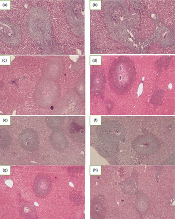
All drug regimens: PZQ, early and late BCA resulted in significantly higher percentage (P < 0.001) of degenerated ova within the liver granulomas compared to the infected untreated group. Regarding the diameter of the hepatic granuloma among the infected groups, both early and late BCA treatment displayed significantly reduced diameter compared to the infected untreated group (P < 0.001) with reduction rates of 18.3% while PZQ treatment displayed hepatic granuloma diameter comparable to the infected untreated group with no statistical difference (table 2, fig. 1).
Effect of administration of biochanin A (BCA) and praziquantel (PZQ) on the histopathological examinations of the liver sections of different mice groups of the study (Masson's trichrome ×200): (a, b) infected untreated control group showed multiple irregular granulomas surrounding partly or totally degenerated ova and large cellular infiltrate; (c, d) PZQ-treated infected group with lesser degree of cellular granuloma number, though approaching in its size the infected untreated group; (e, f) early BCA-treated infected group showed granulomas with less cellular infiltrate of the liver parenchyma compared to the infected control; and (g, h) late BCA-treated group with relatively less cellular and smaller granulomas.

Liver tissues in all treated groups showed variable minimal to moderate degrees of focal necrosis and hydropic changes. In the PZQ-treated group, 80% of examined specimens were of minimal degree of focal necrosis and hydropic changes. In the BCA-treated group specimens, 60% of specimens were mild while 40% of specimens were considered moderate focal necrosis and hydropic changes. In the infected untreated group, focal necrosis was of moderate degree in most examined specimens. Regarding the inflammatory cellular infiltrates, in the infected untreated group, 60% of the examined specimens displayed a moderate degree of inflammatory infiltrate and 40% displayed marked inflammatory cellular infiltrate. In the PZQ-treated group, most of the examined liver tissue sections ranged from mild to moderate degree of inflammatory cellular infiltrates. In both the early and late BCA treated groups, most of the examined specimens (about 80% of the liver sections) showed mild inflammatory cellular infiltrates with a few numbers of moderate cellular inflammatory infiltrates.
Portal tract inflammation ranged from moderate to marked in both infected untreated and PZQ-treated group; meanwhile, the portal inflammatory infiltrate was generally a moderate degree in both infected-BCA treatment groups.
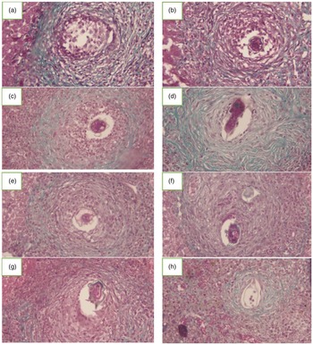
The granulomatous reaction was mainly cellular in the infected untreated group while in all treatment groups, granulomas were mainly fibrocellular. According to the classification of Amaral et al. (Reference Amaral, Silva, Dias, Malta, Rosa, Costa-Neto, Gentile and Melo2017), in the infected untreated group, most of granulomas (80%) were pre-granulomatous exudative, and the exudative–productive type was the main granuloma stage in all treatment groups (fig. 1). Quantification of fibrosis percentage within the granulomas was performed on Masson's trichrome-stained liver sections (fig. 2), whereas the mean percentage of fibrosis was higher in the PZQ-treated group (18.66 ± 2.15) and decreased in the BCA-treated groups (11.56 ± 2.04 and 12.82 ± 1.80) compared to the infected untreated group (13.73 ± 0.57); however, with no statistical difference.
Effect of administration of biochanin A (BCA) and praziquantel (PZQ) on the liver fibrosis of different mice groups of the study (Masson's trichrome ×400): (a, b) infected control group, showed large cellular granuloma surrounding degenerated ova; (c, d) PZQ-treated infected group, granuloma displayed lesser degree of cellular infiltrate and with increased fibrous tissue deposition; and (e–h) early BCA-treated infected (e, f) and late BCA-treated (g, h) showed granulomas surrounding partly degenerated ova with less fibrous tissues’ deposition compared to the PZQ-treated granuloma.

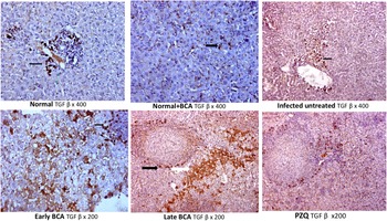
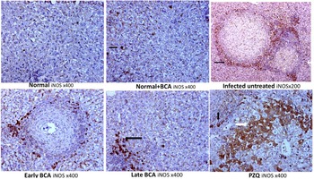
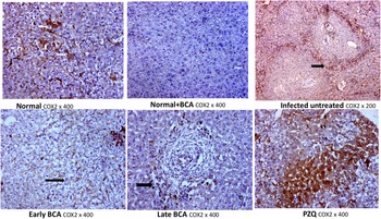
To analyse the changes of TGF-β, iNOS and COX-2 upon BCA treatment, we analysed the baseline expression of each cytokine in normal liver tissue without BCA treatment and in a normal liver tissue treated with BCA, and compared the staining intensity score within the different treated and control positive groups. The results demonstrated that TGF-β and iNOS expression increased significantly after PZQ treatment in both hepatocytes and inflammatory cells; TGF-β expression was 50% in both hepatocytes and inflammatory cells (fig. 3). However, with BCA treatment the score was significantly reduced to 25% and 20% and 20% and 10% in early and late BCA treatment, respectively (fig. 3). PRQ treated liver tissue showed strong positive iNOS expression in hepatocytes (brown staining intensity in 30% of hepatocytes) while BCA late treatment showed mild increase in expression level (25%) (fig. 4). Interestingly, increased COX2 expression was evident in the early BCA treated group: 50% in contrast with late BCA group 10%. However, the late treated group had a lower expression level compared to PZQ treated group: 10% vs. 30% (fig. 5). TGF-β and iNOS expressions in the BCA-treated animals were significantly reduced compared with the PZQ group. At the same time, COX2 expression did not differ significantly between the two treatment arms.
Effect of administration of biochanin A (BCA) and praziquantel (PZQ) on the immunohistochemistry expression of transforming growth factor (TGF-β) in the liver of Schistosoma mansoni-infected mice. Normal liver showed minimal immunohistochemical expression of TGF-β in the cytoplasm of hepatocytes (black arrow). Infected untreated mice showed strong positive expression of TGF β in hepatocytes with mild positive expression in the inflammatory cells (black arrow). Early BCA treatment showed mild positive expression of TGF-β expression in hepatocyte with mild positive expression of TGF-β in the inflammatory cells (black arrow). Late BCA showed mild positive expression of TGF-β in hepatocytes (white arrow) with mild positive expression in the inflammatory cells (black arrow). PZQ treatment showed moderate positivity of TGF-β in hepatocytes.

Effect of administration of biochanin A (BCA) and praziquantel (PZQ) on the immunohistochemistry expression of inducible nitric oxide synthase (iNOS) in the liver of Schistosoma mansoni-infected mice. Normal liver showed negative iNOS expression in hepatocyte and in the inflammatory cells. Normal liver treated with BCA showed negative iNOS expression in hepatocytes with mild positive expression in the inflammatory cells (black arrow). Infected untreated mice showed mild positive iNOS expression in hepatocytes with strong positive expression in the inflammatory cells (black arrow). Early BCA treatment showed negative iNOs expression in hepatocytes with mild positive iNOS expression in the inflammatory cells. Late BCA treatment showed negative iNOS expression in hepatocytes with mild positive expression in the inflammatory cells (black arrow). PZQ treatment showed strong positive iNOS expression in hepatocytes (white arrow) with mild positive expression in the inflammatory cells (black arrow).

Effect of administration of biochanin A (BCA) and praziquantel (PZQ) on the immunohistochemistry expression of cyclooxygenase (COX-2) in the liver of Schistosoma mansoni-infected mice. Normal liver showed mild positive immunohistochemical expression of COX2 in hepatocytes (white arrow) with mild positive expression in the inflammatory cells (black arrow). Normal liver treated with BCA showed negative COX2 expression in hepatocytes and the inflammatory cells. Infected untreated mice showed mild positive COX2 expression in hepatocytes with strong positive expression in the inflammatory cells (black arrow). Early BCA treatment showed negative COX2 expression in hepatocyte with strong expression of Cox2 in the inflammatory cells (black arrow). Late BCA treatment showed negative COX2 expression in hepatocytes with mild positive expression in the inflammatory cells (black arrow). PZQ treatment showed strong positive COX2 expression in hepatocytes (black arrow).

Immunohistochemical expression of TGF-β, iNOS and COX2 receptors on liver tissues
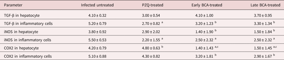
For further evaluation of the BCA effect on the liver fibrosis, IHC of TGF-β in liver sections was done. TGF-β showed cytoplasmic expression in hepatocytes comparable to the infected untreated group sections, while TGF-β expression in inflammatory cells was decreased in the treatment groups (table 3, fig. 3) compared to the infected untreated group at (P < 0.05 in both the early and late BCA-treated groups and at P < 0.001 in the PZQ-treated group). The control uninfected group showed minimal TGF-β expression in the liver tissues while all S. mansoni-infected groups showed a significant increase of TGF-β expression compared to the control uninfected group (P < 0.05). Therefore, the antifibrotic effect of BCA was significant at the inflammatory cells’ expression levels (rather than the tissues) in the S. mansoni-infected mice model treated with either early or late BCA.
Immunohistochemical expression of transforming growth factor (TGF-β), inducible nitric oxide synthase (iNOS) and cyclooxygenase (COX2) in the liver sections on hepatocytes and inflammatory cells (n = 10 for each group).

BCA, biochanin A; PZQ, praziquantel.
Immunohistochemistry parameters were presented as an expression score. Data are presented as mean ± standard error. Statistics: one-way analysis of variance followed by post-hoc multiple comparisons; Games–Howell adjustment.
a P < 0.001 vs. infected control group.
b P < 0.05 vs. infected control group.
c P < 0.001 vs. infected PZQ-treated group.
To evaluate the anti-oxidative effect of BCA on the liver cells, IHC of iNOs in liver sections was done. The iNOS cytoplasmic expression in hepatocytes was evident in comparison to the infected untreated group sections, while its expression in inflammatory cells significantly declined in all treatment groups (table 3, fig. 4) compared to the infected untreated group (P < 0.001). In the hepatic tissues the iNOS expression was also downregulated in the BCA-treated groups (P < 0.005) compared to the infected untreated group. The anti-oxidative properties of BCA were evident in this model.
The IHC of COX2 receptors in the liver sections was carried out to evaluate anti-inflammatory effects of BCA. COX2 showed significantly decreased expression in hepatocytes of both BCA-treated groups (P < 0.001) compared to the infected untreated group and PZQ-treated group (P < 0.05) (table 3, fig. 5). In addition, COX2 expression was downregulated in inflammatory cells in the liver, in both the early and late BCA-treated groups (P < 0.05) compared to the infected untreated group.
Effect of BCA on S. mansoni CYP450 mRNA expression
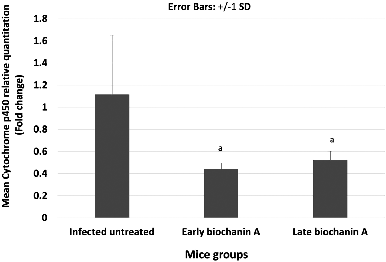
Collected worms were subjected to RT-qPCR to examine the gene expression level of CYP450 in S. mansoni. Early exposure to BCA during the first week and late exposure (60 dpi) significantly inhibited S. mansoni CYP450 mRNA expression in adult stage (P < 0.01 and P < 0.05, respectively) compared to the mRNA expression levels in the infected untreated group adult worms (figs 6, 7). Furthermore, a significantly lower CYP450 mRNA expression was detected during early vs. late BCA treatment (P < 0.05). Total worm burden reduction was evident in the groups treated with BCA: 82.14% reduction in early vs. 77% reduction in late BCA administration.
Schistosoma mansoni cytochrome P450 mRNA relative expressions as quantitative real-time polymerase chain reaction in all studied animal groups: (a) P < 0.001 vs. infected untreated group; (b) P < 0.05 vs. infected untreated group; and (c) P < 0.05 vs. infected late biochanin A-treated group.

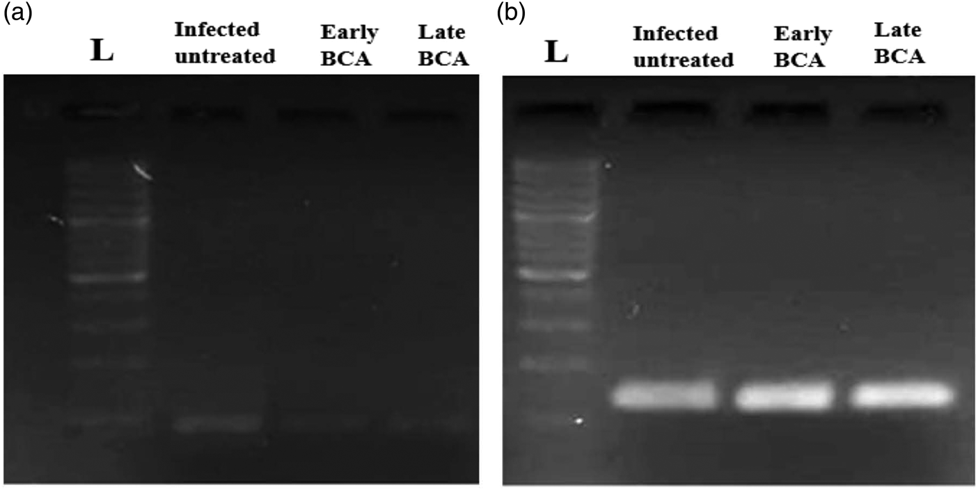
Gel electrophoresis of the real-time quantitative real-time polymerase chain reaction (RT-qPCR) products. Lane 1, 50 base pairs (bp) ladder (L); Lane 2, infected untreated group; Lane 3, early biochanin (BCA)-treated group; and Lane 4, late BCA-treated group: (a) cytochrome P450 RT-qPCR product (100 bp); and (b) alpha tubulin RT-qPCR product (139 bp).

Discussion
In S. mansoni infection, liver affection is a hallmark (Colley et al., Reference Colley, Bustinduy, Secor and King2014; McManus et al., Reference McManus, Dunne, Sacko, Utzinger, Vennervald and Zhou2018). Extensive granulomas could affect the liver tissues, with subsequent hepatic fibrosis, presenting as hepatosplenomegaly and portal hypertension (Andrade, Reference Andrade2009; Chuah et al., Reference Chuah, Jones, Burke, McManus and Gobert2014; Colley et al., Reference Colley, Bustinduy, Secor and King2014). Granulomas which are organized collections of inflammatory cells around deposited eggs in target organs, are considered the most prominent pathological feature of schistosomiasis (Hams et al., Reference Hams, Aviello and Fallon2013; Chuah et al., Reference Chuah, Jones, Burke, McManus and Gobert2014). Although known to be protective, preventing persistent antigenic stimulation from cytokines of trapped schistosome eggs (Pagán & Ramakrishnan, Reference Pagán and Ramakrishnan2018), granulomas themselves are the main pathological lesions in various organs with subsequent fibrosis and scarring (Hams et al., Reference Hams, Aviello and Fallon2013).
Schistosoma liver fibrosis is associated with HSCs’ activation to myofibrobalsts with accumulation of ECM proteins around the eggs. Among the most important pathways involved in hepatic fibrosis is the TGF-β release pathway (Moreira, Reference Moreira2007; Fagone et al., Reference Fagone, Mangano and Mammana2015) and COX-2 which is an essential factor in the progression of hepatic fibrogenesis. In response to these inflammatory changes, oxidative stress response occurs in the host leading to the release of NO and iNOS and other oxidative stress response mediators either directly or indirectly (Kole et al., Reference Kole, Giri, Manna, Pal and Ghosh2011).
Biochanin A is a bioactive isoflavone of soy, peanuts and red clover with versatile pharmacological potential. It has anti-inflammatory, anti-oxidant, neuroprotective, anti-microbial, hepatoprotective and anticancer properties (Yu et al., Reference Yu, Zhang, Lou and Wang2019). BCA exerts its anti-tumorigenic effects by abolition of cellular growth while activating cancer cell apoptosis. BCA displays chemopreventive properties in the liver, prostate, breast and pancreatic cancers (Yu et al., Reference Yu, Zhang, Lou and Wang2019). BCA is metabolized to genistein, BCA conjugates and genistein conjugates (Moon et al., Reference Moon, Sagawa, Frederick, Zhang and Morris2006). In the current study, we investigated BCA's anti-parasitic effects, anti-inflammatory (COX-2 and TGF-β) and antioxidant effects (iNOS) in the context of Schistosoma liver fibrosis. We chose BCA because it has a stronger effect both directly and after conversion to its genistein metabolite, extending its half-life and activity. Investigating its antioxidant, anti-inflammatory, in addition, to its CYP450 inhibitory activity. partially explains its mechanism of action. Hepatic granulomatous inflammation, fibrosis and cytokine production (COX-2 and TGF-β) were monitored.
In our study, BCA reduced S. mansoni egg production, possibly due to reduced worm burden. Oral BCA treatment was more effective than PZQ against immature S. mansoni early infection; however, in chronic infections, BCA was less effective than PZQ. Despite the modest worm and egg burden reductions, BCA was more effective than PZQ against immature parasites (early infection). It was associated with reduced morbidity because schistosome parasites do not multiply in mammals (Guerra et al., Reference Guerra, Silva, Silva, Salvadori, Teixeira, de Oliveira, Rocha, Pinto and de Moraes2019).
Early BCA showed smaller granulomas and more intact ova with less fibrosis than PZQ. In the S. mansoni infection model using red propolis that contains genistein and BCA, Silva et al. (Reference Silva, Silva, Mengarda, Salvadori, Teixeira, Alencar, Luz Filho, Bueno-Silva and de Moraes2021) found that it significantly reduced S. mansoni egg production early in infection compared to late infection. Interfering with egg oviposition might be linked to the schistosome reproductive system changes. Because egg production is essential for schistosome transmission and pathogenesis, the impact on S. mansoni egg-laying is important for natural products with antischistosomal properties (Aleixo de Carvalho et al., Reference Aleixo de Carvalho, Geraldo, de Moraes, Silva Pinto, de Faria Pinto, Pereira Odos and Da Silva Filho2015; Dias et al., Reference Dias, Zuza and Riani2017).
Another study by Mohamed et al. (Reference Mohamed, Hassab El-Nabi, Bayomi and Abdelaal2016) found that propolis reduced the worm and egg burden in S. mansoni-infected mice (chronic infection). However, in another study, Egyptian propolis had poor antischistomal properties on adult worms (chronic infection) in the S. mansoni-mice model (Mahmoud et al., Reference Mahmoud, Rizk, Maghraby and Shaheen2014). The inconsistencies are probably due to variations in propolis and Schistosoma strains. Propolis is a natural resin collected by bees from various plant parts; its chemical composition varies greatly depending on the location and local flora (Rufatto et al., Reference Rufatto, dos Santos, Marinho, Henriques, Roesch Ely and Moura2017).
In accordance with previous reports about the efficiency of BCA as an anti-pathogenic agent, BCA reduced the number of bacterial colonies in the lung tissues and decreased haemolysin secretion in alveolar lavage fluid. It also diminished the degree of pulmonary oedema and protected the host from methicillin-resistant Staphylococcus aureus (MRSA) infection by inhibiting MRSA haemolysin expression, providing experimental evidence for its potential anti-MRSA drug development (Feng et al., Reference Feng, Sun and Wang2021).
Regarding parasitic infections, in vitro studies have shown genistein to affect several enzymatic systems, localized in the tegument and other worm compartments, indicating its multiple molecular targets (Tandon & Das, Reference Tandon and Das2018). In cestodes and trematodes, genistein acts on tegument causing flaccid paralysis in vitro, and causes irreversible structural changes (Tandon et al., Reference Tandon, Pal, Roy, Rao and Reddy1997). In vitro treatment of Echinococcus multilocularis metacestodes by genistein on the metacestode stage of E. multilocularis and Echinococcus granulosus caused profound morphological and structural changes with loss of viability and death (Naguleswaran et al., Reference Naguleswaran, Spicher, Vonlaufen, Ortega-Mora, Torgerson, Gottstein and Hemphill2006). Kar et al. (Reference Kar, Tandon and Saha2002) found that genistein treatment caused death of Fasciolopsis buski.
Similarly, BCA was active against Hymenolepis diminuta, leading to paralysis and death of the worms in a very short duration (Vijaya & Yadav, Reference Vijaya and Yadav2016). In protozoa, several isoflavones showed anti-Cryptosporidium parvum activity in vitro and in vivo (Stachulski et al., Reference Stachulski, Berry, Lilian Low, Moores, Row, Warhurst, Adagu and Rossignol2006). BCA was also active against Leishmania donovani (Tasdemir et al., Reference Tasdemir, Kaiser, Brun, Yardley, Schmidt, Tosun and Rüedi2006) and against trypomastigotes forms of Trypanosoma cruzi with even more efficacy than the standard drug benznidazole (Sartorelli et al., Reference Sartorelli, Carvalho, Reimão, Ferreira and Tempone2009).
Schistosome CYP450 is essential for worm survival and egg development, and thus a druggable target. Potential schistosomiasis treatments which include drugs that target fungal CYP450 were approved for the use in humans (Goswami et al., Reference Goswami, Goswami, Banerjee, Dadhwal, Miglani, Lattif and Kochupillai2006). CYP450 was also a drug target in many parasitic organisms, including T. cruzi, schistosomes, and Opisthorchis felineus. Anthelmintic activity was achieved by CYP450 inhibitors at micromolar to nanomolar concentrations (Mordvinov et al., Reference Mordvinov, Shilov and Pakharukova2017; Pakharukova et al., Reference Pakharukova, Pakharukov and Mordvinov2018).
For better understanding of the activity of BCA as an anti-parasitic agent, we thought to investigate its activity as a CYP450 inhibitor. Given that schistosomes have a single druggable cytochrome gene (Berriman et al., Reference Berriman, Haas and LoVerde2009), we hypothesize that BCA could be an antischistosomal drug via its CYP450 inhibitory action. Humans have 57 CYP450s genes that share 22% sequence identity (Turman et al., Reference Turman, Hatley, Ryder, Ravindranath and Strobel2006) while in schistosomal worm, CYP450 is represented with only one gene. This feature in schistosomes could be used to clear worms from mice at low doses.
In our study, early vs. late BCA treatment resulted in significantly lower CYP450 mRNA expression (P < 0.05) compared to infected untreated adult worms. Although not examined in our study, this could help prevent and control schistosomiasis in endemic areas if used as a prophylactic agent. In agreement with our findings, Ziniel et al. (Reference Ziniel, Karumudi, Barnard, Fisher, Thatcher, Podust and Williams2015) reported that dsRNA silencing of S. mansoni (Sm) CYP450 causes worm death and antifungal azole CYP450 inhibitors kill larval and adult worms at low micromolar concentrations and validate SmCYP450 as a novel drug target for worm survival and egg development. Similarly, slight inhibition of CY450 in the host, allows combined treatment with other effective anti-parasitic drugs to have an additive effect on elimination of schistosomes. Moreover, as an indirect effect, reduced metabolic activation of drugs by the monooxygenase (P450) system depressed the initial formation of reactive oxygen species and some intermediate toxic products, resulting in reduced lipid peroxidation and oxidative stress injury (Breikaa et al., Reference Breikaa, Algandaby, El-Demerdash and Abdel-Naim2013; Saleh et al., Reference Saleh, Abdel Jaleel, El-Awdan, Oraby and Badawi2014).
Regarding fibrosis, granulomas form because of accumulation of ECM proteins such as collagen around eggs causing fibrosis, one of the most severe chronic schistosomiasis consequences. HSCs’ activation to myofibroblast differentiation is a hallmark of liver fibrosis (Moreira, Reference Moreira2007). Among the major transcriptional targets of HSC activation are fibronectin, collagen I, smooth muscle actin (SMA), TGF, tumor necrosis factor, matrix metalloproteinase-2, matrix metallopeptidase 9 and tissue inhibitor of metalloproteinase (Fagone et al., Reference Fagone, Mangano and Mammana2015). Isoflavones including genistein were known to decrease the level of TGF-β, which activates HSCs (Huang et al., Reference Huang, Huang, Zhang, Lin, Wei, He, Zhuo and Lin2013). Although many signalling pathways are involved in liver fibrosis, Cong et al. (Reference Cong, Iwaisako, Jiang and Kisseleva2012) and Lam et al. (Reference Lam, Liang and Peng2021) reported that schisandrin B was effective in ameliorating S. mansoni liver fibrosis through downregulation of TGF-β. BCA acted as an anti-inflammatory agent that regulates TGF-β signalling (Cuiqiong et al., Reference Cuiqiong, Chao, Xinling and Yinyan2020).
The TGF-β promotes ECM formation, mesenchymal cell proliferation, migration and accumulation following inflammatory reactions. As a result, TGF-β is critical in the development of fibrosis in chronic inflammatory disorders. According to the stage of disease and site of action, TGF-β could either exacerbate or ameliorate symptoms. TGF-β was considered as an attractive therapeutic target for severe fibrosis (Pohlers et al., Reference Pohlers, Brenmoehl, Löffler, Müller, Leipner, Schultze-Mosgau, Stallmach, Kinne and Wolf2009). Furthermore, TGF-β could act as either a tumour suppressor or promoter dependent on disease stage (Galliher et al., Reference Galliher, Neil and Schiemann2006; Jakowlew, Reference Jakowlew2006). Inhibition of TGF-β appeared to be a promising strategy for fibrosis relief and cancer prevention. Therefore, there were many clinical trials investigating the use of TGF-β inhibitors targeting both fibrosis and cancer (Prud'homme, Reference Prud'homme2007). In the lungs, BCA treatment showed remarkable anti-inflammatory effects in TGF-β mediated pulmonary fibrosis. BCA inhibited TGF-β mediated epithelial–mesenchymal transition, myofibroblast differentiation and collagen deposition in vitro and in vivo (Andugulapati et al., Reference Andugulapati, Gourishetti, Tirunavalli, Shaikh and Sistla2020). Isoflavones (as genistein) are known to decrease the level of TGF-β, which activates HSCs (Huang et al., Reference Huang, Huang, Zhang, Lin, Wei, He, Zhuo and Lin2013).
The COX-2 is an essential factor in the progression of hepatic fibrogenesis. HSCs that were activated in the liver could hasten the start and progression of liver fibrosis. COX-2 overexpression causes inflammation (Crofford, Reference Crofford1997), which is a key factor in the development of hepatic fibrosis. COX-2 appears to play a role in the major pathophysiology of liver fibrosis, including inflammation, apoptosis and cell senescence. COX-2 expression was changed in people and animal models with cirrhosis or non-alcoholic fatty liver disease (Yang et al., Reference Yang, Xuefeng, Shandong and Jianhua2020).
In our study, hepatic granuloma size was significantly reduced in the group that received BCA in comparison to PZQ treated and infected control groups. Given the importance of liver granuloma in pathology and subsequent fibrosis, a reduction in size and surrounding fibrosis achieved by BCA administration is an important step towards prevention of further damage and development of premalignant lesions. In our results, TGF-β expression in inflammatory cells was decreased in treatment groups compared to the infected untreated group (P < 0.05) in the early and late BCA-treated groups and in the PZQ-treated group. BCA also reduced fibrosis. Treatment with BCA might have reduced fibrosis in part due to its anti-inflammatory properties. Its antioxidant and anti-inflammatory properties had been linked to genistein (Zhao et al., Reference Zhao, Wang, Liu, Wang, Guo, Ji, Wu and Zhou2016). In agreement with our results, Breikaa et al. (Reference Breikaa, Algandaby, El-Demerdash and Abdel-Naim2013) reported that pretreatment with BCA prevented chronic CCl4 induced hepatic damage, oxidative stress, inflammation, and reduced fibrosis markers. BCA's anti-oxidative effects improved remodelling of cardiac tissue after myocardial infarction in diabetic rats (Sangeethadevi et al., Reference Sangeethadevi, Sathibabu Uddandrao, Jansy Isabella, Saravanan, Ponmurugan, Chandrasekaran, Sengottuvelu and Vadivukkarasi2022) which prevented liver fibrosis (Sobhy et al., Reference Sobhy, Mahmoud, El-Sayed, Rizk, Raafat and Negm2018) compared genistein's anti-schistosomal and antifibrotic activity to PZQ in acute and chronic S. mansoni infections. In both the acute and chronic stages, the percentage of collagen decreased, as did the expression of TGF-β in the examined hepatocytes. The findings suggested that genistein, especially in combination with PZQ, might protect against S. mansoni-induced liver damage and fibrosis.
These results corroborate ours, as TGF-β expression in inflammatory cells was decreased in treatment groups compared to the infected untreated group (P < 0.05) in early and late BCA-treated groups and in comparison to the PZQ-treated group.
In schistosomiasis, the cellular antioxidant system is disrupted, and the hepatocyte membrane is damaged (El-Sokkary et al., Reference El-Sokkary, Omar, Hassanein, Cuzzocrea and Reiter2002). Adult parasites with a long life-span inside the mammalian bodies face redox challenges (Song et al., Reference Song, Luo and Fan2016; West et al., Reference West, O'Doherty and Askwith2017). Thus, parasite survival may be dependent on the ability to maintain a balance of oxidation and antioxidation. The number of relevant genes in helminth genomes, their levels of expression, and their presence in the secretome highlight the importance of redox metabolism for parasites (Ershov et al., Reference Ershov, Mordvinov and Prokhortchouk2019). Oxidative processes are involved in pathogenesis and referred to the suggested role of free radicals as major deleterious factors in schistosomiasis (El-Sokkary et al., Reference El-Sokkary, Omar, Hassanein, Cuzzocrea and Reiter2002). Compounds that can effectively inhibit the redox system are promising drugs. Recent research on antioxidants as anthelmintic agents had shown some promising results (Song et al., Reference Song, Luo and Fan2016; Ruan et al., Reference Ruan, Zhang and Tadesse2020; Vale et al., Reference Vale, Gouveia and Gärtner2020). The role of BCA as an antioxidant is well known (Kole et al., Reference Kole, Giri, Manna, Pal and Ghosh2011; Sadri et al., Reference Sadri, Goodarzi, Salemi and Seifi2017; Sarfraz et al., Reference Sarfraz, Javeed and Shah2020). NO has a well-established role in inflammation. Inflammation increases mediators that directly or indirectly induce iNOSs (Kole et al., Reference Kole, Giri, Manna, Pal and Ghosh2011). In schistosomiasis, at the onset of Schistosoma egg laying, the expression of iNOS mRNA was detected in the liver then its levels increased with more eggs’ deposition and accumulation, while the anti-microbicidal activity of iNOS against the S. mansoni eggs’ deposition, led to undesirable deleterious effects in the liver tissue (Abdallahi et al., Reference Abdallahi, Bensalem, Diagana, De Reggi and Gharib2001). There were increased levels of lipid peroxidation and NO in the liver, kidney and spleen of infected mice with S. mansoni, which declined with administration of melatonin resulting in prevention of granuloma formation in the liver, reduction of megakaryocytes’ number in the spleen, and degeneration and necrosis of the kidney cortex (El-Sokkary et al., Reference El-Sokkary, Omar, Hassanein, Cuzzocrea and Reiter2002). BCA inhibited LPS-induced IkB kinase activity, NF-κBa and consequentially iNOS expression (Kole et al., Reference Kole, Giri, Manna, Pal and Ghosh2011). Our results exhibited significant reduction of iNOS either in early or late BCA-treated groups vs. infected untreated mice, supporting the antioxidant and anti-inflammatory activity of BCA.
In our study, BCA-treated hepatocytes and inflammatory cells had lower COX-2 and iNOS expression than PZQ-treated hepatocytes and inflammatory cells. In vitro studies of BCA on macrophage cell lines and mouse peritoneal macrophages documented anti-cell proliferation and anti-inflammatory effects as BCA inhibited LPS-induced NO production in macrophages and iNOS expression, by inhibition of NF-κB binding activity (Kole et al., Reference Kole, Giri, Manna, Pal and Ghosh2011). BCA reduced inflammation and tissue injury via a cascade of reactions that inhibited mitogen-activated protein kinases’ signalling pathways leading to NF-κB-driven inhibition of gene transcription and decreased expression of iNOS and COX-2 (Kole et al., Reference Kole, Giri, Manna, Pal and Ghosh2011; Qiu et al., Reference Qiu, Lin, Lin, Lin, Lin and Yang2012). This matches our findings of decreased iNOS and COX-2 expression in hepatocytes and inflammatory cells.
In conclusion, BCA could be regarded as a potential drug in schistosomiasis treatment. Although the mechanism of its anti-schistosomal action needs further research to be elucidated, we can suggest that considering our results, the effect of BCA could be due to its inhibitory action on CYP450 in the adult worms. Moreover, BCA showed evident anti-inflammatory, antioxidant and anti-fibrotic effects on the host tissues in a schistosomiasis-mice model. Further studies should be conducted to corroborate these results.
Financial support
This research received no specific grant from any funding agency, commercial or not-for-profit sectors.
Conflicts of interest
None.
Ethical standards
This experimental study was approved by the Institutional Review Board Ethical Committee of Mansoura University Faculty of Medicine, code number R.19.04.493.