Lysosomal storage disorders together encompass over 40 rare genetic diseases resulting from defects that impair lysosomal function. Mucolipidosis III, or Pseudo-Hurler Polydystrophy, catalogues as OMIM 252600,Reference Maroteaux and Lamy1 is a rare autosomal recessive lysosomal storage disorder resulting from a reduced concentration of the enzyme UDP-N-acetylglucosamine: lysosomal enzyme N-acetylglucosamine-1-phosphotransferase.Reference Hasilik, Waheed and von Figura2 Absence of this enzyme manifests a more severe phenotype, known as mucolipidosis II, or I-Cell Disease, which is catalogued as OMIM 252500.
Affected individuals demonstrate coarse facial features, have skeletal and joint deformities, learning disabilities and cardiac disease. A suspected diagnosis of mucolipidosis III is supported by the presence of elevated activity of lysosomal enzymes in serum, and decreased activity in cultured fibroblasts. It is confirmed by molecular studies on the GNTAB gene. The aortic and mitral valves are typically affected, similar to the situation in patients with mucopolysaccharidosis.Reference Dangel3 The potential contribution of myocardial involvement to early clinical decompensation is unclear. The penetrance, natural history, and surgical outcomes of valvar disease in this group of patients are largely unknown. In this light, we describe a patient with mucolipidosis III suffering with severe aortic insufficiency, and treated by replacement of the aortic valve, as far as we know the first such report in this population of patients. The patient, who underwent replacement of the valve at the age of 9 years, has no cardiac symptoms 9 years later. As new therapies become available for patients with lysosomal storage disorders, the associated valvar disease will take on greater importance, and will require a disease-specific surgical approach.
Case Report
A 9 year old female with mucolipidosis III presented with exercise intolerance. She was diagnosed with mucolipidosis III at the age of 5 years, having presented with short stature, developmental delay, and limited range of motion in her joints. A skeletal survey demonstrated dysostosis multipex consistent with a lysosomal storage disorder. Diagnosis was confirmed by the presence of markedly elevated level of lysosomal acid hydrolase enzymes in the plasma, with normal activity in the leukocytes. She had previously been seen by a cardiologist at another institution, and diagnosed with mild-to-moderate aortic insufficiency. Interestingly, the father had undergone replacement of a stenotic aortic valve with 2 leaflets at the age of 41 years, and the paternal great-grandfather had also required aortic valvar replacement because of aortic insufficiency.
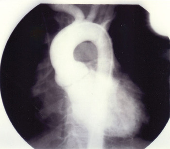
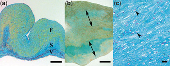
On examination, her blood pressure was 108/36, with a pulse pressure of 72 mmHg. Cardiac examination revealed a harsh systolic ejection murmur, graded at 2 from 6, at the upper right sternal border, and a blowing diastolic murmur, graded at 3 from 6, which radiated to the apex. The electrocardiogram was normal. Echocardiographic examination demonstrated severe aortic and mild mitral valvar insufficiency. The leaflets of the aortic valve appeared thickened and dysplastic, with the diameter of the valvar orifice measured at 18 mm, corresponding with a z score of 1.4. The leaflets of the mitral valve demonstrated trivial prolapse. The left ventricular end diastolic dimension was enlarged at 4.8 cm, the normal values for age ranging from 2.97 to 4.19 cm, and the left ventricular shortening fraction was normal. The ventricular septum was 5.0 mm thick, while the parietal wall was measured at 6.0 mm, both values being normal. Cardiac catheterization confirmed severe aortic valvar insufficiency (Fig. 1). The patient underwent replacement of the aortic valve with a 16 mm aortic homograft. Intraoperative inspection revealed a severely thickened and redundant valve with 3 leaflets that was not amenable to surgical repair. Pathologic study demonstrated marked thickening of the nodular leaflets, due primarily to accumulation of proteoglycans (Fig. 2). There was marked disorganization of the extracellular matrix, with prominent expansion of the spongy layer and disarray of collagen fibers (Fig. 2b), the leaflets being markedly thickened, at 6580 μm, when compared with an age-matched control valve measuring 360 μm obtained from a patient who died of noncardiac causes (Fig. 2a). In addition, there were swollen cytoplasmic lysosomes throughout the thickness of the leaflet (Fig. 2c). Staining with Alizarin red failed to demonstrate any valvar calcification.
Angiography in the lateral projection demonstrating severe aortic regurgitation.

Histopathology of the excised aortic valve, demonstrating unique findings. The normal trilaminar organization of the matrix is demonstrated in a control aortic valve from a child using pentachrome staining (panel a, scale bar 100 μm). In addition to marked thickening, there is disorganization of the extracellular matrix, with expanded areas of the spongy layer (double arrow heads), disarray of collagen fibers and increased density of interstitial cells in the diseased valve, also stained using pentachrome (panel b, scale bar 1000 μm). Alcian blue staining demonstrates intracellular swollen lysosomal bodies (arrow heads), marked proteoglycan accumulation, and separated collagen fibers (panel c, scale bar 100 μm). F Fibrous layer, S Spongy layer, V Ventricular layer. The fibrous layer is oriented upward. Movat’s pentachrome stain identifies elastin fibers as black, collagens as yellow, proteoglycans as blue/green, muscle as red, and cell nucleuses as purple.

Since the valvar replacement, the patient has had no cardiac symptoms over a period of 9 years. Serial echocardiograms over this period have demonstrated normal aortic valvar function without recurrent insufficiency or stenosis, the Doppler velocity across the valve being measured at 1.4 m/sec. There has been no progression in the degree of mitral insufficiency, which has remained mild. The left ventricular diastolic dimension, measured at 4.6 cm, is within normal limits for age, these varying from 3.47 to 4.91 cm. The left ventricular mural thickness and systolic function continue to be normal.
Discussion
Mucolipidosis III is a lysosomal storage disorder with cardiac involvement, typically manifesting as aortic and mitral valvar insufficiency. We present, to our knowledge, the first report of aortic valvar replacement in a patient with mucolipidosis III and severe aortic insufficiency. There is a paucity of literature about surgical valvar replacement in this population, despite the prevalence of cardiac disease. Replacement has been successfully reported in other lysosomal storage disorders, such as Maroteaux-Lamy syndrome,Reference Tan, Schaff, Miller, Edwards and Karnes4 and the attenuated form of mucopolysaccharidosis I.Reference Minakata, Konishi, Matsumoto and Miwa5 Mitral valvoplasty was successfully accomplished in a patient with mucolipidosis II.Reference Daimon and Yamagishi6 An understanding of the pathology in these disorders should be considered when seeking the optimal surgical approach.
We chose to insert an aortic homograft. Given the appearance of the valve at surgery, and the underlying metabolic abnormality, primary repair was not entertained. The Ross procedureReference Ross7 was also excluded because of the underlying metabolic process. As a result, the viable options included replacement with either a mechanical or bioprosthetic valve. A recent reports of long term outcomes in patients who have undergone aortic valvar replacement with homografts suggests positive results for up to 25 years.Reference Lund, Chandrasekaran and Grocott-Mason8 An aortic homograft was chosen as it avoided the need for long term anticoagulation in a young individual facing future orthopaedic surgeries.
The histopathologic findings in the valvar tissue obtained from our patient differed from histopathology in traditional aortic valvar disease. Both demonstrated a loss of trilaminar extracellular matrix stratification and interstitial cell compartmentalization, hallmarks of valvar disease.Reference Hinton, Lincoln and Deutsch9 The marked increase in thickness of the leaflets noted in our patient, however, was due primarily to accumulation of proteoglycans rather than the typical accumulation of collagen. Given the high content of water in proteoglycans, this finding may explain the observation that the valve was dilated and incompetent. Valvar histopathology in rare diseases may inform fundamental research efforts, and provide insights for novel therapeutic approaches, including improved design of bioprostheses.
Our experience highlights the need for cardiovascular surveillance in patients with mucolipidosis III. As novel therapeutic strategies emerge, and longevity is extended for individuals with lysosomal storage disorders, the cardiac disease takes on increasing importance. Even though enzyme replacement therapy is not currently available for mucolipidosis III, allogeneic transplantation of haematopoietic stem cells has been employed with success in patients with mucolipidosis II, and may represent a future option for patients with mucolipidosis III.Reference Martin, Carter and Kernan10 Increasingly, enzyme replacement therapies are being developed for metabolic disorders, and the impact of these treatments on the natural history of the valvar disease will be important to determine. Despite the fact that consensus guidelines do not exist for the treatment of the majority of inborn errors of metabolism, including mucolipidosis III, medical management needs to be directed to the individual patient. As longevity increases for such patients, surgical decision making needs to take into consideration the complex medical issues of this group of patients.
Acknowledgements
We thank Nancy Leslie, from our Department of Human Genetics, and Peter B. Manning, from our Department of Cardiothoracic Surgery, for their clinical expertise and review of the manuscript.
Financial support:
This work was supported by Cincinnati Children’s Research Foundation and the National Institutes of Health HL085122 (RBH).