Background
The neurobiological underpinnings of psychosis are thought to rely on abnormal connectivity between and within neuronal networks (Fornito, Zalesky, Pantelis, & Bullmore, Reference Fornito, Zalesky, Pantelis and Bullmore2012). The thalamus is thought to be an essential hub in many of these networks, as connections stemming from cortical areas are relayed to the region from the basal ganglia and cerebellum. The thalamus then returns projections to the cortex in feedback loops known as striato-thalamo-cortical and cerebello-thalamo-cortical loops (Halassa & Kastner, Reference Halassa and Kastner2017; Hwang, Bertolero, Liu, & D'Esposito, Reference Hwang, Bertolero, Liu and D'Esposito2017). Furthermore, although the thalamus was initially characterized as merely a filter relay of sensory information heading to cortical areas, recent evidence suggests the area is involved in higher order cognitive processes via connections with a fronto-parietal network that regulates a broad range of cognitive processes (Niendam et al., Reference Niendam, Laird, Ray, Dean, Glahn and Carter2012).
Functional magnetic resonance imaging during resting state (rs-fMRI) is an established method for studying intrinsic brain connectivity unrelated to any specific cognitive domain. As recently reviewed (Giraldo-Chica & Woodward, Reference Giraldo-Chica and Woodward2017), previous research using this methodology in schizophrenia has found a mixed pattern of differences in thalamic connectivity, with hyperconnectivity between the thalamus and sensorimotor and temporal regions as well as hypoconnectivity between the thalamus and prefrontal cortex (PFC) and cerebellum. Although most studies have focused on schizophrenia spectrum disorders, the few studies that have included individuals with bipolar disorder (BD) have shown a similar pattern of aberrant thalamic connectivity, particularly for individuals with psychosis (Anticevic et al., Reference Anticevic, Yang, Savic, Murray, Cole, Repovs and Glahn2014b; Woodward & Heckers, Reference Woodward and Heckers2016). These findings have been replicated using different ROIs, including individually delineated thalamus seeds (Anticevic et al., Reference Anticevic, Cole, Repovs, Murray, Brumbaugh, Winkler and Glahn2014a), atlas-based seeds (Ferri et al., Reference Ferri, Ford, Roach, Turner, van Erp, Voyvodic and Mathalon2018), functionally-defined parcellation-based seeds (Woodward & Heckers, Reference Woodward and Heckers2016) or independent component analysis-extracted functional components (Skåtun et al., Reference Skåtun, Kaufmann, Brandt, Doan, Alnæs, Tønnesen and Westlye2018). Regardless of the method, findings from these studies have converged on disrupted thalamocortical connectivity as a cross-diagnostic biomarker in psychotic disorders.
Although most studies of thalamocortical connectivity in schizophrenia have been conducted using chronic samples, previous work in first-episode psychosis (FEP) (Woodward & Heckers, Reference Woodward and Heckers2016) as well as individuals at high risk for psychosis (Anticevic et al., Reference Anticevic, Haut, Murray, Repovs, Yang, Diehl and Cannon2015) have also reported a similar pattern of abnormalities, suggesting these changes may occur before the brain is fully mature (Giraldo-Chica & Woodward, Reference Giraldo-Chica and Woodward2017). Such findings are consistent with the neurodevelopmental hypothesis of schizophrenia, in which pathological changes in brain function occur early in development and then are relatively stable (compared to healthy individuals). Although the preferred method to test this hypothesis is to examine connectivity longitudinally in recent-onset patients, to the best of our knowledge, no studies have been published using such an approach.
To that end, the goal of this study was to examine thalamic connectivity longitudinally over a 1-year period in individuals with recent-onset psychosis. Based on previous work, we hypothesized hypoconnectivity between the thalamus and frontal regions in patients as well as hyperconnectivity between the thalamus and sensorimotor/temporal regions. Consistent with the neurodevelopmental hypothesis, we also hypothesized these changes would be stable over the 1-year follow-up period. We also examined the relationship between resting-state functional connectivity (FC) and clinical symptomatology and medication history.
Methods
Subjects
One hundred and twenty-nine subjects with recent-onset psychosis were recruited from outpatient services from the Early Diagnosis and Preventative Treatment Clinic at the University of California, Davis (for extended details, see https://health.ucdavis.edu/psychiatry/specialties/edapt/index.html). Patients were 16–30 years old and presented with a DSM-IV-TR diagnosis of psychosis (either schizophrenia, brief psychotic disorder, schizoaffective disorder, BD with psychotic features or schizophreniform disorder). All individuals were within 2 years of illness onset (based on SCID-IV or K-SADS interview). Eighty-seven control subjects were recruited in parallel and evaluated using the SCID to rule out past or present Axis I or Axis II DSM-IV-TR diagnosis. Exclusion criteria were substance abuse or dependence during the 3 months prior to entering the study, a positive urine test for drug use (cannabis, cocaine and amphetamines), history of head trauma, presenting any contraindications (e.g. metal in the body) to enter the MRI scanner, or scoring below 70 in the Wechsler Abbreviated Scale of Intelligence (WASI). Forty-three of the recent-onset psychosis participants and 40 of the controls scanned at baseline were reassessed and re-scanned after 12 months. The study received approval from the University of California, Davis Institutional Review Board. Written informed consent and assent (in the case of minors) was obtained from all participants. All procedures contributing to this work complied with the ethical standards of the relevant national and institutional committees on human experimentation and with the Helsinki Declaration of 1975 as revised in 2008.
Clinical evaluations
Patients were clinically evaluated using the Scale for the Assessment of Positive Symptoms (SAPS) (Andreasen, Reference Andreasen1984), the Scale for the Assessment of Negative Symptoms (SANS) (Andreasen, Reference Andreasen1989), the Brief Psychiatric Rating Scale (BPRS) (Lukoff, Nuechterlein, & Ventura, Reference Lukoff, Nuechterlein and Ventura1986) and the Global Assessment of Functioning (GAF) (Aas, Reference Aas2010). Detailed antipsychotic intake was obtained from clinical records and subject interview. This information was used to compute an accumulated dose of chlorpromazine equivalents at scan time. Duration of untreated psychosis and time since psychosis onset was measured as the time from onset of psychotic symptoms (based on medical records and retrospective clinical interview with the patient or relatives) to the initiation of treatment and evaluation of the study, respectively.
Image acquisition
All participants underwent MRI scanning in a 3T Siemens Tim Trio scanner. Scans included structural images and 6 min of resting-state functional images with eyes open. Structural (T1 weighted) MPRAGE image parameters were TR = 2.530 ms, echo time = 3.5 ms, flip-angle = 7°, field of view = 256 mm, 1 mm isotropic voxels. Functional imaging parameters were TR = 2 s, ascending interleaved acquisition, matrix = 64 × 64 × 32, original voxel dimensions: 3.75 × 3.75 × 4.55, 180 volumes. A subset of 43 patients and 40 controls underwent a second clinical evaluation and MRI acquisition after 12 months.
Image processing
Structural images were normalized using the unified segmentation and normalization procedure in SPM12, which generated white matter (WM) and cerebrospinal fluid (CSF) probability images in normalized MNI space. These images were used in the denoising step to extract the principal components from WM and CSF areas. In addition, structural images were segmented with Free-surfer v5.3 (http://surfer.nmr.mgh.harvard.edu/) using the Desikan/Killiany atlas (Desikan et al., Reference Desikan, Ségonne, Fischl, Quinn, Dickerson, Blacker and Killiany2006) to produce individualized parcellation maps in subject space. We extracted the bilateral thalamus region as a mask to use as a seed in the functional seed-to-voxel analysis.
Functional images were preprocessed using the CONN toolbox v18.a (Whitfield-Gabrieli & Nieto-Castanon, Reference Whitfield-Gabrieli and Nieto-Castanon2012) with the following steps: removal of the initial four volumes, slice-timing correction, realignment and direct normalization of functional images to the EPI template in MNI space. No field maps were available and previous attempts using indirect normalization resulted unsatisfactorily. Final voxel size was 3 × 3 × 3 mm. Second, to extract seed BOLD signal from the thalamus mask generated by Free-surfer in subject space, we generated functional volumes in subject space. To this purpose, we implemented an ‘indirect coregistration’ by applying the inverse normalization transformation of the structural files to the normalized functional images. We chose this method, over normalizing the thalamus mask, to avoid the risk of inaccurate normalization of the thalamus. Finally, the normalized functional images were smoothed using a full-width half-maximum (FWHM) of 10 mm. Thus, preprocessing steps resulted in a set of smoothed and normalized functional images, and a set of unsmoothed functional images coregistered to single subject Free-surfer-based parcellation maps.
Great care was taken to minimize the effects of subject motion while maintaining data integrity and preserving statistical power. To this purpose, we determined the most appropriate framewise displacement (FD) threshold in our sample. FD was calculated using Artifact Detection Tools (ART) (https://www.nitrc.org/projects/artifact_detect/) using the formula FD = ${\rm max}\left({\sqrt {{\rm d}x^2{\rm} + {\rm d}y^2{\rm} + {\rm d}z^2} } \right)$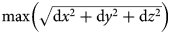 , where dx, dy and dz represent the displacement in mm in the three axes of six reference voxels placed at the centre of the default MNI brain's bounding box. The best balance between eliminating movement and minimizing data loss, both in terms of excluded subjects and volumes, corresponded to an FD value of 0.9, which coincides with the ‘intermediate level’ according to ART developers.
Both sets of functional images were then denoised using aCompCor in CONN. Specifically, aCompCor removes from each subject time-series images: (1) five components of WM signal and CSF signal in the MNI space, (2) the components associated with motion parameters during the realignment process (including their first and second derivatives), and (3) the components of the volumes flagged as excessive motion in the previous scrubbing step (and its first derivative). This strategy, in contrast to global signal regression (GSR), does not introduce artifactual negative correlations while still protecting against the potential biases of subject-motion and physiological effects (Chai, Castañón, Ongür, & Whitfield-Gabrieli, Reference Chai, Castañón, Ongür and Whitfield-Gabrieli2012; Murphy, Birn, Handwerker, Jones, & Bandettini, Reference Murphy, Birn, Handwerker, Jones and Bandettini2009). Due to the existing controversy in the literature regarding the best choice of these two methods, however, an alternative analysis using GSR instead of aCompCor was also performed as reported in Supplementary Materials.
Connectivity analysis
Using the CONN toolbox, BOLD time series were averaged and extracted from the bilateral thalamus of the second set of functional images (unsmoothed, in subject space), and bivariate correlations (i.e. connectivity values) calculated to each voxel of the denoised, smoothed and normalized images. Realignment parameters and adjacent volumes with excessive motion (i.e. FD > 0.9) were added here as first-level covariates. This procedure generated a connectivity map for each subject. A Fisher's transformation was then applied to transform the Pearson correlation coefficients of these maps to z-scores.
Two separate second-level analyses were computed. First, a general linear model was created using z-score correlation maps of the baseline participants. Second, a repeated measures model was created with those subjects whose follow-up images were available. For both analyses, subject group (recent-onset psychosis or control), mean motion and the number of scrubbed volumes for each subject were entered as nuisance covariates. Using the baseline general linear model, correlation maps at baseline were calculated separately for each group to display the group thalamic connectivity pattern. Then, contrasts comparing the two groups at baseline were calculated. For the repeated measures analysis, the contrast comparing decreasing connectivity over time between groups was calculated.
All results were corrected for multiple comparisons using a threshold of p < 0.001 (voxelwise uncorrected), false discovery rate cluster-corrected for multiple comparisons (FDR) p < 0.05 (Friston, Worsley, Frackowiak, Mazziotta, & Evans, Reference Friston, Worsley, Frackowiak, Mazziotta and Evans1994). The resulting statistical maps were displayed and overlaid onto an average structural image. To ease interpretation, significant clusters were labelled using the Harvard-Oxford atlas (Desikan et al., Reference Desikan, Ségonne, Fischl, Quinn, Dickerson, Blacker and Killiany2006), the AAL atlas (Tzourio-Mazoyer et al., Reference Tzourio-Mazoyer, Landeau, Papathanassiou, Crivello, Etard, Delcroix and Joliot2002) or by Brodman's area using CONN and XJVIEW toolboxes (http://www.alivelearn.net/xjview).
Exploratory associations between rs-FC and clinical symptoms were examined by extracting the average thalamic-connectivity values at each of the significant clusters from the between-group comparison at baseline and correlating these values with clinical measures of interest (total SANS score, total SAPS score, cumulative antipsychotic intake and mean FD). These analyses were performed using R (Rstudio v1.1.423© 2009–2018 RStudio, Inc.).
Results
After removing five recent-onset psychosis participants for excessive movement, the final sample at baseline consisted of 124 patients and 87 HC individuals. One additional participant with recent-onset psychosis from the follow-up acquisition was discarded due to excessive motion. Between-group comparisons of motion correction-related parameters are available in online Supplementary Material S1. The results of the denoising process with aCompCor or GSR are shown in online Supplementary Material S2. The socio-demographic and clinical characteristics of recent-onset psychosis and HC are shown in Table 1. Participants with recent-onset psychosis with and without follow-up data did not show significant differences in any of the socio-demographic or clinical characteristics (see online Supplementary Material S3).
Socio-demographic and clinical characteristics of recent-onset psychosis and healthy controls at baseline and after 1 year

ROP, recent-onset psychosis; HC, healthy controls; GAF, Global Assessment of Function; BPRS, Brief Psychiatric Rating Scale; SAPS, Scale for the Assessment of Positive Symptoms; SANS, Scale for the Assessment of Negative Symptoms.
a Affective psychosis includes bipolar disorder and major depressive disorder with psychotic features.
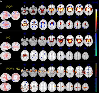
Figure 1 shows thalamic connectivity patterns separated by group. Participants with recent-onset psychosis displayed positive connectivity in the thalamus, insula, temporal regions (including bilateral parahippocampal gyrus, hippocampus, lingual gyrus, Heschl's gyrus and superior temporal gyrus), medial PFC, the anterior and posterior lobe of the cerebellum, as well as negative thalamic connectivity in occipital regions extending to posterior regions of the parietal cortex and bilateral middle frontal gyrus (Fig. 1a). In comparison, control subjects displayed a more constrained pattern both of positive and negative connectivity (Fig. 1b). In comparison to controls, participants with recent-onset psychosis showed increased thalamic connectivity in several regions of the bilateral temporal cortex and bilateral precentral and postcentral gyri, as well as decreased connectivity in the right middle frontal gyrus, bilateral thalamus and bilateral cerebellum (see Table 2 and Fig. 1c). Average thalamic connectivity extracted from all significant clusters from the contrast ‘recent-onset psychosis > HC’ inversely correlated to average thalamic connectivity extracted from all significant cluster from the contrast ‘HC > recent-onset psychosis’, both in the whole sample (r = −0.45, p < 0.001), and in the group of recent-onset psychosis (r = −0.23, p = 0.011).
Significant clusters showing within-group patterns and between-group differences in thalamic connectivity at baseline. ROP, recent-onset psychosis; HC, healthy controls. Within-group thalamic connectivity at baseline in ROP (a) and controls (b). Red range colour scale displays a positive correlation with thalamic activity. Blue range colour scale represents negative correlations with thalamic activity. (c) Between-group significant clusters in thalamic connectivity. Red range scale displays higher thalamic connectivity in ROP compared to controls. Blue range scale displays lower connectivity in ROP compared to controls. All results are corrected using an initial cluster-defining threshold of p < 0.001 at voxel-level and a subsequent cluster-extent FDR correction at p < 0.05.

Clusters showing between-group significant differences in thalamic connectivity

ROP, recent-onset psychosis; HC, healthy controls; BA, Brodmann area; STG, superior temporal gyrus; G, gyrus; sup, superior; MTG, middle temporal gyrus; MFG, middle frontal gyrus; MeFG, medial frontal gyrus; L, lobule.
Results are corrected using an initial cluster-defining threshold of p < 0.001 at voxel-level and a subsequent cluster-extent FDR correction at p < 0.05.
The equivalent analysis using GSR instead of aCompCor showed a similar pattern of results (see online Supplementary Material S4). A subanalysis using only those subjects with available baseline and follow-up data is also shown in online Supplementary Material S5. The same between-group differences in the pattern of thalamic connectivity were observed
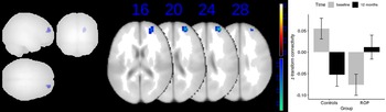
Longitudinal results are shown in Fig. 2 and Table 3. Participants with recent-onset psychosis showed increased connectivity over time (follow-up higher than baseline) relative to controls in the anterior part of the right superior and middle frontal gyrus. The equivalent analysis using GSR instead of aCompCor showed similar results with the same cluster of increased connectivity over time in the anterior part of the right superior and middle frontal gyrus, with the addition of a cluster of decreased connectivity over time in the right hippocampus (see details in online Supplementary Material S9).
Significant clusters in the group per time interaction contrast. Left: Red range scale displays decrease over time of thalamic connectivity in recent-onset psychosis (ROP) compared to controls (no suprathreshold clusters). Blue range scale displays increase over time of thalamic connectivity in ROP compared to controls. All results are corrected using an initial cluster-defining threshold of p < 0.001 at voxel-level and a subsequent cluster-extent FDR correction at p < 0.05. Right: Boxplot showing z-transformed averaged connectivity values per group and time (baseline or 12 months) from the significant cluster of the left figure (peak at x, y, z = +28, +56, +22).

Clusters showing between-group significant differences in baseline higher than 12 months thalamic connectivity

ROP, recent-onset psychosis; HC, healthy controls; BA, Brodmann area; R, right; L, left; Lob, lobule; G, gyrus.
Results are corrected using an initial cluster-defining threshold of p < 0.001 at voxel-level and a subsequent cluster-extent FDR correction at p < 0.05.
Clinical correlates
Average thalamic connectivity in the group of recent-onset psychosis extracted from all significant clusters from the contrast ‘recent-onset psychosis > HC at baseline’ was significantly correlated with SANS Total score [t (119) = 2.011, p = 0.04], although it did not survive correction for multiple comparisons. No other significant correlations in the group of patients between connectivity extracted from clusters from both direction contrasts and average FD, number of scrubbed volumes, symptoms or cognition were observed. Individual cluster correlations across groups with symptoms, medication and motion parameters are shown in detail in online Supplemental Material S6 and S7. No significant correlations with change in symptoms or cognition over time were found (see details in online Supplementary Material S8).
Discussion
In this first longitudinal study to investigate changes in thalamic connectivity at rest in recent-onset psychosis, we confirm an aberrant pattern of thalamic connectivity in individuals with recent-onset psychosis, including increased connectivity in the somatosensory, motor and temporal regions as well as decreased connectivity in PFC and cerebellum. Additionally, we report a ‘normalizing’ increase in connectivity over time in recent-onset psychosis in some of the PFC regions where decreased connectivity was found at baseline (as compared to controls).
Our finding of abnormal thalamic connectivity at baseline is in agreement with previous studies in schizophrenia samples and subjects at risk of psychosis (Anticevic et al., Reference Anticevic, Cole, Repovs, Murray, Brumbaugh, Winkler and Glahn2014a, Reference Anticevic, Yang, Savic, Murray, Cole, Repovs and Glahn2014b; Anticevic et al., Reference Anticevic, Haut, Murray, Repovs, Yang, Diehl and Cannon2015; Cheng et al., Reference Cheng, Skosnik, Pruce, Brumbaugh, Vollmer, Fridberg and Newman2014; Ferri et al., Reference Ferri, Ford, Roach, Turner, van Erp, Voyvodic and Mathalon2018; Giraldo-Chica & Woodward, Reference Giraldo-Chica and Woodward2017; Woodward & Heckers, Reference Woodward and Heckers2016; Woodward, Karbasforoushan, & Heckers, Reference Woodward, Karbasforoushan and Heckers2012) and confirms the presence of abnormal FC as early as the onset of psychosis. This pattern of aberrant connectivity, with both increases and decreases, may suggest a blurring of the normal connectivity pattern and may be linked to previous robust findings in schizophrenia.
On the one hand, hypoconnectivity between the thalamus and prefrontal regions is in line with several resting-state FC studies (Karcher, Rogers, & Woodward, Reference Karcher, Rogers and Woodward2019; Viher et al., Reference Viher, Docx, Van Hecke, Parizel, Sabbe, Federspiel and Morrens2019) and WM structural connectivity studies (Levitt et al., Reference Levitt, Nestor, Levin, Pelavin, Lin, Kubicki and Rathi2017), which, beyond thalamic connectivity, have consistently found striatal-prefrontal disconnectivity in schizophrenia. In this line, previous research has reported extended cortico-thalamic hypoconnectivity to basal ganglia, one component of the well-known cortico-striato-pallido-thalamo-cortical circuits (Avram, Brandl, Bäuml, & Sorg, Reference Avram, Brandl, Bäuml and Sorg2018). A review of functional studies has shown a non-specific association between striato-prefrontal disconnectivity and performance on distinct cognitive domains, suggesting that it may underlie mechanisms that are shared across high order cognitive processes (Sheffield & Barch, Reference Sheffield and Barch2016), such as loss of top-down inhibition of sensory processing (Avram et al., Reference Avram, Brandl, Bäuml and Sorg2018). Moreover, we and others (Avram et al., Reference Avram, Brandl, Bäuml and Sorg2018) have found an inverse correlation in terms of thalamic connectivity between clusters that showed increased and decreased connectivity in recent-onset psychosis compared to controls, suggesting that they are interrelated findings.
Interestingly, a significant correlation was observed between increased thalamic connectivity in somatosensory/temporal regions and negative symptoms, although it did not survive correction for multiple comparisons. These clinical connections are in partial agreement with some previous work (Cheng et al., Reference Cheng, Skosnik, Pruce, Brumbaugh, Vollmer, Fridberg and Newman2014). Overall findings related to this relationship are mixed (Giraldo-Chica & Woodward, Reference Giraldo-Chica and Woodward2017). No relationships, furthermore, were observed between change in negative symptoms at baseline or follow-up and change in connectivity, suggesting the relationship(s) (if they truly exist) are unstable. Indeed, it is possible that many different processes mediate the relationship between thalamic hyperconnectivity and the symptomatic presentation, making it difficult to parse out. The study of longitudinal correlations may run into sample size reduction, clinically biased subject dropout and additional heterogeneity in treatment-related variables, and therefore be even more difficult to test. Consequently, future longitudinal studies using larger samples are likely necessary to more clearly understand these associations.
On the other hand, hypoconnectivity was also observed between the thalamus and cerebellum in individuals with recent-onset psychosis, highlighting the role of the cerebellum in schizophrenia, which remains poorly understood. Andreassen, Paradiso, and O'Leary (1998) introduced the concept of ‘cognitive dysmetria’, wherein the pathophysiology of schizophrenia is associated with abnormal connectivity within the cortico-cerebellar-thalamo-cortical circuit, inducing difficulty with coordination and fine-tuning of motor activity as well as many other cognitive processes. This idea is in line with previous studies reporting abnormalities in WM fibre tracts communicating the thalamus and the cerebellum (Liu, Fan, Xu, & Wang, Reference Liu, Fan, Xu and Wang2011; Magnotta et al., Reference Magnotta, Adix, Caprahan, Lim, Gollub and Andreasen2008).
Bearing in mind the evidence of these findings across different phases of the illness, from high-risk subjects (Anticevic et al., Reference Anticevic, Haut, Murray, Repovs, Yang, Diehl and Cannon2015) to relatives (Cho et al., Reference Cho, Kim, Yoon, Lee, Lee and Kwon2019), recent-onset psychosis and chronic patients, these findings may reflect abnormal development of brain networks during brain maturation (Woodward et al., Reference Woodward, Karbasforoushan and Heckers2012). However, no longitudinal studies in thalamic connectivity had been previously reported to test for the presence of a progressive change in this pattern. In our longitudinal study, we have shown that baseline thalamic hypoconnectivity to the anterior part of the superior and middle frontal gyri (the frontal pole) shifts to a more heightened connectivity 12 months later. The plotted connectivity values within the significant cluster in the interaction effect shows an increase over time in thalamic connectivity in ROC, but quite surprisingly, also a decrease in connectivity in controls (see Fig. 2 in the Results section). A plausible explanation may emerge from studies describing the maturation of thalamo-cortical connectivity across age. This maturation process terminates typically during adolescence and early adulthood, after an increase in thalamo-frontal connectivity and a decrease in thalamo-temporal connectivity (Fair et al., Reference Fair, Bathula, Mills, Costa Dias, Blythe, Zhang and Nagel2010). Interestingly, young subjects with mental health conditions show a delayed pattern of maturation in brain connectivity (Kaufmann et al., Reference Kaufmann, Alnæs, Doan, Brandt, Andreassen and Westlye2017). They present changes in the slope of maturation at a different age than controls, especially in fronto-parietal and subcortical regions, which could, eventually, result in opposite directions in connectivity growth. Neurodevelopmentally, the frontal pole is one of the last brain regions to fully mature, with continued myelin formation and synaptic pruning even after early adulthood (Dumontheil, Burgess, & Blakemore, Reference Dumontheil, Burgess and Blakemore2008; Miller et al., Reference Miller, Duka, Stimpson, Schapiro, Baze, McArthur and Sherwood2012). Increase in connectivity to this region in patients may therefore represent resilience or partial recovery, suggesting that in addition to being non-degenerative, the abnormal connectivity phenotype may (at least partially) improve during the first years after psychosis onset.
In fact, most patients in the study showed symptomatic improvement over the course of the follow-up period (see Table 1) and previous reports have shown recovery of connectivity between striatal and PFC related to clinical improvement through the use of antipsychotic drugs (Sarpal et al., Reference Sarpal, Robinson, Lencz, Argyelan, Ikuta, Karlsgodt and Malhotra2015). These findings are in conceptual agreement with previous reports in recent-onset illness demonstrating ongoing functional PFC development (Gold, Arndt, Nopoulos, O'Leary, & Andreasen, Reference Gold, Arndt, Nopoulos, O'Leary and Andreasen1999; Niendam et al., Reference Niendam, Ray, Iosif, Lesh, Ashby, Patel and Carter2018). Recently, PFC activation during proactive control has been reported as a predictor of clinical improvement in recent-onset psychosis (Smucny, Lesh, & Carter, Reference Smucny, Lesh and Carter2019), which, together with our longitudinal findings, rise the idea that PFC functionality and plasticity is preserved at least in some individuals with recent-onset psychosis subjects and is critical for a better outcome.
To our knowledge, this is the first longitudinal resting-state thalamic connectivity study in recent-onset psychosis. Although several studies have focused on thalamic connectivity in schizophrenia, most of them have studied older, chronic patients (~30 years) (Anticevic et al., Reference Anticevic, Yang, Savic, Murray, Cole, Repovs and Glahn2014b; Ferri et al., Reference Ferri, Ford, Roach, Turner, van Erp, Voyvodic and Mathalon2018; Woodward et al., Reference Woodward, Karbasforoushan and Heckers2012). Only a few of them comprised mixed samples of FEP and chronic schizophrenia (Woodward & Heckers, Reference Woodward and Heckers2016), and only one included subjects at high risk of psychosis (Anticevic et al., Reference Anticevic, Haut, Murray, Repovs, Yang, Diehl and Cannon2015). The mean age of our sample was <20 years old, significantly lower than most of the previous studies. Bearing in mind a critical period of illness progression which has been described as the first 2–3 years since illness onset (Birchwood, Todd, & Jackson, Reference Birchwood, Todd and Jackson1998), the study of young samples with recent-onset psychosis is critical.
Due to its potentially deleterious confounding effects on resting-state connectivity (Power, Barnes, Snyder, Schlaggar, & Petersen, Reference Power, Barnes, Snyder, Schlaggar and Petersen2012), we made a concerted effort to eliminate the effect of motion from our analysis. First, we discarded subjects and volumes with excessive motion. However, in agreement with other studies in thalamic connectivity (Anticevic et al., Reference Anticevic, Cole, Repovs, Murray, Brumbaugh, Winkler and Glahn2014a, Reference Anticevic, Yang, Savic, Murray, Cole, Repovs and Glahn2014b; Cheng et al., Reference Cheng, Palaniyappan, Li, Kendrick, Zhang, Luo and Feng2015; Woodward & Heckers, Reference Woodward and Heckers2016), we still found significant between-group differences in terms of motion parameters. The fact that excessive motion may be an intrinsic characteristic of psychosis in comparison to controls may make it unfeasible to eliminate between-group differences. To further address the effect of motion on our results, motion parameters were introduced in the main analysis as covariates, and finally, we correlated motion parameters with thalamic connectivity in the significant clusters. No significant effects were found in patients or in controls except for one cluster in the group of controls (see online Supplementary Data S6).
In summary, in this first study to longitudinally examine abnormalities in intrinsic thalamocortical connectivity in recent-onset psychosis, we not only confirmed a pattern of aberrant connectivity previously observed in schizophrenia but also show that these changes show no evidence of deterioration and, in fact, partially improve over time. These results are in agreement with a neurodevelopmental hypothesis of schizophrenia and a non-degenerative model of the illness in which functional changes occur early in development and do not deteriorate over time, consistent with other recent work from our laboratory using the AX CPT tasks and task-related fMRI (Niendam et al., Reference Niendam, Ray, Iosif, Lesh, Ashby, Patel and Carter2018; Smucny et al., Reference Smucny, Lesh, Zarubin, Niendam, Ragland, Tully and Carter2020).
Supplementary material
The supplementary material for this article can be found at https://doi.org/10.1017/S0033291720004808
Financial support
The first author received funds from the grant ‘Movilidad de profesionales sanitarios e investigadores del SNS (M-BAE)’ from the ISCIII-Ministry of health, Spain, and from the grant ‘Short-term visiting fellowships’ from the Alicia Kolpowitz Foundation. This work was supported by the National Institutes of Mental Health (grant number 5R01MH059883 to C.S.C., fellowship number F32-MH114325 to J.S.).
Conflict of interest
None.