Introduction
The genus Rhizoplaca (Lecanoraceae, Lecanorales) was established based on R. opaca (Ach.) Zopf (= R. melanophthalma (DC.) Leuckert & Poelt; Zopf Reference Zopf1905) and until recently included c. 10 species, distinguished from Lecanora by a mainly peltate-umbilicate thallus attached with an umbilicus, the lack of distinctly radiating marginal lobes, the loose medulla, and thick lower cortex (Ryan Reference Ryan, Nash, Ryan, Gries and Bungartz2002). Subsequent molecular phylogenetic studies have greatly modified the concept of this genus, in particular by transferring Rhizoplaca peltata Ramond ex Leuckert & Poelt to Protoparmeliopsis and including a number of placodioid Lecanora species (Zhao et al. Reference Zhao, Leavitt, Zhao, Zhang, Arup, Grube, Pérez-Ortega, Printzen, Śliwa and Kraichak2016). Kondratyuk et al. (Reference Kondratyuk, Lőkös, Jang, Hur and Farkas2019) proposed a further transfer of some Rhizoplaca species to the genera Omphalodina and Sedelnikovaea; however, these proposals require verification as the phylogenetic trees justifying them do not indicate branch support values, so there is no way to assess the reliability of the proposed tree topology. According to Zhang et al. (Reference Zhang, Yin, Wang, Printzen, Wang and Wang2024), the genus Rhizoplaca s. str. currently includes c. 25 species distributed worldwide.
While investigating the lichenicolous fungi of Mongolia, the author found a number of rare, poorly known or apparently undescribed species growing on Rhizoplaca s. lat. sensu Ryan (Reference Ryan, Nash, Ryan, Gries and Bungartz2002), species of which are common there on rock (Zhurbenko et al. Reference Zhurbenko, Enkhtuya and Javkhlan2019, Reference Zhurbenko, Enkhtuya and Javkhlan2020). A subsequent survey of Rhizoplaca s. lat. specimens mainly from some other countries of Central Asia revealed a number of additional interesting specimens of lichenicolous fungi. The aims of this paper are to present the results of the revision of this material, including the description of four new species and one new variety, and to provide a first identification key to the species of lichenicolous fungi and lichens growing on Rhizoplaca s. lat.
Material and Methods
The study is based on 65 specimens of lichenicolous fungi and lichens growing on Rhizoplaca chrysoleuca (Sm.) Zopf, R. melanophthalma, R. subdiscrepans (Nyl.) R. Sant. and Protoparmeliopsis peltata Ramond ex Arup et al. (≡ Rhizoplaca peltata), mainly from Kazakhstan, Kyrgyzstan, Mongolia, Russia and Turkmenistan, deposited in the herbarium of the Komarov Botanical Institute of RAS, St Petersburg, Russia (LE). The age of the specimens ranges from 1–72 years and can be easily calculated from the cited labels.
Microscopy was carried out and images were captured using a Zeiss Axio Zoom V16 stereomicroscope fitted with an AxioCam 712 colour digital camera, and a Zeiss Axio Imager A1 compound microscope equipped with Nomarski differential interference contrast optics and fitted with an AxioCam 807 colour digital camera. Microscopic characters were studied using sections hand-cut with a razor blade and mounted in water, 10% potassium hydroxide (K), Lugol's iodine directly (I) or after K pretreatment (K/I), or 50% aqueous solution of nitric acid (N). Measurements and descriptions refer to water mounts unless otherwise indicated. When the number of measurements (n) > 10, the dimensions of ascomata, conidiomata, asci, ascospores, conidia and some other microstructures, as well as the length/width ratio (l/w) of the ascospores and conidia, are given as (min‒)(x̄ – SD) ‒ (x̄ + SD)(‒max), where min and max are the extreme values observed, x̄ the arithmetic mean, and SD the corresponding standard deviation. Dimensions of the microstructures, both newly obtained and taken from the literature, were rounded to the nearest 0.5 μm. Standard statistical tests (Mann-Whitney U test and Student's t-test) were used to assess the reliability of differences in ascospore size. Colours are named according to Kornerup & Wancher (Reference Kornerup and Wanscher1978).
The Taxa
Arthonia clemens (Tul.) Th. Fr. var. clemens
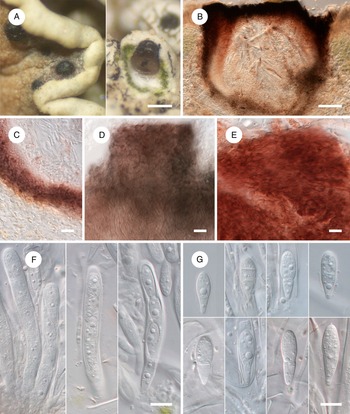
Epihymenium light brown to brown, usually with an additional grey shade, viz. greyish brown throughout or brown above, grey below (Fig. 1D). Ascospores (10‒)11.5‒13(‒15) × (5‒)5.5‒6.5 μm, l/w = (1.6‒)1.9‒2.3(‒2.6) (n = 68), which agrees with the dimensions given by Grube (Reference Grube, Nash, Gries and Bungartz2007), viz. 10‒15 × 4‒7 μm.
Arthonia clemens var. peltatae (A & B, holotype; C, LE 310337). A, habitus of fused ascomata. B, ascomata in cross-section. C, epihymenium. Arthonia clemens var. clemens (LE 310307). D, epihymenium. B–D = in water. Scales: A = 500 μm; B = 100 μm; C & D = 10 μm. In colour online.

New to Kazakhstan, Kyrgyzstan and the Republic of Sakha (Yakutia) in Russia.
Selected specimens examined (all except LE 310304 on apothecial discs of Rhizoplaca chrysoleuca)
Kazakhstan: Karaganda Region: 25 km SW of Kenistas settlement, Konurtiube (Konyrtobe) Mts, 49°41ʹ40ʺN, 73°03ʹ27ʺE, 13 vii 1967, E. Leont'eva (LE 310305).—Kyrgyzstan: Terskey Alatau, Altyn-Arashan Canyon, Kel'dyne River, 17 ix 1954, A. N. Sobolev (LE 310311); southern slope of the Kyrgyz Alatau Ridge, Shamsi River valley, 30 km NW of Kochkorki settlement, 2100 m, 1972, L. I. Bredkina 1441b (LE 310323b). Issyk-Kul’ Region: 40 km E of Karakol town, Terskei Alatau Ridge, Turgen’-Aksu Canyon, 42°34ʹ48ʺN, 78°53ʹ15ʺE, 1979, L. I. Bredkina 2821a (LE 310302a); Alai Ridge, 1979, L. I. Bredkina 3425b (LE 310310).—Mongolia: Arkhangai Region: Tevshrulekh District, 15 km NW of Tevshrulekh, Urd’-Tamir River valley, rocks in mountain steppe, on Rhizoplaca subdiscrepans (apothecial discs), 13 vii 1970, L. G. Biazrov (LE 310304) [Identification is somewhat uncertain as the epihymenium lacks grey shade]; Khotont District, foot of Mt Tsagaan Uul, 47°13ʹ19.8ʺN, 102°18ʹ51.6ʺE, 1690 m, 2023, M. P. Zhurbenko 23104 (LE 310307). Bayan Khongor Region: Zhargalant District, NE of Zhargalant settlement, near Egiin Davaa Pass, 47°12ʹ26.3ʺN, 99°50ʹ33.5ʺE, 2560 m, 2023, M. P. Zhurbenko 2385 (LE 310308).—Russia: Republic of Sakha (Yakutia): Oimyakon District, near Ust’-Nera settlement, right bank of Indigirka River, 64°30ʹN, 143°10ʹE, 1000 m, 1992, M. P. Zhurbenko 92571c (LE 310316c); Moma District, 56 km NNW of Tiubeliakh settlement, right bank of Indigirka River, 65°52ʹN, 143°01ʹE, 250 m, 1992, M. P. Zhurbenko 92570 (LE 310309).
Arthonia clemens (Tul.) Th. Fr. var. peltatae Zhurb. var. nov.
MycoBank No.: MB 854767
Distinguished from Arthonia clemens var. clemens mainly by the brown versus greyish brown epihymenium, and a different host selection, Protoparmeliopsis versus Rhizoplaca (both in Lecanoraceae).
Type: Mongolia, Zavkhan Region, 10 km NE of Erdene Hairkhan District, Kotlovina Ozer (Lake Basin), between Gurvan Bulak Ula and Tsakhir Ula Mts, upper Khungui River, on Protoparmeliopsis peltata (apothecial discs), 20 August 1979, U. Tsogt 1089 (LE 310318—holotype).
Ascomata apothecia, erumpent, without a distinct margin, disc blackish, matt, ±rounded to irregularly elongated in surface view, slightly convex, up to 1 mm diam., smooth or eventually finely fissured, arising singly or in groups, sometimes confluent. Exciple indistinct. Epihymenium conspicuous, light to medium brown, 10‒15 μm thick, usually with deposits of orange-white crystalline granules penetrating the hymenium. Hymenium subhyaline to pale brownish orange, 30‒40 μm tall, with widely spaced asci, hymenial gel I+ red, K/I+ blue; subhymenium rather indistinct, the same colour as the hymenium, up to 30 μm tall. Paraphysoids branched, anastomosed, apices light brown to brown, occasionally branched, often somewhat enlarged, 2.5‒5 μm diam. Asci clavate, mainly stalked, (32‒)33.5‒39.5(‒43) × 15‒18(‒19.5) μm (n = 15), 8-spored. Ascospores hyaline, 1-septate, narrowly obovate with wider and often longer upper cell, (9.5‒)11‒13(‒14) × (4‒)4.5‒5(‒5.5) μm, l/w = (1.8‒)2.3‒2.9(‒3.3) (n = 55, in water or I), smooth-walled, without a gelatinous sheath, irregularly biseriate in the ascus.
Asexual morph not observed.
Etymology
The epithet refers to the host lichen Protoparmeliopsis peltata.
Distribution and host
The new variety is known from two collections in Central Asia (Kyrgyzstan and Mongolia), growing on apothecial discs of Protoparmeliopsis peltata. Deleterious effects on the host were not observed.
Notes
Arthonia clemens was described on Rhizoplaca chrysoleuca (Tulasne Reference Tulasne1852) and according to Grube (Reference Grube, Nash, Gries and Bungartz2007) is probably confined to this host. The new variety described on Protoparmeliopsis peltata is mainly distinguished by the absence of grey shade in the epihymenium coloration. It may also have narrower ascospores, mostly 4.5‒5 μm versus mostly 5.5‒6.5 μm wide as in the Arthonia clemens var. clemens specimens presented above. This difference is confirmed by a Student's t-test with a probability of 99%. However, this assumption needs to be checked since in Arthonia clemens var. peltatae, spores were measured only in the asci, whereas in the type variety they were measured mainly outside the asci.
A report on Protoparmeliopsis peltata from Greenland (Alstrup & Hawksworth Reference Alstrup and Hawksworth1990: 16, based on Fries (Reference Fries1871)) probably belongs to the same variety.
Additional specimen examined
Kyrgyzstan: Terskey Alatau Ridge, on Protoparmeliopsis peltata (apothecial discs), 17 viii 1953, A. N. Sobolev (LE 310337).
Caeruleoconidia ahtii Zhurb. sp. nov.
MycoBank No.: MB 854768
Distinguished from Caeruleoconidia ochrolechiae Zhurb. & Diederich mainly by the hyaline to occasionally pale greyish turquoise versus greenish blue, larger conidia, (6.5–)7.5–9.5(–11) × (5–)5.5–7(–8) μm versus (4–)5–7(–10.5) × (3–)4–5.5(–6.5) μm, and a different host selection, Rhizoplaca (Lecanorales) versus Ochrolechia (Pertusariales).
Type: Mongolia, Khovd Region, Must District, Bodonch Gol River valley, 46°31ʹ53ʺN, 92°23ʹ28ʺE, 2400 m, sandstone boulders in steppe, on Rhizoplaca chrysoleuca (thallus), 14 July 2019, M. P. Zhurbenko 19364 (LE 310278—holotype).
(Fig. 2)
Caeruleoconidia ahtii (holotype). A, habitus of conidiomata. B–D, conidiomata in cross-section. E, basal stroma giving rise to conidiophores (in K). F, basal stroma giving rise to conidiophores, conidiogenous cells and conidia. G, conidia. B–D, F & G = in water. Scales: A = 100 μm; B–D = 50 μm; E–G = 10 μm. In colour online.

Conidiomata initially immersed in the host thallus, then erumpent, becoming superficial, (45–)60–95(–145) μm diam. (n = 27), mostly 35–70 μm tall, initially almost closed, pycnidioid, then saucer-shaped, sporodochial, with a widely exposed conidiogenous layer, irregularly circular to somewhat elongated in surface view, with a greyish green (best visible at ≥ ×100) flat centre surrounded by a thick, black, elevated margin, arising singly or in groups, sometimes contiguous to confluent up to eight together. Lateral wall of mature conidiomata 7–26 μm thick, greyish turquoise combined with brown, K−, composed of irregularly orbicular to somewhat elongated cells, (3.5–)5–7.5(–10.5) × (3–)4–6 (–7) μm (n = 35), with walls 0.5–1 μm thick. Basal stroma hyaline to greyish turquoise, K−, composed of suborbicular to somewhat elongated cells, c. 4–9 × 3–6 μm, located in the centre, and of smaller, tangentially elongated cells, (3.5–)4–6.5(–9) × (1.5–)2–3(–4) μm (n = 28), located on the periphery. Conidiophores originating from the basal stroma, poorly differentiated from the underlying stromatic cells, hyaline to greyish turquoise, K−, composed of suborbicular to somewhat elongated cells, (3.5–)5–8(–10) × (2.5–)3.5–5.5(–7) μm (n = 31). Conidiogenous cells integrated, poorly differentiated. Conidia hyaline to occasionally pale greyish turquoise, with rounded edges, angular rounded, ellipsoid (sometimes asymmetric), ovate, occasionally oblong, reniform, cuneiform, ampulliform, triangular or irregular, (6.5–)7.5–9.5(–11) × (5–)5.5–7(–8) μm, l/w = (1.0–)1.2–1.6(–1.8) (n = 44), aseptate, solitary, dry, with a smooth wall c. 0.5 μm thick, sometimes with a few small guttules.
Sexual morph not observed.
Etymology
The species is dedicated to the eminent Finnish lichenologist Teuvo Ahti, on the occasion of his 90th birthday.
Distribution and host
The new species is known only from the type collection in Central Asia (Mongolia), growing on the thallus of Rhizoplaca chrysoleuca, not visibly damaging the host.
Notes
With its mainly sporodochial conidiomata, hyaline to greyish turquoise conidiophores aggregated in a compact basal stroma, surrounded by a lateral wall, integrated conidiogenous cells, and hyaline to occasionally pale greyish turquoise, aseptate, smooth-walled, rounded or slightly elongated conidia of various shapes, the new species fits well the current concept of Caeruleoconidia (Zhurbenko et al. Reference Zhurbenko, Frisch, Ohmura and Thor2015). It is well distinguished from the two previously known species of the genus, viz. C. biazrovii Zhurb. (growing on Cladonia; Zhurbenko & Pino-Bodas Reference Zhurbenko and Pino-Bodas2017) and C. ochrolechiae (growing on Ochrolechia; Zhurbenko et al. Reference Zhurbenko, Frisch, Ohmura and Thor2015), by the partially brown pigmentation of the lateral wall of the conidiomata, the mainly hyaline versus greenish blue or bluish green conidia, and the host preference. Additionally, conidia of the compared species are smaller, (3–)3.5–5(–6.5) × (2.5–)3–4.5(–5.5) μm and (4–)5–7(–10.5) × (3–)4–5.5(–6.5) μm, respectively.
Carbonea cf. aggregantula (Müll. Arg.) Diederich & Triebel
The material examined fits well the species description presented in Cannon et al. (Reference Cannon, Malíček, Ivanovich, Printzen, Aptroot, Coppins, Sanderson, Simkin and Yahr2022) except for the smaller apothecia, 80‒270 μm versus 200‒400 μm diam., and wider ascospores, (8‒)9‒11(‒13) × (4‒)4.5‒5(‒5.5) μm, l/w = (1.5‒)1.8‒2.4(‒2.7) (n = 36) versus 9.5‒12 × 3‒4 μm.
Specimens examined (both on lobe underside and stalk of Rhizoplaca chrysoleuca)
Kyrgyzstan: Terskey Alatau Ridge, 16 vii 1953, A. N. Sobolev (LE 310282a).—Mongolia: Bayan Khongor Region: Gurvan Bulag District, NW of Buga settlement, near Khukh nuur Lake, 47°31ʹ32.5ʺN, 98°31ʹ19.8ʺE, 2630 m, 2023, M. P. Zhurbenko 2398d (LE 310283).
Cercidospora cf. crozalsiana (H. Olivier) Nav.-Ros., Cl. Roux & Casares
Ascomata perithecioid, externally black, mainly subglobose, 170‒320 μm diam., protruding in the ostiolar region to semi-immersed, ±dispersed. Exciple in the upper part green, greenish grey, greyish turquoise or olive, sometimes brown on the outside, rarely with violet speckles throughout, medium coloured internally, darkly coloured externally and near the ostiole, colourless to pale coloured (brown, orange-grey or dull green) at the base. Paraphysoids abundant, filiform, occasionally branched, septate, with many guttules. Asci (sub)cylindrical, (2‒)4(‒8)-spored. Ascospores hyaline, distinctly heteropolar, clavate, with a wider upper (relative to the position in the ascus) part to occasionally almost homopolar, fusiform, ends rounded to rather acute or occasionally attenuated, (17.5‒)22‒29(‒38) × (5.5‒)6.5‒8(‒9) μm, l/w = (2.4‒)3.0‒4.2(‒5.5) (n = 183) (according to the Mann-Whitney U test, the difference in spore length between samples on different hosts is not significant with a 99% probability), (0‒)1-septate, septum sometimes slightly to strongly displaced towards the narrow or wide end of the spore, not or slightly constricted at the septum, guttulate, smooth-walled, rarely halonate, mainly diagonally uniseriate in the ascus. Pycnidiospores hyaline, short bacilliform, 3.5‒4.5 × 1‒1.5 μm.
This description probably refers to more than one taxon, but clear morphological differences are difficult to articulate. In the description of Cercidospora crozalsiana presented in Calatayud et al. (Reference Calatayud, Navarro-Rosinés and Hafellner2013), the exciple is characterized as blue-green in its upper part and colourless at the base, the ascospores as more elongated (main range of l/w = 3.7‒5.5), only slightly heteropolar, and mainly with a distinct halo, and the host genus is Squamarina (Stereocaulaceae) versus Rhizoplaca (Lecanoraceae, both Lecanorales). The examined material is also reminiscent of C. macrospora (Uloth) Hafellner & Nav.-Ros., reliably reported from Lecanora, Protoparmeliopsis and Rhizoplaca (R. novomexicana (H. Magn.) S.D. Leav. et al. and R. phaedrophthalma (Poelt) S.D. Leav. et al.) (Calatayud et al. Reference Calatayud, Navarro-Rosinés and Hafellner2013). However, Cercidospora macrospora is characterized by smaller (mostly 20‒25 × 4‒6 μm) and more elongated ascospores (main range of l/w = 3.8‒5.4), that are only rarely slightly heteropolar (Calatayud et al. Reference Calatayud, Navarro-Rosinés and Hafellner2013).
Specimens examined
India: Jammu and Kashmir State: near Khardung-La Pass, 34°19.890ʹN, 77°38.810ʹE, 4650 m, mountain desert, on Rhizoplaca melanophthalma (discs and margins of apothecia, thallus), 2013, M. P. Zhurbenko 1359 (LE 260918) (previously published as Cercidospora melanophthalmae Nav.-Ros. et al. (Zhurbenko Reference Zhurbenko2013)).—Mongolia: Arkhangai Region: Bulgan District, Khul Sayayn Davaa Pass, 46°49ʹ52ʺN, 100°48ʹ45ʺE, 2800 m, on R. chrysoleuca (thallus), 2019, M. P. Zhurbenko 19358 (LE 310056); Taryat District, NW of Taryat settlement, near Khorgo Uul Volcano, 48°11ʹ28ʺN, 99°49ʹ47ʺE, 2080 m, on R. subdiscrepans (thallus), 27 vii 2019, S. Javkhlan (LE 310151) (previously published as Cercidospora cf. macrospora (Zhurbenko et al. Reference Zhurbenko, Enkhtuya and Javkhlan2020)). Khovd Region: Must District, Bodonch Gol River valley, 46°31ʹ53ʺN, 92°23ʹ28ʺE, 2400 m, on R. chrysoleuca (apothecia, thallus), 2019, M. P. Zhurbenko 19360 (LE 310058), S. Javkhlan (LE 310060).—Russia: Republic of Adygeya: Caucasian Biosphere Reserve, north-eastern spur of Mt Tybga, 43°52ʹ48ʺN, 40°15ʹ59ʺE, 2480 m, on R. melanophthalma (apothecial discs, rarely margins), 2014, M. P. Zhurbenko 14188 (LE 264366) (previously published as Cercidospora cf. macrospora (Zhurbenko & Kobzeva Reference Zhurbenko and Kobzeva2016)). Krasnodar Territory: Caucasian Biosphere Reserve, northern spur of Mt Armovka, 43°52ʹ28ʺN, 40°39ʹ20ʺE, 2250 m, on R. melanophthalma (apothecial discs, rarely margins), 2014, M. P. Zhurbenko 14189 (LE 264336) (previously published as Cercidospora cf. macrospora (Zhurbenko & Kobzeva Reference Zhurbenko and Kobzeva2016)).
Cercidospora melanophthalmae Nav.-Ros., Calat. & Hafellner
The material examined differs slightly from the species protologue (Calatayud et al. Reference Calatayud, Navarro-Rosinés and Hafellner2013). Ascomata are not always completely immersed in the host but also often 1/8 to rarely 1/2 protruding. Exciple medium to dark greyish turquoise, bluish grey or dull blue inside, brown with occasional violet stripes outside in the upper part, pale greyish brown in the lower part versus ‘blue-green in its upper part, sometimes violaceous brown in the outermost part, colourless towards its base’. Ascospores (13‒)17.5‒22.5(‒26.5) × (3‒)5‒7 μm, l/w = (2.4‒)2.9‒3.9(‒4.6) (n = 74) versus (16‒)18‒22(‒24) × (4‒)5‒6.5(‒7) μm, not always (0‒)1-septate but rarely also 3-septate.
New to Kazakhstan, Kyrgyzstan and Mongolia.
Specimens examined (all on apothecia, occasionally thallus of Rhizoplaca melanophthalma)
Kazakhstan: Zailiyskiy Alatau Ridge, 1971, L. I. Bredkina 1203 (LE 310293).—Kyrgyzstan: Terskey Alatau Ridge, 17 viii 1953, A. N. Sobolev (LE 310285); Chu River valley, 1971, L. I. Bredkina 268a (LE 310286).—Mongolia: Arkhangai Region: Tevshrulekh District, watershed of Khukh-Sumein-Gol and Tsetserleg-Gol Rivers, Mt Khairkhan, 47°15ʹN, 101°50ʹE, 2400 m, 3 viii 1979, L. G. Biazrov (LE 310284).
Cercidospora mongolica Zhurb. & Cl. Roux sp. nov.
MycoBank No.: MB 854769
Distinguished from Cercidospora barrenoana Calat. & Nav.-Ros. by the mainly reddish brown versus mainly violaceous exciple, often strongly heteropolar, shorter ascospores, mostly 23‒28.5 μm versus mostly 30‒38 μm long, and a different host selection, Rhizoplaca versus Protoparmeliopsis (both in Lecanoraceae).
Type: Mongolia, Khovd Region, Must District, Bodonch Gol River valley, 46°31ʹ53ʺN, 92°23ʹ28ʺE, 2400 m, sandstone boulders in steppe, 14 July 2019, on Rhizoplaca chrysoleuca (thallus), M. P. Zhurbenko 19359a (LE 310057—holotype).
(Fig. 3)
Cercidospora mongolica (A, LE 309872, LE 310153; B, LE 310323a; C, LE 310153; D, LE 310312; E, holotype; F, holotype, LE 309872; G, LE 309872, LE 310153). A, habitus of ascomata. B, ascoma in cross-section. C, exciple in cross-section. D, exciple in surface view (in K). E, exciple in surface view (in N). F, asci. G, ascospores. B, C, F & G = in water. Scales: A = 200 μm; B = 50 μm; C–G = 10 μm. In colour online.

Ascomata perithecioid, externally black, subglobose to ampulliform, usually flattened at the top, occasionally shortly papillate, sometimes flattened at the base, (195‒)200‒330(‒360) μm diam. (n = 17), with an ostiole 15‒40 μm diam., protruding in the ostiolar region to rarely semi-immersed, dispersed or occasionally loosely aggregated. Exciple in the upper part reddish brown, occasionally greyish brown on the outside, K−, N+ intensifying the red hue, medium coloured internally, darkly coloured externally and near the ostiole, in the lower part pale brownish grey to colourless, in surface view of textura intricata combined with textura globulosa, in cross-section of textura globulosa, 10–20 μm thick at the base, up to 50 μm thick near the ostiole. Paraphysoids abundant, growing along the entire inner surface of the ascoma from the base to the ostiole, filiform, occasionally branched, septate, mostly 2‒3 μm thick, varying slightly in thickness within a single filament, with many guttules. Asci (sub)cylindrical, short-stalked, (75‒)87‒108(‒120) × (10‒)11‒12(‒13) μm (n = 16), (2‒)4(‒8)-spored, I−, K/I− except for the ascoplasm turning orange-brown. Ascospores hyaline, slightly to mainly strongly heteropolar, more or less clavate, with a wider upper (relative to the position in the ascus) part, and an attenuated, narrower lower part, (19‒)23‒28.5(‒36.5) × (4.5‒)6.5‒8(‒9) μm, l/w = (2.5‒)3.1‒4.3(‒5.6) (n = 165), (0‒)1(‒rarely 2)-septate, septum often much shifted towards the narrower end, sometimes constricted at the septum, smooth-walled, often with a halo up to 1.5 μm thick, with many guttules, diagonally uniseriate in the ascus.
Asexual morph not observed.
Etymology
The epithet refers to Mongolia, where the holotype and most other specimens were collected.
Distribution and hosts
The new species is known from six collections in Central Asia (Kyrgyzstan and Mongolia), growing on the apothecia and thallus of Rhizoplaca chrysoleuca (mostly) and R. subdiscrepans. Deleterious effects on the host were not observed.
Notes
The new species fits well the concept of Cercidospora presented in Navarro-Rosinés et al. (Reference Navarro-Rosinés, Calatayud, Hafellner, Nash, Ryan, Diederich, Gries and Bungartz2004). It may also be confused with a species of Zwackhiomyces (type Z. coepulonus (Norman) Grube & R. Sant.) but differs from this genus in such features as the non-uniformly coloured exciple, partly of textura intricata, without a distinct cloudy granulate pigmentation, only occasionally branched versus regularly branched and anastomosed paraphysoids, and (sub)cylindrical versus clavate asci; halonate, non-verruculose ascospores are also not typical for the species of Zwackhiomyces (Grube & Hafellner Reference Grube and Hafellner1990; Roux et al. Reference Roux, Gonnet, Gonnet and Poumarat2023; specimen of Zwackhiomyces coepulonus examined for comparison).
According to the recent treatment of Cercidospora species growing on Lecanora s. lat., Protoparmeliopsis, Rhizoplaca and Squamarina (Calatayud et al. Reference Calatayud, Navarro-Rosinés and Hafellner2013), only three species of this genus growing on these hosts are characterized by asci mostly with 4 spores that are similar in size, viz. C. barrenoana (on Protoparmeliopsis), C. crozalsiana (on Squamarina) and C. macrospora (on Lecanora, Protoparmeliopsis and Rhizoplaca). Two other similar species, Cercidospora tyanshanica Zhurb. & Cl. Roux (on Protoparmeliopsis and Rhizoplaca) and C. cf. crozalsiana (on Rhizoplaca), are described herein. Of these, Cercidospora barrenoana differs in the predominantly violaceous exciple, larger asci, 90‒145 × 10‒15 μm, and only slightly heteropolar, narrowly ellipsoid-fusiform to almost cylindrical, longer ascospores, (27‒)30‒38(‒40) × (5‒)6‒8 μm, l/w = (3.6‒)4.3‒5.8(‒7.2). Cercidospora crozalsiana is distinct in the blue-green exciple and the only slightly heteropolar, somewhat longer ascospores, mostly 24.5‒31.5 μm long; C. cf. crozalsiana can be distinguished by its mainly green, greyish turquoise or olive upper part of the exciple. Cercidospora tyanshanica differs in having more submerged ascomata, a uniformly grey, evenly coloured exciple, the occurrence of strong swellings at paraphysoids, rare formation of a gelatinous perispore, and the gall induction. Cercidospora macrospora differs in having smaller ascomata, 150‒220 μm diam., a green-blue exciple, and somewhat smaller ascospores, (19‒)20‒25(‒30) × (4‒6(‒7) μm, that are usually homopolar with a median septum.
Additional specimens examined (all except LE 310153 on apothecia and thallus of Rhizoplaca chrysoleuca)
Kyrgyzstan: southern slope of the Kyrgyz Alatau Ridge, Shamsi River valley, 30 km NW of Kochkorki settlement, 2100 m, 1972, L. I. Bredkina 1441a (LE 310323a).—Mongolia: Arkhangai Region: Tevshrulekh District, 16 km N of Tevshrulekh, 1610 m, 22 viii 1978, L. G. Biazrov (LE 310312). Bayan-Ulgii Region: Tolbo District, 15 km E of Tolbo settlement, Mt Sairyn Uul, 48°22ʹ39ʺN, 90°29ʹ29ʺE, 3050 m, 2019, M. P. Zhurbenko 19279a (LE 310059a). Khovd Region: Altai District, near Barlag settlement, 45°53ʹ42ʺN, 93°12ʹ24ʺE, 1850 m, on Rhizoplaca subdiscrepans (apothecia, thallus), 2019, M. P. Zhurbenko 19315 (LE 310153). Khuvsugul Region: Ulaan-Uul District, Khugiin Gol River, S slope of Mt Khara-Khabo, 50°59ʹ15ʺN, 99°01ʹ37ʺE, 1650 m, 2018, M. P. Zhurbenko 18145 (LE 309872) (previously published as Cercidospora sp. (Zhurbenko et al. Reference Zhurbenko, Enkhtuya and Javkhlan2019)).
Specimen of Zwackhiomyces coepulonus examined for comparison
Austria: Steiermark: on Rusavskia elegans, 2001, W. Obermayer 13627 (LE F-342129) (Hafellner, Lichenicolous Biota no. 240).
Cercidospora tyanshanica Zhurb. & Cl. Roux sp. nov.
MycoBank No.: MB 854770
Distinguished from Cercidospora crozalsiana by the grey versus mainly blue-green exciple, and a different host selection, Protoparmeliopsis and Rhizoplaca (both in Lecanoraceae) versus Squamarina (Stereocaulaceae).
Type: Kazakhstan, Zailiyskiy Alatau Ridge, Syugoty Mts, on Protoparmeliopsis peltata (apothecia, thallus), 5 July 1970, L. I. Bredkina 40a (LE 310287a—holotype).
(Fig. 4)
Cercidospora tyanshanica (A, LE 310289; B–D, LE 310324; E, LE 310291; F, LE 310291, LE 310324). A, infection inducing galls on thallus of Protoparmeliopsis peltata. B, ascoma in cross-section. C, upper, central and basal parts of exciple. D, exciple in K (above) and N (below). E, paraphysoids. F, asci with spores, in water and K/I (far right). B, C & E = in water. Scales: A = 1000 μm; B = 50 μm; C–F = 10 μm. In colour online.

Ascomata perithecioid, externally black, subglobose, usually shortly papillate, (120‒)200‒300 μm diam. (n = 11), protruding only in the ostiolar region, dispersed or occasionally loosely aggregated. Exciple uniformly light to dark grey, sometimes tinged with olive or brown, evenly coloured or occasionally paler to rarely almost colourless at the base, K+ brown to greyish brown, N+ reddish/violet-brown, greyish red or light brown, in surface view of textura intricata combined with textura globulosa, in cross-section of textura globulosa, 15–30 μm thick at the base and laterally, 25–50 μm thick near the ostiole. Subhymenium colourless, up to 30 μm tall. Paraphysoids abundant, growing along the entire inner surface of the ascoma from the base to the ostiole, filiform, occasionally branched and anastomosed, septate, mostly 2 μm thick, varying in thickness within a single filament, occasionally locally swollen up to 7 μm diam, without distinct guttules. Asci (sub)cylindrical, short-stalked, (82‒)92‒116(‒132) × (11‒)12‒15 μm (n = 30), (2‒)4(‒8)-spored, mostly with 4 mature spores, occasionally with 2 or 6 mature spores or 8 immature spores, I−, K/I− except for the ascoplasm turning orange-brown. Ascospores hyaline, homopolar to slightly heteropolar, narrowly ellipsoid-fusiform or with a slightly wider upper (relative to the position in the ascus) part, and slightly attenuated, narrower lower part (sometimes located in the ascus with the narrow end facing upwards), (19.5‒)25.5‒31.5(‒34.5) × (5.5‒)6.5‒7.5(‒9) μm, l/w = (2.3‒)3.5‒4.5(‒5.3) (n = 186), (0‒)1(‒rarely 2)-septate, septum median or slightly shifted towards the wider end, not constricted at the septum, smooth-walled, rarely with a halo up to 1.5 μm thick, without distinct guttules, diagonally uniseriate in the ascus.
Pycnidiospores hyaline, short bacilliform, 4.5‒5 × 1 μm.
Etymology
The epithet is derived from Tyan’-Shan’, a vast mountain system in Central Asia, where most specimens of the new species were collected.
Distribution and hosts
The new species is known from seven collections in Central Asia (Kazakhstan, Kyrgyzstan and Turkmenistan), growing on the apothecia and thallus of Protoparmeliopsis peltata and Rhizoplaca chrysoleuca. On Protoparmeliopsis peltata, the parasite induces gall-formation in the form of conspicuous swellings of the host thallus up to 3 mm diam.
Notes
Cercidospora tyanshanica is similar to C. crozalsiana, a species known from many collections in Europe (mainly in the Mediterranean region) and one in Western Asia (Turkey), always growing on Squamarina species, and characterized by the following coloration of the exciple: greenish blue/blue-green near the ostiole and colourless at the base/pale brownish red in the lower part, blue-green throughout or reddish in some of the older ascomata (Navarro-Rosinés et al. Reference Navarro-Rosinés, Roux and Casares1995; Candan & Halıcı Reference Candan and Halıcı2011; Calatayud et al. Reference Calatayud, Navarro-Rosinés and Hafellner2013). The new species differs from Cercidospora crozalsiana in the uniformly grey exciple, less elongated ascospores (main range of l/w = 3.5‒4.5 versus 3.7‒5.5), gall induction, and different host genera. A comparison of Cercidospora tyanshanica with C. mongolica has been given above under the latter species. Cercidospora barrenoana, also growing on Protoparmeliopsis peltata, differs in the violaceous, sometimes with a greenish shade exciple, longer ascospores, mostly 30‒38 μm long, and the absence of gall induction (Calatayud et al. Reference Calatayud, Navarro-Rosinés and Hafellner2013).
Zwackhiomyces zareii S.Y. Kondr. described from Iran growing on Rhizoplaca melanophthalma (type) and Protoparmeliopsis peltata (Kondratyuk et al. Reference Kondratyuk, Zarei-Darki and Khajeddin2011), is morphologically similar to the new species. However, Cercidospora tyanshanica differs from the species of Zwackhiomyces by the same features given above for C. mongolica, except for the uniformly coloured exciple. In addition, ascomata of Zwackhiomyces zareii have been characterized as ‘black to black eruginose …. pale brown to dirty hyaline in the immersed parts, pigment extracellular’ and the asci as (6‒)8-spored (Kondratyuk et al. Reference Kondratyuk, Zarei-Darki and Khajeddin2011: 837).
Additional specimens examined
Kazakhstan: Zailiyskiy Alatau Ridge, Syugoty Mts, on Protoparmeliopsis peltata (apothecia, thallus), 1970, L. I. Bredkina 32b (LE 310288).—Kyrgyzstan: Terskey Alatau Ridge, mountain station, on Rhizoplaca chrysoleuca (thallus), 15 vii 1953, A. N. Sobolev (LE 310291); same ridge, Altyn-Arashan Canyon, Kel'dyne River, on R. chrysoleuca (thallus), 17 ix 1954, A. N. Sobolev (LE 310292); Atbashi Ridge, 492 km from Frunze to Torugart Ridge, 3250 m, on P. peltata (thallus), 1973, L. I. Bredkina 2082 (LE 310289); same ridge, Karasu River valley, on R. chrysoleuca (thallus), 1971, L. I. Bredkina 1209 (LE 310290).—Turkmenistan: North-West Kopetdag, foothills of Kurendag Ridge, Danata gorge, near Danata spring, 39°06ʹN, 55°06ʹE, on P. peltata (thallus), 2 xi 1952, A. D. Pochaeva & I. G. Gringof (LE 310324).
Didymocyrtis rhizoplacae Y. Joshi & K. Bisht
This species has been described from India growing on Rhizoplaca chrysoleuca (Joshi et al. Reference Joshi, Tripathi, Bisht, Upadhyay, Kumar, Pal, Gaira, Pant, Rawat and Bisht2018) and subsequently reported from Mongolia on the same host (Zhurbenko et al. Reference Zhurbenko, Enkhtuya and Javkhlan2019, Reference Zhurbenko, Enkhtuya and Javkhlan2020). Analysis of new and previously published specimens revealed a number of differences from the species protologue: 1) interascal filaments 1.5‒4.5 μm thick, branched, occasionally anastomosed; 2) ascospores larger, (7.5‒)10.5‒13.5(‒17) × (4.5‒)5‒6(‒7) μm, l/w = (1.6‒)1.8‒2.4(‒3.5) (n = 125) versus 10‒12 × 4‒5 μm, 1(rarely 2‒3)-septate versus 1-septate, apparently sometimes with a halo c. 2 μm thick; 3) conidia (4‒)5‒7.5(‒9) × (3.5‒)4‒5.5(‒6.5) μm, l/w = (1.0‒)1.1‒1.5(‒2.1) (n = 60) versus (4‒)5 × 3 μm.
New to Russia. Rhizoplaca subdiscrepans is a new host species.
Specimens examined
Mongolia: Bayan Khongor Region: Gurvan Bulag District, NW of Buga settlement, near Khukh-Nuur Lake, 47°31ʹ32.5ʺN, 98°31ʹ19.8ʺE, 2630 m, on Rhizoplaca chrysoleuca (thalline margin of apothecia), 2023, M. P. Zhurbenko 2398b (LE 310315).—Russia: Karachaevo-Cherkesiya Republic: 6 km E of Teberda town, Dzhemagatskoe Canyon, right bank of Goralykol River, 43°27ʹ13ʺN, 41°49ʹ11ʺE, 2130 m, on R. chrysoleuca (apothecia), 2012, M. P. Zhurbenko 1276 (LE 261151) (anamorph); Teberda town, left bank of Teberda River, 1.5 km upstream of Teberdinskii Reserve office, 43°25ʹ28ʺN, 41°43ʹ50ʺE, 1350 m, on R. subdiscrepans (apothecial disc, thallus), 2012, M. P. Zhurbenko 12181a (LE 261275a).
Katherinomyces cetrariae Khodos.
Vegetative hyphae immersed, well developed, branched, pale brown, 3‒7(‒12) μm diam. Conidiomata sporodochial (Fig. 5), erumpent, cupulate, black, (45‒)55‒85(‒105) μm diam. (n = 30), up to 70 μm tall, crowded up to 100 together, forming blackish patches of infection up to 2 mm diam. Conidiomatal wall composed of 2‒3 layers of poorly differentiated, tangentially compressed brown cells. Conidiophores, conidiogenous cells and form of conidiogenesis are hard to interpret. Conidia initially subhyaline, then brownish orange, greyish brown and eventually brown, oblong, ellipsoid, occasionally subglobose, reniform, cuneiform, triangular, square or irregular, with rounded ends, without a basal scar, (5‒)6.5‒10(‒12.5) × (4.5‒)5‒6.5(‒7.5) μm, l/w = (1.0‒)1.1‒1.7(‒2.2) (n = 180), varying markedly in shape and size between specimens, 0(‒2)-septate, wall c. 0.5‒1 μm thick, granulate, arising singly or sometimes joined in twos when young.
Katherinomyces cetrariae (A, LE 310053; B, LE 310319). A, habitus of conidiomata. B, conidioma in cross-section (in water). Scales: A = 50 μm; B = 10 μm. In colour online.

The material examined revealed some differences from the species protologue where vegetative hyphae were given as 3‒4 μm diam., conidiomata 30‒50 μm diam., ‘sometimes resembling pycnidia’, and conidia aseptate and narrower, (4.5‒)6.5‒10.5(‒16.5) × (3‒)3.5‒4.5(‒6) μm (Khodosovtsev et al. Reference Khodosovtsev, Gavrylenko and Klymenko2016). This fungus is morphologically very close to species of Coniambigua (Etayo & Diederich Reference Etayo, Diederich, Daniels, Schulz and Peine1995), from which it might be distinguished by its well-developed brown mycelium.
Specimen examined
Mongolia: Arkhangai Region: Khotont District, foot of Mt Tsagaan Uul, 47°13ʹ19.8ʺN, 102°18ʹ51.6ʺE, 1690 m, on Rhizoplaca chrysoleuca (damaged thalline margins of apothecia), 2023, M. P. Zhurbenko 23109 (LE 310319).
Leptosphaeria sp.
Vegetative hyphae not observed. Ascomata perithecioid, black, obpyriform (somewhat attenuated above), 100–170 μm diam., with an ostiole c. 20 μm diam., 1/3–1/4 protruding, loosely aggregated (Fig. 6). Exciple medium reddish brown throughout, K+ brown, thin, in surface view of textura angularis. Periphyses present. Interascal filaments well developed, 1.5–2 μm diam. Asci subcylindrical to somewhat clavate, short-stalked, apex not or slightly thickened up to 2 μm, sometimes with a poorly visible tiny indentation 0.5 μm tall, 8-spored, K/I−. Ascospores medium brown, narrowly ellipsoid to fusiform, sometimes slightly narrower below, (14–)14.5–16(–16.5) × (4.5–)5–5.5(–6) μm, l/w = (2.5–)2.7–3.1(–3.2) (n = 17), 3-septate, not or slightly constricted at median septum, smooth-walled, non-halonate, often with one large guttule in each cell, diagonally uniseriate to biseriate in the ascus.
Leptosphaeria sp. (LE 310338). A, squashed ascoma (in water). B, asci with spores in K/I. Scales: A = 20 μm; B = 10 μm. In colour online.

Specimen examined
Turkmenistan: North-West Kopetdag, foothills of Kurendag Ridge, Danata gorge, near Danata spring, 39°06ʹN, 55°06ʹE, bottom of dry stony bed, on Protoparmeliopsis peltata (thallus), 2 xi 1952, A. D. Pochaeva & I. G. Gringof (LE 310338).
Lichenoconium lecanorae (Jaap) D. Hawksw.
Infections were always associated with damaged areas of host apothecia, rarely thallus.
New to Kyrgyzstan.
Selected specimens examined
Kyrgyzstan: Terskey Alatau Ridge, on Rhizoplaca chrysoleuca, 16 vii 1953, A. N. Sobolev (LE 310282b). Issyk-Kul’ Region: 40 km E of Karakol town, Terskei Alatau Ridge, Turgen’-Aksu Canyon, 42°34ʹ48ʺN, 78°53ʹ15ʺE, on R. chrysoleuca, 1979, L. I. Bredkina 2821b (LE 310302b).—Mongolia: Arkhangai Region: Ikhtamir District, upper reaches of Khoid Tamir Gol River, near Khokh Nuur Lake, 47°05ʹ47.4ʺN, 102°28ʹ49.7ʺE, 2670 m, on R. chrysoleuca, 29 vii 2023, O. Enkhtuya & S. Javkhlan (LE 310296b). Bayan Khongor Region: Zhargalant District, NE of Zhargalant settlement, near Egiin Davaa Pass, 47°12ʹ26.3ʺN, 99°50ʹ33.5ʺE, 2560 m, on R. chrysoleuca, 2023, M. P. Zhurbenko 23108 (LE 310297). Dzabkhan Region: Tsagan-Chuluta District, top of unnamed Mt 2653, on R. subdiscrepans, 1978, L. G. Biazrov 8837 (LE 310295).—Russia: Krasnoyarsk Territory: Putorana Plateau, N of Ayan Lake, 69°20ʹN, 93°30ʹE, 800 m, on R. subdiscrepans (apothecia), 1984, M. P. Zhurbenko 84146b (LE 310299b); Taimyr Peninsula, S of Levinson-Lessing Lake, 74°24ʹN, 98°49ʹE, 100 m, on R. melanophthalma, 1995, M. P. Zhurbenko 95621 (LE 310300). Republic of Sakha (Yakutia): Oimyakon District, near Ust’-Nera settlement, right bank of Indigirka River, 64°30ʹN, 143°10ʹE, 1000 m, on R. chrysoleuca, 1992, M. P. Zhurbenko 92571b (LE 310316b).
Lichenostigma cf. chlaroterae (F. Berger & Brackel) Ertz & Diederich
Conidiomata stromatic, superficial, black, irregularly discoid, flattened, sometimes centrally depressed, (25‒)35‒79(‒120) μm diam. (n = 56), arising singly or occasionally aggregated to confluent; entirely composed of spherical or occasionally elongated cells; external cells brown, mostly 5‒8 μm diam., granulate, internal cells hyaline to pale brown, mostly 3‒5 μm diam., smooth (Fig. 7). Conidiophores absent. Conidiogenous cells subhyaline to light grey or pale brownish grey, clavate/shortly subcylindrical (6.5‒8 × 3‒4 μm) or indistinguishable from conidial cells, forming conidia by budding. Conidia originating in the interior part of the stroma, typically cross-shaped, (8‒)9.5‒11.5(‒12) × (6.5‒)7.5‒9(‒10) μm (n = 63), composed of (2‒)4(‒5) cells (including a conidiogenous cell); conidial cells light grey to pale brownish grey, angularly rounded, broadly ellipsoid, trapezoid or occasionally triangular, (3.5‒)4‒5(‒5.5) μm diam. (n = 77), with a somewhat darker, greyish brown wall, 0.5‒1 μm thick, smooth to indistinctly finely granulate; cells in a conidium often varying significantly in size. Ascomata not observed. No change in coloration of the infected apothecial discs was noted, but the host thallus was sometimes slightly darkened under heavy infections.
Lichenostigma cf. chlaroterae (LE 310325). A, habitus of conidiomata. B & C, squashed conidiomata. D, conidia. B, C & D = in water. Scales: A = 50 μm; B–D = 10 μm. In colour online.

The material studied differs from typical Lichenostigma chlaroterae, mainly growing on corticolous Lecanora species, by the (2‒)4(‒5)-celled versus (3‒)4‒9(‒16)-celled conidia (Berger & Brackel Reference Berger and Brackel2011; Ertz et al. Reference Ertz, Lawrey, Common and Diederich2014). Perhaps it represents a separate taxon but the data obtained are insufficient to test this hypothesis.
Specimens examined
Kazakhstan: Zailiyskiy Alatau Ridge, Syugoty Mts, on Protoparmeliopsis peltata (apothecia, thallus), 1970, L. I. Bredkina 40b (LE 310287b).—Kyrgyzstan: Susamyrtau Ridge, Kekemeren River valley, 25 km NW of Aral settlement, 1800 m, on P. peltata (apothecia, thallus), 1973, L. I. Bredkina 1935 (LE 310326).—Mongolia: Khovd Region: Must District, Bodonch Gol River valley, 46°31ʹ53ʺN, 92°23ʹ28ʺE, 2400 m, sandstone boulders in steppe, on Rhizoplaca chrysoleuca (apothecia, thallus), 2019, M. P. Zhurbenko 19367 (LE 310325).
Muellerella erratica (A. Massal.) Hafellner & V. John
Ascomata 135‒190 μm diam. (n = 14). Asci 32‒64-spored. Ascospores 6.5‒7.5(‒8) × (3.5‒)4‒4.5(‒5) μm, l/w = (1.4‒)1.6‒1.8(‒2) (n = 41). The examined material fits well the species description presented in Triebel (Reference Triebel1989).
New to Kyrgyzstan.
Specimens examined (both on thallus of Rhizoplaca chrysoleuca)
Kyrgyzstan: Terskey Alatau Ridge, 16 vii 1953, A. N. Sobolev (LE 310280a); Alai Ridge, 1979, L. I. Bredkina 3425d (LE 310279).
Pyrenidium actinellum Nyl. s. lat.
Ascomata c. 400 μm diam., without blue-green flecks in the ostiolar region. Asci 8-spored. Ascospores 3-septate, (34‒)35.5‒42.5(‒46) × (11.5‒)12‒13.5(‒14) μm, l/w = (2.7‒)2.8‒3.2(‒3.5) (n = 18). Induction of galls not observed.
Understood broadly, this species has been reported from many distantly related lichen genera (Brackel Reference Brackel2014; Huanraluek et al. Reference Huanraluek, Ertz, Phukhamsakda, Hongsanan, Jayawardena and Hyde2019) but is documented for the first time here on Protoparmeliopsis.
Specimen examined
Turkmenistan: Badkhyz Nature Reserve, 3 km SE from Akar-Cheshme cabin, 35°42ʹN, 61°49ʹE, on Protoparmeliopsis peltata (thallus), 27 iv 1964, A. A. Yunatov (LE 310313).
Stigmidium pseudosquamariae Zhurb. sp. nov.
MycoBank No.: MB 854771
Distinguished from Stigmidium squamariae (B. de Lesd.) Cl. Roux & Triebel mainly by the consistently immersed versus semi-immersed ascomata and the induction of brown galls.
Type: Kyrgyzstan, Alai Ridge, on Protoparmeliopsis peltata (apothecial discs, thallus), 10 August 1979, L. I. Bredkina 3425a (LE 3103333—holotype).
Stigmidium squamariae (LE 310328). A & B, infection on apothecium of Protoparmeliopsis peltata. Stigmidium pseudosquamariae (holotype). C, infection inducing galls on thallus of Protoparmeliopsis peltata. D, ascomata submerged in host thallus in cross-section. E, ascoma in cross-section. F, pseudoparaphyses of type b sensu Roux & Triebel (Reference Roux and Triebel1994) (in I). G, asci (in I). H, ascospores. E & H = in water. Scales: A = 500 μm; B = 200 μm; C = 1000 μm; D = 100 μm; E = 50 μm; F–H = 10 μm. In colour online.

Distinct vegetative hyphae not observed. Ascomata perithecioid, immersed with only the black ostiolar region visible on the outside, subglobose, (55‒)57‒85(‒105) μm diam. (n = 19), with an ostiole 5‒10 μm diam., dispersed. Exciple medium brown above, colourless at the base, 10‒15 μm thick (thicker above), in cross-section composed of 2‒4 layers of tangentially elongated cells, in surface view of textura angularis, composed of cells 4‒12 × 3‒7 μm. External periphyses rather inconspicuous, pale to medium brown, 5‒10 × 1‒3 μm, 0(‒1)-septate. Internal periphyses intermingled with pseudoparaphyses of type b sensu Roux & Triebel (Reference Roux and Triebel1994) and difficult to distinguish from each other, well developed along the entire length of the exciple from the ostiole to the base, 8‒35 × 1‒4 μm, occasionally branched, 1‒5-septate. Asci ellipsoid, ovoid or broadly clavate, stalk short or indistinct, apex thickened up to 3 μm, with an internal apical beak c. 2‒3 μm wide and 1‒1.5 μm tall, (35‒)37‒47(‒50) × 14‒20(‒22) (n = 14, in water or I), I−, K/I− except for the ascoplasm turning orange-brown. Ascospores hyaline, 1-septate, narrowly obovate (usually with wider upper cell relative to the position in the ascus), (8.5‒)11‒13(‒14.5) × (4.5‒)5.5‒6.5(‒7) μm, l/w = (1.5‒)1.7‒2.2(‒2.5) (n = 78), not or slightly constricted at the septum, non-halonate, rarely with a large guttule in a cell, irregularly biseriate in the ascus.
Asexual morph not observed.
Etymology
The epithet reflects the morphological similarity of the ascoma structure with Stigmidium squamariae.
Distribution and host
The new species is known from three collections in Central Asia (Kyrgyzstan), growing on the apothecia and thallus of Protoparmeliopsis peltata. The parasite induces gall-formation, infected parts of apothecial discs and thallus become darker (light brown to brown), swollen, cerebriform, up to 7 mm diam. However, in the infected host hymenium perithecia of the parasite are adjacent to host asci with mature spores.
Notes
Stigmidium pseudosquamariae is morphologically very similar to S. squamariae s. lat. described below, but the infection symptoms are strikingly different, viz. the formation of large brown cerebriform galls on the host apothecia and thallus versus the formation of blackish spots on its apothecial discs or no symptoms at all. Additionally, Stigmidium pseudosquamariae differs from S. squamariae s. lat. in the consistently immersed versus often semi-immersed ascomata, somewhat longer pseudoparaphyses, and slightly wider ascospores (according to the Student's t-test, difference in this last parameter from the examined specimens of S. squamariae s. lat. is significant with a 99% probability). In two (LE 3103333, LE 310336) of the three collections examined, both species grew together on different individuals of Protoparmeliopsis peltata and were clearly distinct macroscopically.
Additional specimens examined (both on apothecial discs of Protoparmeliopsis peltata)
Kyrgyzstan: Terskey Alatau Ridge, 16 vii 1953, A. N. Sobolev (LE 310335); 17 viii 1953, A. N. Sobolev (LE 310336).
Stigmidium squamariae (B. de Lesd.) Cl. Roux & Triebel s. lat.
Presumed vegetative hyphae conspicuous, brown, 4‒7 μm diam., septate, constricted at the septa (toruloid), immersed, at least sometimes attached to the lower part of the ascoma; often causing blackening of infected parts of the host hymenium (Fig. 8A & B). Ascomata perithecioid, black, subglobose, (50‒)61‒91(‒102) μm diam. (n = 65), with an ostiole 5‒10 μm diam., mainly semi-immersed, aggregated, sometimes contiguous. Exciple brown, medium to dark coloured above, pale at the base, 10‒15 μm thick, in surface view of textura angularis, composed of cells 6‒12 × 4.5‒10 μm. Periphyses hyaline to pale brown (when exterior), 5‒12 × 1.5‒3 μm, 0‒1-septate. Pseudoparaphyses of type b sensu Roux & Triebel (Reference Roux and Triebel1994) 10‒25 × 1‒2 μm (10‒15 × 2‒3.5 μm fide Roux & Triebel (Reference Roux and Triebel2005)), 0‒3-septate. Asci ellipsoid, ovoid or broadly clavate, stalk short or indistinct, (27‒)31‒42(‒44) × (11‒)12‒17(‒19) μm (n = 36). Ascospores hyaline, 1-septate, narrowly obovate (usually with wider upper cell relative to the position in the ascus), (8‒)9.5‒11.5(‒13.5) × (4‒)4.5‒5.5(‒6.5) μm, l/w = (1.6‒)1.9‒2.3(‒2.5) (n = 100), not or slightly constricted at the septum, not pseudotetrablastic, rarely with an inconspicuous halo c. 0.5 μm thick, occasionally with 1‒3 large guttules in a cell, irregularly biseriate in the ascus.
The material examined differs from specimens of this species from Europe and North America studied by Roux & Triebel (Reference Roux and Triebel1994) in the characteristics of the vegetative hyphae. In the latter specimens they are colourless, not visible without staining. However, the upper part of their ascoma sometimes bears brown hyphoid appendages (Roux & Triebel Reference Roux and Triebel1994: fig. 3). Further studies are needed to clarify the nature and taxonomic significance of the brown toruloid hyphae observed here.
Stigmidium squamariae has not previously been reported from Kazakhstan, Kyrgyzstan or Turkmenistan.
Specimens examined (all on apothecial discs of Protoparmeliopsis peltata)
Kazakhstan: Zailiyskiy Alatau Ridge, Syugoty Mts, 1970, L. I. Bredkina 32a (LE 310329).—Kyrgyzstan: Terskey Alatau Ridge, 17 viii 1953, A. N. Sobolev (LE 310328); Susamyrtau Ridge, Kekemeren River valley, 25 km NW of Aral settlement, 1800 m, 1973, L. I. Bredkina 2317 (LE 310331); Alay Valley, 1979, L. I. Bredkina 3388 (LE 310327); Alay Ridge, 1979, L. I. Bredkina 3425c (LE 310330).—Turkmenistan: North-West Kopetdag, foothills of Kurendag Ridge, Danata gorge, near Danata spring, 39°06ʹN, 55°06ʹE, 2 xi 1952, A. D. Pochaeva & I. G. Gringof (LE 310334).
A key to the species of lichenicolous fungi and lichens growing on Rhizoplaca s. lat.
The key does not include some species, evidently from the genera Lichenostigma and Sphaerellothecium, that the author encountered but could not identify. A poorly understood Sphaerellothecium ‘rhizoplacae’ ined. (Noell & Hollinger Reference Noell and Hollinger2019: 82) is also not included. References are given to the main publications on parasite taxonomy and their occurrence on the specified hosts. Lichenized species are denoted by ‘L’.
1 Conidiomata or ascomata absent; presence of sterile, erumpent, pastel red, subspherical, ellipsoid or irregular bulbils, 80‒250 μm diam., composed of subspherical to elongate, catenate cells; on Rhizoplaca chrysoleuca and R. subdiscrepans (Diederich Reference Diederich2003; Diederich et al. Reference Diederich, Millanes, Wedin and Lawrey2022) ……… Marchandiomyces corallinus
Conidiomata or ascomata present ……… 2
2(1) Conidiomata present ……… 3
Ascomata present ……… 12
3(2) Conidiomata superficial, subspherical, discoid or elongate, closed, entirely composed of subspherical or occasionally elongated stromatic cells with grey to brown conidia developing between them ……… 4
Conidiomata different ……… 5
4(3) Conidia 4‒22-celled; on Protoparmeliopsis peltata (Halıcı et al. Reference Halıcı, Hawksworth and Aksoy2007; Ertz et al. Reference Ertz, Lawrey, Common and Diederich2014) ……… Lichenostigma alpinum
Conidia (2‒)4(‒5)-celled; on Protoparmeliopsis peltata and Rhizoplaca chrysoleuca (Berger & Brackel Reference Berger and Brackel2011; Ertz et al. Reference Ertz, Lawrey, Common and Diederich2014; present paper) ……… Lichenostigma cf. chlaroterae
5(3) Conidiomata initially sometimes pycnidioid, eventually sporodochial ……… 6
Conidiomata true pycnidia ……… 7
6(5) Conidiomata in groups of up to 100, forming blackish infection patches up to 2 mm diam.; conidia initially subhyaline, then brownish orange, greyish brown, and eventually brown; on Rhizoplaca chrysoleuca (Khodosovtsev et al. Reference Khodosovtsev, Gavrylenko and Klymenko2016; Zhurbenko et al. Reference Zhurbenko, Enkhtuya and Javkhlan2020; present paper) ……… Katherinomyces cetrariae
Conidiomata arising singly or in smaller groups; conidia hyaline to pale greyish turquoise; on Rhizoplaca chrysoleuca (present paper) ……… Caeruleoconidia ahtii
7(5) Conidia hyaline ……… 8
Conidia brown ……… 9
8(7) Conidia orbicular, broadly oblong or broadly ellipsoid; on Rhizoplaca chrysoleuca and R. subdiscrepans (Joshi et al. Reference Joshi, Tripathi, Bisht, Upadhyay, Kumar, Pal, Gaira, Pant, Rawat and Bisht2018; Zhurbenko et al. Reference Zhurbenko, Enkhtuya and Javkhlan2019, Reference Zhurbenko, Enkhtuya and Javkhlan2020; present paper) ……… Didymocyrtis rhizoplacae
Conidia Y-shaped; on Rhizoplaca chrysoleuca (Hawksworth Reference Hawksworth1976, Reference Hawksworth1981) ……… Spirographa lichenicola agg.
9(7) Conidia aseptate ……… 10
Conidia septate ……… 11
10(9) Conidiogenous cells (4‒)5‒7(‒8) μm long; on Rhizoplaca chrysoleuca, R. melanophthalma and R. subdiscrepans (Hawksworth Reference Hawksworth1977, Reference Hawksworth1981; present paper) ……… Lichenoconium lecanorae
Conidiogenous cells (5‒)7‒9(‒11) μm long; on Rhizoplaca melanophthalma (Hawksworth Reference Hawksworth1977; Olech & Alstrup Reference Olech and Alstrup1996) ……… ……… Lichenoconium usneae
11(9) Conidia 1-septate; on Rhizoplaca chrysoleuca and R. melanophthalma (Hawksworth & Dyko Reference Hawksworth and Dyko1979; Alstrup & Hawksworth Reference Alstrup and Hawksworth1990). Note: probably an asexual stage of Muellerella lichenicola (Muggia et al. Reference Muggia, Kopun and Ertz2015) ……… ……… Lichenodiplis lecanorae
Conidia (1‒)3-septate; on Rhizoplaca chrysoleuca (Calatayud & Etayo Reference Calatayud and Etayo2001) ……… Lichenohendersonia varians
12(2) Ascomata superficial, subspherical, discoid or elongate, entirely composed of subspherical, dark brown and verrucose (external) to hyaline and smooth (internal) stromatic cells multipluing by budding, with asci, containing eight 1- septate, hyaline spores developing between them; on Protoparmeliopsis peltata (Halıcı et al. Reference Halıcı, Hawksworth and Aksoy2007; Ertz et al. Reference Ertz, Lawrey, Common and Diederich2014) ……… ……… Lichenostigma alpinum
Ascomata different ……… 13
13(12) Ascomata apothecia ……… 14
Ascomata perithecia ……… 22
14(13) Ascospores hyaline ……… 15
Ascospores brown ……… 21
15(14) Ascospores aseptate ……… 16
Ascospores 1-septate ……… 18
16(15) Lichenized thallus present; on Rhizoplaca melanophthalma (Øvstedal Reference Øvstedal1986: 67, as ‘Lecidea’ oroantarctica Øvstedal; Cannon et al. Reference Cannon, Malíček, Ivanovich, Printzen, Aptroot, Coppins, Sanderson, Simkin and Yahr2022) ……… Carbonea vorticosaL
Lichenized thallus absent ……… 17
17(16) Apothecia mainly densely aggregated, disc strongly convex; ascospores 4‒5.5 μm wide, apices rounded; on Rhizoplaca subdiscrepans (Rambold & Triebel Reference Rambold and Triebel1992; Cannon et al. Reference Cannon, Malíček, Ivanovich, Printzen, Aptroot, Coppins, Sanderson, Simkin and Yahr2022; present paper) ……… Carbonea cf. aggregantula
Apothecia dispersed to loosely aggregated, disc concave to flat; ascospores 4.5‒7 μm wide, apices attenuated; on Rhizoplaca subdiscrepans (Santesson et al. Reference Santesson, Moberg, Nordin, Tønsberg and Vitikainen2004; Cannon et al. Reference Cannon, Malíček, Ivanovich, Printzen, Aptroot, Coppins, Sanderson, Simkin and Yahr2022) ……… Carbonea supersparsa
18(15) Lichenized thallus present; apothecia with blackish to occasionally red margin; ascospores polarilocular; on Protoparmeliopsis peltata, Rhizoplaca chrysoleuca, R. melanophthalma and R. subdiscrepans (Hansen et al. Reference Hansen, Poelt and Søchting1987; Rambold & Triebel Reference Rambold and Triebel1992; Santesson et al. Reference Santesson, Moberg, Nordin, Tønsberg and Vitikainen2004) ……… Caloplaca epithallinaL
Lichenized thallus absent; apothecia without blackish or red margin; ascospores not polarilocular ……… 19
19(18) Ascomata convex, subhymenium brownish, apical cells of paraphyses with black caps; on Rhizoplaca melanophthalma (Alstrup & Hansen Reference Alstrup and Hansen2001) ……… Arthonia glacialis
Ascomata slightly convex, subhymenium hyaline, apical cells of paraphyses without black caps ……… 20
20(19) Epihymenium greyish brown throughout or brown above, grey below; on Rhizoplaca chrysoleuca, R. melanophthalma (?) and R. subdiscrepans (?) (Santesson et al. Reference Santesson, Moberg, Nordin, Tønsberg and Vitikainen2004; Grube Reference Grube, Nash, Gries and Bungartz2007; present paper) ……… Arthonia clemens var. clemens
Epihymenium light brown to brown, without grey shade; on Protoparmeliopsis peltata (present paper) ……… ……… Arthonia clemens var. peltatae
21(14) Apothecia 0.2‒0.5 mm diam., margin always distinct; ascospores 9‒12.5 × 5‒6.5 μm; on Rhizoplaca melanophthalma and Rhizoplaca sp. (Hafellner Reference Hafellner1979; Alstrup et al. Reference Alstrup, Hansen and Daniels2000; Roux et al. Reference Roux, Coste, Bricaud and Masson2006) ……… Sclerococcum rimulicola
Apothecia 0.4‒1 mm diam., margin sometimes indistinct; ascospores 16‒20 × 7‒10 μm; on Rhizoplaca melanophthalma (Calatayud & Barreno Reference Calatayud and Barreno1995) ……… Buellia vouauxii
22(13) Ascospores hyaline or occasionally pale yellowish in Placocarpus americanus ……… 23
Ascospores brown ……… 33
23(22) Ascospores aseptate or occasionally 1-septate in Placocarpus americanus; juvenile parasites, eventually developing an independent lichenized thallus ……… 24
Ascospores persistently or mostly 1-septate; not lichenized ……… 25
24(23) Ascospores 12.5‒22.5 × 5‒9 μm; on Rhizoplaca chrysoleuca, R. melanophthalma (?) and R. novomexicana (?) (Knudsen et al. Reference Knudsen, Breuss and Kocourková2009, Reference Knudsen, Harding and Hoines2013; Zhurbenko & Notov Reference Zhurbenko and Notov2015; Noell & Hollinger Reference Noell and Hollinger2019) ……… Placocarpus americanusL
Ascospores 15.5‒27 × 6‒12 μm; on Rhizoplaca melanophthalma (Roux & Gueidan Reference Roux and Gueidan2011; Roux & Bertrand Reference Roux and Bertrand2016) ……… ……… Placocarpus melanophthalmosusL
25(23) Ascomata mostly up to 100 μm diam.; ascospores up to 14.5 μm long ……… 26
Ascomata larger than 150 μm diam.; ascospores longer ……… 27
26(25) Induces formation of brown cerebriform galls up to 7 mm diam. on the host apothecia and thallus, ascomata consistently immersed; on Protoparmeliopsis peltata (present paper) ……… Stigmidium pseudosquamariae
Galls absent, ascomata often semi-immersed; on Protoparmeliopsis peltata, Rhizoplaca melanophthalma, R. novomexicana and R. phaedrophthalma (Roux & Triebel Reference Roux and Triebel1994, Reference Roux and Triebel2005; Zhurbenko et al. Reference Zhurbenko, Himelbrant, Kuznetsova and Stepanchikova2012; Noell & Hollinger Reference Noell and Hollinger2019; present paper) ……… ……… Stigmidium squamariae s. lat.
27(25) Asci mostly 8-spored ……… 28
Asci mostly 4-spored ……… 29
28(27) Ascospores mostly 24–31 × 7–8.5 μm; on Protoparmeliopsis peltata and Rhizoplaca melanophthalma (Kondratyuk et al. Reference Kondratyuk, Zarei-Darki and Khajeddin2011) ……… Zwackhiomyces zareii
Ascospores mostly 17.5‒22.5 × 5‒7 μm; on Rhizoplaca melanophthalma (Calatayud et al. Reference Calatayud, Navarro-Rosinés and Hafellner2013; present paper) ……… ……… Cercidospora melanophthalmae
29(27) Ascospores mostly 30‒38 μm long; exciple violaceous, sometimes with a greenish shade; on Protoparmeliopsis peltata (Calatayud et al. Reference Calatayud, Navarro-Rosinés and Hafellner2013) ……… Cercidospora barrenoana
Ascospores mostly up to 31.5 μm long; exciple differently coloured ……… 30
30(29) Exciple mainly green or green-blue ……… 31
Exciple differently coloured ……… 32
31(30) Ascospores mostly 20‒25 μm long; exciple green-blue; on Rhizoplaca melanophthalma, R. novomexicana and R. phaedrophthalma (Calatayud et al. Reference Calatayud, Navarro-Rosinés and Hafellner2013; Noell & Hollinger Reference Noell and Hollinger2019) ……… Cercidospora macrospora
Ascospores mostly 22‒29 μm long; exciple mainly green, greenish grey, greyish turquoise or olive; on Rhizoplaca chrysoleuca, R. melanophthalma and R. subdiscrepans (present paper) ……… Cercidospora cf. crozalsiana
32(30) Ascospores mostly 23‒28.5 μm long; exciple reddish brown above, brownish grey to colourless below; on Rhizoplaca chrysoleuca and R. subdiscrepans (present paper) ……… Cercidospora mongolica
Ascospores mostly 25.5‒31.5 μm long; exciple uniformly grey; on Protoparmeliopsis peltata and Rhizoplaca chrysoleuca (present paper) ……… Cercidospora tyanshanica
33(22) Asci 8-spored ……… 34
Asci polyspored ……… 37
34(33) Ascospores 3-septate ……… 35
Ascospores mostly/consistently 1-septate ……… 36
35(34) Ascomata c. 400 μm diam.; ascospores 34‒46 × 11.5‒14 μm; on Protoparmeliopsis peltata (present paper) ……… ……… Pyrenidium actinellum s. lat.
Ascomata 100‒170 μm diam.; ascospores 14‒16.5 × 4.5‒6 μm; on Protoparmeliopsis peltata (present paper) ……… ……………… Leptosphaeria sp.
36(34) Ascomata 40‒80 μm diam.; asci ovoid to broadly ellipsoid or almost spherical; ascospores 1-septate, smooth-walled, irregularly arranged in the ascus; on Rhizoplaca melanophthalma (Triebel Reference Triebel1989; Triebel et al. Reference Triebel, Rambold and Nash1991) ……… ……… Sphaerellothecium contextum
Ascomata 90‒150 μm diam.; asci cylindrical; ascospores 1(‒3)-septate, verruculose, ±uniseriate; on Rhizoplaca chrysoleuca and R. subdiscrepans (Joshi et al. Reference Joshi, Tripathi, Bisht, Upadhyay, Kumar, Pal, Gaira, Pant, Rawat and Bisht2018; Zhurbenko et al. Reference Zhurbenko, Enkhtuya and Javkhlan2019, Reference Zhurbenko, Enkhtuya and Javkhlan2020; present paper) ……… Didymocyrtis rhizoplacae
37(33) Asci c. 100-spored; ascomata mostly 100‒125 μm diam.; ascospores pale brown, mostly 5‒6 × 2.5‒3 μm; on Rhizoplaca melanophthalma (Triebel Reference Triebel1989; Alstrup Reference Alstrup2002) ……… Muellerella lichenicola
Asci c. 20‒64-spored; ascomata larger; ascospores larger and mainly darker ……… 38
38(37) Asci 32‒64-spored; ascomata mostly 125‒200 μm diam.; ascospores pale to medium brown, mostly 6‒8 × 3‒4.5 μm; on Rhizoplaca chrysoleuca and R. melanophthalma (Triebel Reference Triebel1989; Joshi et al. Reference Joshi, Falswal, Tripathi, Upadhyay, Bisht, Chandra, Bajpai and Upreti2016; present paper) ………Muellerella erratica
Asci c. 20‒32-spored; ascomata mostly 175‒250 μm diam.; ascospores medium to dark brown, mostly 8‒10 × 4‒5 μm; on Protoparmeliopsis peltata and Rhizoplaca melanophthalma (Triebel Reference Triebel1989; Hafellner & John Reference Hafellner and John2006; Hawksworth & Iturriaga Reference Hawksworth and Iturriaga2006) ……… Muellerella pygmaea
Discussion
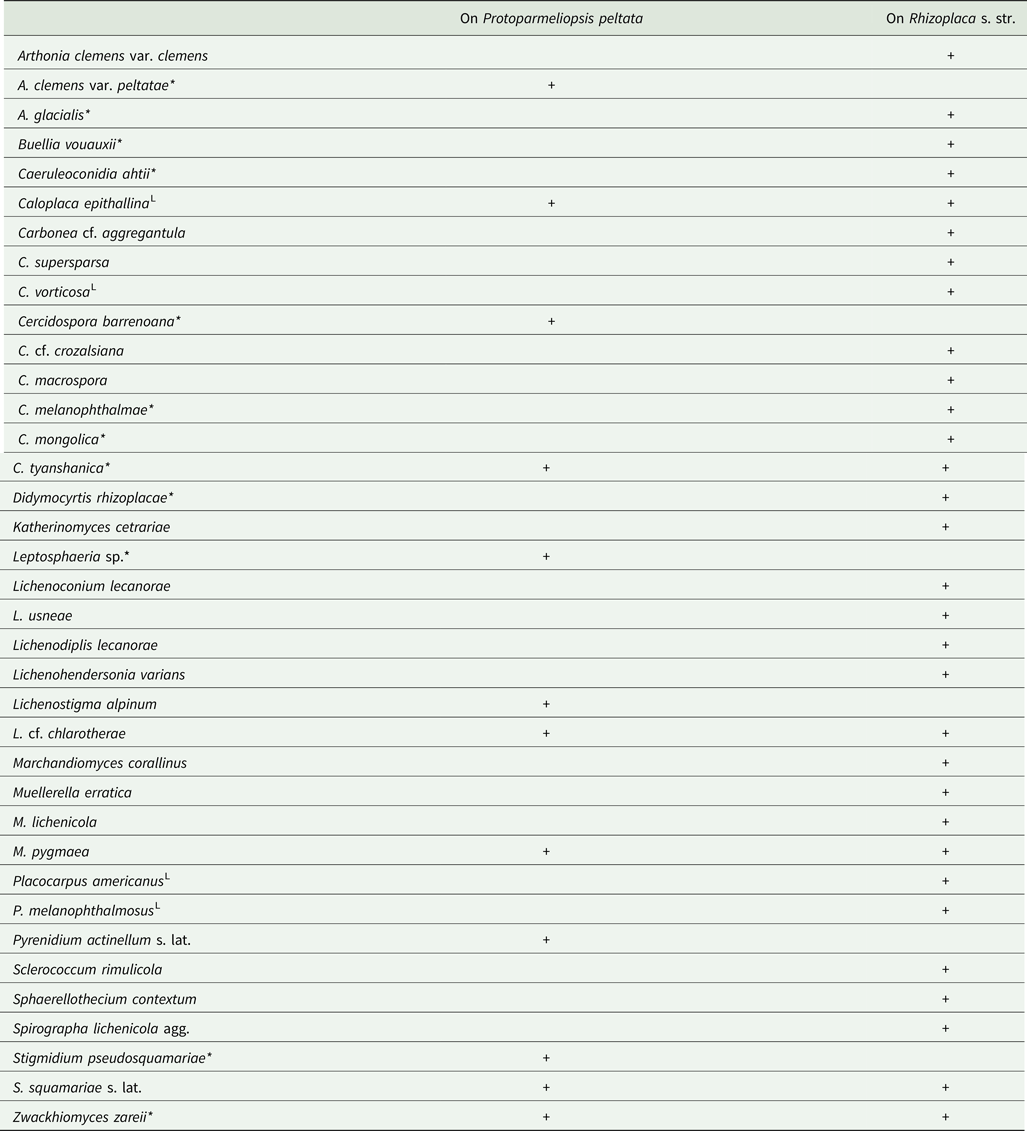
As follows from the data presented in the key above, a total of 32 species of lichenicolous fungi and four species of lichenicolous lichens have so far been recorded on Rhizoplaca s. lat.; of these, three species and one variety were recorded only on Protoparmeliopsis peltata, six species only on Rhizoplaca s. str. and another two species are confined to both of these host genera (Table 1).
Taxa of lichenicolous fungi recorded on Protoparmeliopsis peltata and/or Rhizoplaca s. str. Based on data from the key. Parasite species known only on these host genera are shown with an asterisk (*). Lichenized species are denoted by ‘L’.

Of the c. 25 known species of Rhizoplaca s. str., lichenicolous fungi and lichens were observed on five species: R. chrysoleuca (17 species of parasites), R. melanophthalma (19), R. novomexicana (3), R. phaedrophthalma (2) and R. subdiscrepans (8). A total of 25 parasite species have been observed on Rhizoplaca s. str., thus the ratio of the number of parasite species to the number of host species for this lichen genus is 1.0 (compare similar data in Zhurbenko & Ohmura (Reference Zhurbenko and Ohmura2020)).
In this study, we were able to identify 16 species of lichenicolous fungi growing on Rhizoplaca s. lat., representing half of their known species diversity. The most frequently collected species in the study area were Arthonia clemens (10 finds), Lichenoconium lecanorae (8), Cercidospora cf. crozalsiana (7), C. mongolica (6) and Stigmidium squamariae (6). Four (25%) of these 16 species are described as new to science, which corresponds to the level of taxonomic novelty in recent studies of lichenicolous fungi on such host groups as Siphula-like lichens (38% spp. nov.; Motiejūnaitė et al. Reference Motiejūnaitė, Zhurbenko, Suija and Kantvilas2019), Sphaerophoraceae (33% spp. nov.; Zhurbenko Reference Zhurbenko2023) or Thamnolia (30% spp. nov.; Zhurbenko Reference Zhurbenko2012).
Acknowledgements
The research of M. P. Zhurbenko was carried out within the framework of the research project of the Komarov Botanical Institute of the Russian Academy of Sciences ‘Taxonomic, ecological and structural-functional diversity of fungi and fungus-like protists’ (124013100829-3) using the equipment of its Core Facility Center ‘Cell and Molecular Technologies in Plant Science’. The author's field research in Mongolia was supported by the joint Russian-Mongolian complex biological expedition of the Russian Academy of Sciences and the Academy of Sciences of Mongolia. The author is grateful to Lev Byazrov and Lyudmila Bredkina, who kindly made their lichen collections available to him. Wolfgang von Brackel, Paul Diederich, Javier Etayo, Martin Grube, Josef Hafellner, James Hollinger and Claude Roux are thanked for valuable comments and assistance with taxonomic decisions.
Author ORCID
Mikhail P. Zhurbenko, 0000-0002-9839-4698.