New molecular/pharmacological and genetic approaches
Novel experimental approaches in neuroscience have recently identified promising molecular, pharmacological, and genetic avenues for amblyopia therapy. The common goal of these therapies is to harness the brain’s inherent ability to restructure itself by tapping into specific brain circuits and cellular mechanisms that promote plasticity. Such targets include, for example, excitatory and inhibitory synaptic components, neuromodulators, and epigenetic regulators. Commonly used pharmacological agents, transcranial direct current and magnetic stimulation (tDCS/TMS), and behavioral therapies may act through these or other cellular pathways yet to be discovered. Understanding the precise cellular mechanisms that promote circuit changes in experimental animals promises to guide new therapeutic approaches for treating amblyopia in humans.
Targeting inhibitory and excitatory synapses
Monocular visual deprivation during a critical period in early life remodels excitatory synapses extensively, inducing a rapid loss of dendritic spines and elimination of many axonal branches of geniculocortical afferents serving the deprived eye (Antonini & Stryker, Reference Antonini and Stryker1993; Mataga et al., Reference Mataga, Mizaguchi and Hensch2004). These losses of input are followed by a progressive expansion of axons and potentiation of responses from the open eye (Antonini et al., Reference Antonini, Fagiolini and Stryker1999; Frenkel & Bear, Reference Frenkel and Bear2004). Because these modifications are assumed to underlie the development of amblyopia, excitatory synapses represent strong candidate targets for its treatment. Indeed, recent reports have revealed that changes in the levels of the excitatory postsynaptic density protein PSD-95 govern the duration of the critical period for ocular dominance (OD) plasticity in the visual cortex, independent of changes in inhibitory circuits (Huang et al., Reference Huang, Stodieck, Goetze, Cui, Wong, Wenzel, Hosand, Dong, Löwel and Schlüter2015). PSD-95 expression increases in the visual cortex during the critical period for OD plasticity and promotes the progressive maturation of the so-called “silent” synapses that contain only NMDA-type glutamate receptors and that lack AMPA receptors. Genetic loss of PSD-95 function leads to the persistence of silent synapses, allowing the juvenile form of OD plasticity to be maintained lifelong. Strikingly, using a viral gene silencing approach to reduce PSD-95 in the visual cortex of adult mice rejuvenates excitatory synapses by reinstating silent synapses like those in the immature cortex and reopens a critical period for visual cortical plasticity (Huang et al., Reference Huang, Stodieck, Goetze, Cui, Wong, Wenzel, Hosand, Dong, Löwel and Schlüter2015).
Converging studies also point to intracortical inhibitory synapses as key regulatory sites of critical period plasticity (reviewed in Takesian & Hensch, Reference Takesian and Hensch2013; see also Hensch & Quinlan, this volume). Reducing the inhibitory synapse function by intracortical microperfusion of a GABA synthesis inhibitor or GABAA receptor antagonist can enhance plasticity in the rodent visual cortex during adulthood (Harauzov et al., Reference Harauzov, Spolidoro, Dicristo, Pasquale, Cancedda, Pizzorusso, Viegi, Berardi and Maffei2010). However, this manipulation does not produce plasticity like that in the critical period, where responses in the deprived eye are dramatically reduced. Instead, it accelerates or enhances the adult form of plasticity seen in rodents, which increases the response to the open fellow eye with little or no effect on the deprived-eye responses. In contrast, transplantation of specific types of embryonic inhibitory neurons into a postnatal visual cortex creates a second critical period of OD plasticity that follows the end of the normal one and is of similar duration (Southwell et al., Reference Southwell, Froemke, Alvarez-Buylla, Stryker and Gandhi2010; Tang et al., Reference Tang, Stryker, Alvarez-Buylla and Espinosa2014). The most prominent feature of this second critical period is the reduction of deprived-eye responses, exactly as in the normal critical period. Future work is needed to elucidate how the interplay of the excitatory and inhibitory synaptic function across cortical cell types may control cortical network plasticity.
Targeting neuromodulatory systems
Evidence has accumulated that neuromodulatory systems are also key targets for inducing plasticity to improve amblyopia. Neuromodulators such as serotonin and acetylcholine are released in the visual cortex from projections arising from the raphe nuclei and basal forebrain. These inputs are normally activated by salient stimuli and specific behavioral states, such as reward acquisition, punishment, and exercise (Fu et al., Reference Fu, Tucciarone, Espinosa, Sheng, Daracy, Nicoll, Huang and Stryker2014; Hangya et al., Reference Hangya, Ranade, Lorenc and Kepecs2015). However, these neuromodulatory systems can also be pharmacologically targeted by drugs commonly used to treat depression, such as selective serotonin reuptake inhibitors (SSRIs), or Alzheimer’s disease, such as cholinesterase inhibitors. Interestingly, it has been found that these pharmacological agents promote recovery from amblyopia in rodent models. For example, chronic treatment with SSRIs to enhance serotonergic signaling reopens a period of plasticity in the visual cortex of adult amblyopic rats, allowing for the recovery of visual acuity (Maya Vetencourt et al., Reference Maya Vetencourt, Sale, Viegi, Baroncelli, De Pasquale, O’Leary, Castrén and Maffei2008). Likewise, boosting acetylcholine signaling with a cholinesterase inhibitor enables recovery from amblyopia in the adult visual cortex (Morishita et al., Reference Morishita, Miwa, Heintz and Hensch2010). How do neuromodulators act within visual cortical circuits? Recent studies have uncovered a specific set of cortical inhibitory neurons that respond robustly to neuromodulators to enhance cortical plasticity (Letzkus et al., Reference Letzkus, Wolff, Meyer, Tovote, Courtin, Herry and Lüthi2011; Pi et al., Reference Pi, Hangya, Kvitsiani, Sanders, Huang and Kepecs2013; Fu et al., Reference Fu, Tucciarone, Espinosa, Sheng, Daracy, Nicoll, Huang and Stryker2014; Fu et al., Reference Fu, Kaneko, Tang, Alvarez-Buylla and Stryker2015). These GABAergic cells reside in the outermost layers of the cortex and are identified by the selective expression of the vasoactive intestinal peptide (VIP). Optogenetic activation of VIP cells directly drives plasticity in the primary visual cortex of the adult mouse (Fu et al., Reference Fu, Kaneko, Tang, Alvarez-Buylla and Stryker2015). VIP cells are thought to augment cortical activity and plasticity through inhibition of other cortical GABAergic interneurons (Letzkus et al., Reference Letzkus, Wolff, Meyer, Tovote, Courtin, Herry and Lüthi2011; Pfeffer et al., Reference Pfeffer, Xue, He, Huang and Scanziani2013; Pi et al., Reference Pi, Hangya, Kvitsiani, Sanders, Huang and Kepecs2013; Donato et al., Reference Donato, Rompani and Caroni2013; Fu et al., Reference Fu, Tucciarone, Espinosa, Sheng, Daracy, Nicoll, Huang and Stryker2014; Fu et al., Reference Fu, Kaneko, Tang, Alvarez-Buylla and Stryker2015). Other cell types in the visual cortex may also have a role in the plasticity induced by neuromodulators in adult mice. Pairing acetylcholine release in the visual cortex with specific visual stimuli enhances stimulus-selective responses of the cortical neurons, by engaging astrocyte-dependent strengthening of excitatory synapses (Chen et al., Reference Chen, Sugihara, Sharma, Perea, Petravicz, Le and Sur2012).
Ongoing clinical trials with drugs targeting these neuromodulatory systems highlight this approach as a promising avenue for amblyopia treatment in adult patient populations. SSRI treatment has been shown to augment visually-evoked potentials (VEPs) in normal human subjects (Normann et al., Reference Normann, Schitz, Fürmaier, Döing and Bach2007). In a few adult patients with amblyopia, SSRI (citalopram) enhanced visual acuity improvements when combined with two weeks of occlusion therapy, but effects in the population were not significantly different from placebo. (Thompson et al., Reference Thompson, Lagas, Stinear, Byblow, Russel and Kydd2014). Another study pairing SSRIs with video game training demonstrated that while video games improved visual acuity, no added value of the SSRI treatment was observed (Uusitalo, Reference Uusitalo2013). It is possible that such behavioral and pharmacological manipulations reach a ceiling effect if they engage similar neuromodulatory pathways. Likewise, an ongoing clinical study at Boston Children’s Hospital is using donepezil, a cholinesterase inhibitor that is typically used to treat Alzheimer’s disease, to boost cholinergic signaling and recover vision in amblyopic patients (T. Hensch, personal communication).
Targeting epigenetic regulation using histone deacetylase (HDAC) inhibitors
Brain circuits respond to environmental signals via dynamic changes in DNA methylation and histone modifications that control gene transcription (Fagiolini et al., Reference Fagiolini, Jensen and Champagne2009). Visual stimulation during early life induces histone acetylation in the mouse visual cortex, but the same stimulation in adulthood has little effect. This age-related decline in the capacity for experience-dependent regulation of histone acetylation makes it a candidate to underlie the developmental reduction in visual cortical plasticity (Putignano et al., Reference Putignano, Lonetti, Cancedda, Ratto, Costa, Maffei and Pizzorusso2007). In fact, increasing histone acetylation by inhibition of HDACs can reinstate plasticity in the adult visual cortex of rodents to allow recovery from amblyopia (Putignano et al., Reference Putignano, Lonetti, Cancedda, Ratto, Costa, Maffei and Pizzorusso2007; Silingardi et al., Reference Silingardi, Scali, Belluomini and Pizzorusso2010). Thus, HDAC inhibitors may represent yet another class of drugs with the potential to improve visual acuity beyond early life.
The HDAC inhibitor valproate (VPA) has already been found in a clinical study to reopen a period of plasticity to learn absolute pitch (Gervain et al., Reference Gervain, Vines, Chen, Seo, Hensch, Werker and Young2013). Absolute pitch, the ability to produce or identify a musical pitch without a reference sound, is possessed by only about 0.01% of the general population and acquired during a critical period in early life. Generally, absolute pitch is learned through musical training before 6 years of age and is rarely, or perhaps never, acquired during adulthood (Van Hedger et al., Reference Van Hedger, Heald, Koch and Nusbaum2015). However, administration of VPA, a commonly used mood stabilizer, opened a window of opportunity for adults to learn absolute pitch. These findings suggest that epigenetic actions of VPA may reset cortical circuitry to allow for juvenile-like plasticity. Future work will be required to reveal the cellular and circuit mechanisms underlying HDAC inhibitors such as VPA.
Transcranial direct current stimulation (tDCS)/transcranial magnetic stimulation (TMS)
Pharmacological interventions aimed to stimulate cortical plasticity pose risks of side effects. Therefore, there is a great deal of interest in discovering novel and less invasive alternatives to activate endogenous plasticity mechanisms. One promising strategy is to use transcranial direct current or magnetic stimulation, noninvasive brain stimulation techniques that can transiently alter neural excitability in targeted brain regions. Ongoing work is attempting to exploit this technique in adult patients with amblyopia to open a brief window of opportunity to improve visual function (B. Thompson, personal communication). A recent study found that a single session of tDCS can temporarily increase VEPs and contrast sensitivity driven by amblyopic eyes of adult patients (Ding et al., Reference Ding, Li, Spiegel, Chen, Chan, Luo, Yuan, Deng, Yu and Thompson2016), paving the way for future studies that will combine this stimulation technique with visual training for long-term improvements. Intriguingly, tDCS may increase excitability by a reduction in GABAergic inhibition (Stagg et al., Reference Stagg, Best, Stephenson, O’Shea, Wylezinska, Kincses, Morris, Matthews and Johansen-Berg2009), a mechanism known to regulate adult visual cortical plasticity. It should be mentioned, however, that these current/magnetic stimulation protocols may have a problem of pathway specificity. It is, therefore, essential that measures be taken to ensure that changes are restricted to target circuits.
Combining pharmacological interventions and behavioral training
Across both human and animal studies, it is evident that pharmacological intervention alone is not generally sufficient for successful treatment of amblyopia. Instead, the research strongly supports the need to combine a pharmacological approach with personalized behavioral training, with the goal of targeting plasticity within specific brain regions or specific cortical circuits. Interestingly, VPA treatment improved absolute pitch, but not other measures of auditory function (T. Hensch, personal communication), suggesting that VPA may not induce widespread effects, but may instead induce focal plasticity in response to targeted training paradigms. In the mouse visual system, recovery of closed-eye responses following long-term monocular deprivation is preferentially enhanced to the particular visual stimuli presented during VIP cell activation induced by running (Kaneko & Stryker, Reference Kaneko and Stryker2014). Can these pharmacological approaches be used to shorten or enhance behavioral treatment? The next section will discuss promising behavioral treatments that could be used alone or in combination with pharmacological approaches.
Environmental and behavioral treatments
In an attempt to find novel noninvasive methods of stimulating visual plasticity in adulthood, a number of behavioral interventions have emerged that may help to treat amblyopia in humans. These include manipulations of the environment, such as exposure to an enriched environment, or complete visual deprivation, both of which reactivate robust plasticity in the visual cortex. Similarly, engaging humans and animals in voluntary physical exercise (e.g., running wheels) and visuomotor tasks has led to remarkable increases in neuronal plasticity. Finally, novel vision training paradigms may result in more practical, less expensive therapies, and faster recovery. Here, we highlight some of these novel treatments and discuss their success in both animals and humans.
Environmental enrichment (EE)/running
It is now evident that increased levels of environmental stimulation have a profound impact on experience-dependent plasticity within both the developing and adult brain. Experimental animals are generally raised in small cages with only their littermates, strongly limiting social interactions and physical exercise. Recent studies have found significant effects of environmentally-enriched cages in which the animals are housed in large groups, have access to running wheels, and are exposed to a complex environment that elicits social and exploratory behaviors. Notably, adult amblyopic rats housed in enriched environments recovered from long-term monocular deprivation (Sale et al., Reference Sale, Maya Vetencourt, Medini, Cenni, Baroncelli, De Pasquale and Maffei2007). Moreover, raising mice in an enriched environment extended the critical period for a juvenile form of OD plasticity into adulthood (Greifzu et al., Reference Greifzu, Pielecka-Fortuna, Kalogeraki, Krempler, Favaro, Schlüter and Löwel2014) and allowed an adult form of OD plasticity to persist even throughout life (Greifzu et al., Reference Greifzu, Kalogeraki and Löwel2016). Remarkably, placing standard-cage-reared mice into an enriched environment as adults restored OD plasticity, apparently rejuvenating the visual cortex (Greifzu et al., Reference Greifzu, Pielecka-Fortuna, Kalogeraki, Krempler, Favaro, Schlüter and Löwel2014; Greifzu et al., Reference Greifzu, Kalogeraki and Löwel2016). These effects of EE on plasticity seem largely to be due to the reduction of GABAergic inhibition to juvenile levels (Greifzu et al., Reference Greifzu, Pielecka-Fortuna, Kalogeraki, Krempler, Favaro, Schlüter and Löwel2014) accompanied by decreased peri-neuronal nets in the visual cortex (Sale et al., Reference Sale, Maya Vetencourt, Medini, Cenni, Baroncelli, De Pasquale and Maffei2007). These studies highlight EE as a noninvasive means of harnessing known plasticity mechanisms (reduced intracortical inhibition) to promote visual recovery. Surprisingly, the use of just one of the components of EE has recently been shown to preserve plasticity to older ages, namely a running wheel allows adult mice raised in standard cages to express OD-plasticity into adulthood (Kalogeraki et al., Reference Kalogeraki, Greifzu, Haack and Löwel2014). Furthermore, even short-term running, just during the 7-day monocular deprivation period, restored OD-plasticity to adult standard cage-raised mice. Notably, it is important to distinguish the extension into adult life or the enhancement of the effects of deprivation (e.g., MD) from the enhancement of recovery of the visual function, because the underlying mechanisms may be different.
However, the human environment is generally much more ‘enriched’ than that of any experimental animal, raising the question of whether EE in rodents may be translated into a treatment protocol for humans. Social interactions, novelty, exercise, and engagement of the visuomotor systems are all components of EE that may engage distinct brain regions, circuits, and cellular mechanisms. Researchers using animal models or human subjects should strive toward identifying the key aspects of an EE that can be implemented to promote plasticity.
Dark exposure
It has long been known that dark rearing from birth retains the visual cortex in an immature state and prolongs the critical period for OD plasticity (e.g., Cynader, Reference Cynader1983; Mower et al., Reference Mower, Caplan, Christen and Duffy1985; Fox et al., Reference Fox, Daw, Sato and Czepita1991). Recent work has shown that periods of total darkness that eliminate all visually-driven activity can reactivate robust plasticity in the adult visual cortex of rats (He et al., Reference He, Hodos and Quinlan2006; He et al., Reference He, Ray, Dennis and Quinlan2007) and mice (Stodieck et al., Reference Stodieck, Greifzu, Goetze, Schmidt and Löwel2014), and in juvenile kittens (Duffy & Mitchell, Reference Duffy and Mitchell2013). The ability to promote synaptic plasticity in the adult visual cortex through dark exposure was predicted by the BCM sliding threshold theory of synaptic plasticity, which predicts that the loss of patterned visual experience would lower the value of the synaptic modification threshold and enable recovery of weakened deprived-eye inputs (Cooper & Bear, Reference Cooper and Bear2012). Indeed, dark exposure in adulthood stimulates the expression of the NMDA receptor, a molecular switch known to lower the threshold for synaptic modification (Yashiro et al., Reference Yashiro, Corlew and Philpot2005; He et al., Reference He, Hodos and Quinlan2006; Philpot et al., Reference Philpot, Cho and Bear2007).
The plasticity that is reactivated by dark exposure can be harnessed to promote the recovery from amblyopia. For example, rats rendered amblyopic by chronic monocular deprivation initiated at eye opening recovered visual acuity when 10 days of dark exposure in young adulthood (P70-100) were followed by binocular vision or reverse occlusion (He et al., Reference He, Ray, Dennis and Quinlan2007). Remarkably, short-term dark exposure (10 days) can reinstate visual cortical plasticity even in very old mice (P535; Stodieck et al., Reference Stodieck, Greifzu, Goetze, Schmidt and Löwel2014). However, the effects of dark exposure on adult plasticity may vary across species; while 10 days of darkness enhanced OD plasticity in juvenile kittens, this treatment failed to restore OD plasticity in adult cats (Duffy et al., Reference Duffy, Lingley, Holman and Mitchell2016). The recovery from amblyopia following dark exposure was rapid, occurring in kittens within just a week after removal from the darkness (Duffy & Mitchell, Reference Duffy and Mitchell2013). Importantly, repetitive performance of a visual task following dark exposure further improved the recovery of acuity, but delaying the visual stimulation for several weeks following the dark exposure prevented the recovery of visual acuity (Eaton et al., Reference Eaton, Sheehan and Quinlan2016). This finding suggests that a period of darkness opens a limited window of plasticity during which the cortex is more receptive to visual training and may guide the design of new therapies.
It may be puzzling that manipulations that on the surface appear to be very different—putting animals either in complete darkness or in an enriched environment—both stimulate robust plasticity in the adult cortex. It is possible that distinct neural mechanisms lead to a common outcome, possibly through a common substrate. A reduction in inhibitory synaptic transmission by dark exposure may be a feature in common with EE. As with environmental enrichment (Greifzu et al., Reference Greifzu, Pielecka-Fortuna, Kalogeraki, Krempler, Favaro, Schlüter and Löwel2014), dark exposure may work through a rejuvenation of intracortical inhibition, including a reduction of the excitatory drive onto fast-spiking interneurons (Huang et al., Reference Huang, Gu, Quinlan and Kirkwood2010; Gu et al., Reference Gu, Tran, Murase, Borrell, Kirkwood and Quinlan2016) and a decrease in the number of parvalbumin-expressing inhibitory cells and surrounding peri-neuronal nets (Stodieck et al., Reference Stodieck, Greifzu, Goetze, Schmidt and Löwel2014). Indeed, the adult recovery from long-term monocular deprivation can be stimulated by a reduction in the activity of inhibitory neurons (Kaneko & Stryker, Reference Kaneko and Stryker2014). Dark exposure also reduces neurofilament protein levels, which is hypothesized to destabilize the neuronal cytoskeleton (Duffy & Mitchell, Reference Duffy and Mitchell2013), and promotes the recovery of thalamocortical synaptic transmission and the density of dendritic spines on pyramidal neurons in the primary visual cortex (Montey & Quinlan, Reference Montey and Quinlan2011).
An exciting pilot study is now evaluating whether adult amblyopic humans will also recover visual function following a brief period in complete darkness (B. Backus & E. Quinlan, personal communication). Preliminary results suggest that 5 days of darkness is well tolerated, with no reports of anxiety or changes in physical health. An ongoing study is now evaluating the effects of 10 days of darkness on adult amblyopic patients. To exploit the transient period of plasticity immediately following darkness, the patients will undergo intensive visual training in the weeks following the dark exposure. The practicality of using this protocol in humans as a treatment for amblyopia must be addressed, as studies in rodents and kittens suggested that shorter periods of dark exposure are ineffective, and even brief periods of light exposure prevented the effects (He et al., Reference He, Ray, Dennis and Quinlan2007; Mitchell et al., Reference Mitchell, MacNeill, Crowder, Holman and Duffy2016). Thus, access to completely dark environments for a sufficient duration of time with proper support to guarantee safety and well-being may pose challenges for widespread use. Interestingly, visual deprivation for several days by binocular intravitreal injections of the pufferfish toxin tetrodotoxin promotes similar fast visual recovery in amblyopic cats (Fong et al., Reference Fong, Mitchell, Duffy and Bear2016), raising the possibility of developing novel pharmacological techniques to transiently block vision, if adequate safety measures can reliably be ensured. Blindfolding may offer another solution—one study suggested that blindfolding normally sighted adults for 5 days leads to rapid changes in experience-dependent functional neural connectivity (Merabet et al., Reference Merabet, Hamilton, Schlaug, Swisher, Kiriakopoulos, Pitskel, Kauffman and Pascual-Leone2008). However, it has been shown in amblyopic kittens that binocular visual deprivation by lid-suture does not recapitulate the recovery-promoting effects of darkness (Duffy et al., Reference Duffy, Bukhamseen, Smithen and Mitchell2015). If it is established that 10 days of complete darkness promotes visual recovery in amblyopic humans, future studies in both humans and animal models may identify methods to shorten, segment, or facilitate binocular occlusion. A further understanding of the neural mechanisms underlying dark exposure may allow us to predict which combination of treatments will induce robust visual cortical plasticity. Dark exposure offers a novel and promising noninvasive approach for the recovery of vision.
Exercise and visuomotor engagement
Recent research links physical activity to profound adult plasticity in the visual cortex, providing another promising behavioral intervention for recovery from amblyopia. Remarkably, allowing adult mice to run on a treadmill potently enhances visual cortical activity (Niell & Stryker, Reference Niell and Stryker2010) and promotes recovery of vision following monocular deprivation (Kaneko & Stryker, Reference Kaneko and Stryker2014). Moreover, visual stimulation or running alone did not improve visual function, suggesting that plasticity is facilitated only in activated neural circuits during running (Kaneko & Stryker, Reference Kaneko and Stryker2014). Recent studies have identified the key cellular mechanism underlying the effects of locomotion—VIP cells (Fu et al., Reference Fu, Tucciarone, Espinosa, Sheng, Daracy, Nicoll, Huang and Stryker2014). In fact, genetic silencing of VIP cells in amblyopic adult mice prevents the recovery of the visual function by running, suggesting that the activity of these cells is necessary for the enhanced plasticity (Fu et al., Reference Fu, Kaneko, Tang, Alvarez-Buylla and Stryker2015).
The potential of physical activity to promote amblyopic recovery has caught the attention of the clinical field. Adult subjects who intermittently cycled on a stationary bicycle while watching a movie showed enhanced effects of transient eye patching compared to those subjects who watched the movie while sitting still (Lunghi & Sale, Reference Lunghi and Sale2015). Moreover, tasks that directly engage both visual and motor circuits have achieved great success in reversing amblyopia. For example, recovery from amblyopia is expedited by tasks requiring coordination of hand and eye movements, such as having patients manipulate objects during visual training (reviewed in Daw, Reference Daw2013). Patients with amblyopia exhibit impairments in oculomotor performance, including saccadic eye movements (Niechwiej-Szwedo et al., Reference Niechwiej-Szwedo, Goltz, Chandrakumar, Hirji and Wong2010; Niechwiej-Szwedo et al., Reference Niechwiej-Szwedo, Chandrakumar, Goltz and Wong2012; McKee et al., Reference McKee, Levi, Schor and Movshon2016; Perdziak et al., Reference Perdziak, Witkowska, Gryncewicz and Ober2016), smooth pursuit (Raashid et al., Reference Raashid, Liu, Blakeman, Goltz and Wong2016), fixation stability (González et al., Reference González, Wong, Niechwiej-Szwedo, Tarita-Nistor and Steinbach2012; Chung et al., Reference Chung, Kumar, Li and Levi2015), hand-eye coordination (Niechwiej-Szwedo et al., Reference Niechwiej-Szwedo, Goltz, Chandrakumar, Hirji and Wong2011, Reference Niechwiej-Szwedo, Goltz, Chandrakumar and Wong2014), and execution of grasping movements (Grant et al., Reference Grant, Melmoth, Morgan and Finlay2007; Suttle et al., Reference Suttle, Melmoth, Finlay, Sloper and Grant2011). Therefore, targeting visuomotor circuits during treatment may help to alleviate some of these deficits.
Novel visual training procedures
There is no doubt that conventional patch therapy, directed toward improving the visual function of the amblyopic eye while occluding the fellow eye, is effective in the majority of cases if initiated at the appropriate age. However, it is not always effective, and it has additional limitations: patching interferes with binocular input, which can lead to poor binocular outcome, and it often has poor compliance (reviewed in Birch, Reference Birch2012; Hess & Thompson, Reference Hess and Thompson2015). When the patch therapy is ineffective, there is no consensus on effective treatment. Since the 1970s, a number of alternative procedures, many inspired by the animal research literature, have been proposed. None of these have attained the degree of acceptance that would make them a new standard of care. We note a number of them here not as an endorsement but to foster a critical examination of the relationship between laboratory research findings and approaches to therapy for human patients.
Some alternative strategies use binocular exposure approaches to treat amblyopia. For example, a set of training paradigms called “monocular fixation in a binocular field” (MFBF), in which both eyes remain open while a vision task is accomplished by a single eye, have been reported to have some success (Brock, Reference Brock1963; Cohen, Reference Cohen1981). One MFBF procedure places a red filter over the dominant eye such that both eyes receive light, but only the amblyopic eye can see the markings of a red pen used to perform various tasks (reviewed in Daw, Reference Daw2013). The Cambridge Vision Stimulator treatment combined a short-term occlusion, visual stimulation using contours of all orientations, and training exercises, although efficacy was not demonstrated in a randomized clinical trial (Campbell et al., Reference Campbell, Hess, Watson and Banks1978). Indeed, there are many reports of successful results of active vision therapy with only minimal amounts of occlusion (reviewed in Garzia, Reference Garzia1987). Some reports suggest that this approach may be particularly successful in older patients; for example, a group of older children and adult patients with anisometropic amblyopia achieved long-lasting improvements in visual acuity and a binocular function following a treatment that combined active vision therapy with occlusion of only 2–5 h per day, although the role, if any, of active vision was not demonstrated using a control group (Wick et al., Reference Wick, Wingard, Cotter and Scheiman1992).
Video game therapy has emerged as a form of active vision training aimed at improving both acuity and stereopsis in both amblyopic children and adults (reviewed in Hess & Thompson, Reference Hess and Thompson2015; Levi et al., Reference Levi, Knill and Bavelier2015). Anaglyphic video games were developed for vision therapy more than 30 years ago (Press, Reference Press1981) and used as an MFBF approach for the treatment of amblyopia (Ludlam, Reference Ludlam and Press1992). The clinical finding that suppression—a reduced contribution of the amblyopic eye during binocular viewing—is an important part of the amblyopia syndrome along with a greater understanding of the biological underpinnings of amblyopia has spurred new dichoptic approaches to promote “binocular rebalancing”. These approaches may reactivate latent binocular pathways by reducing inhibitory interactions, boosting attenuated excitatory function, and/or shifting the synaptic modification threshold in favor of potentiation (reviewed in Hess & Thompson, Reference Hess and Thompson2015). For example, recent studies have found that both amblyopic preschool children and adults can show enhanced improvements in visual and motor function by playing dichoptic iPad or iPod games (Hess et al., Reference Hess, Mansouri and Thompson2011; Birch et al., Reference Birch, Li, Jost, Morale, De La Cruz, Stager, Dao and Stager2015; Vedamurthy et al., Reference Vedamurthy, Nahum, Huang, Zheng, Bayliss, Bavelier and Levi2015; Webber et al., Reference Webber, Wood and Thompson2016). Since the lack of binocular function is a key risk factor for persistent amblyopia, the development of new binocular training strategies may have a substantial impact on improving acuity and recovering stereopsis (reviewed in Levi et al., Reference Levi, Knill and Bavelier2015). Unfortunately, recent randomized clinical trials have failed to provide evidence that video games are any better than patching in older children or that they improve binocular function (Holmes et al., Reference Holmes, Manh, Lazar, Beck, Birch, Kraker, Crouch, Erzurum, Khuddus, Summers and Wallace2016; Kelly et al., Reference Kelly, Jost, Dao, Beauchamp, Leffler and Birch2016). Future work will be required to elucidate how monocular and dichoptic experience contribute to amblyopia recovery across distinct patient populations.
Elaborate visual training regimes have been shown in some cases by others (Paul Harris, unpublished observation) to improve vision in amblyopic humans but are expensive and time intensive. They have not yet been adopted widely because of a lack of controlled trials that demonstrate both efficacy and safety. Therefore, there is a continued need to seek novel, faster training approaches. For example, a study in adult amblyopic macaques suggested that implementing a ‘global’ training paradigm may lead to visual improvements that generalize beyond the trained stimulus (Kiorpes & Mangal, Reference Kiorpes and Mangal2015). Moreover, training strategies that engage attentional and emotional processes, including movies and action video games, appear in some reports to be particularly successful (reviewed in Levi & Li, Reference Levi and Li2009, and in Bavelier et al., Reference Bavelier, Levi, Li, Dan and Hensch2010). Such training is likely to stimulate neuromodulatory systems that promote plasticity and to activate circuits encoding higher-order visual functions that are impaired in patients with amblyopia (reviewed in Kiorpes, Reference Kiorpes2006).
Use of animal models to find treatments for amblyopia
With the advent of new technologies in neuroscience, the field continues to rely heavily on animal models to uncover the neural mechanisms underlying human pathologies such as amblyopia. The mouse in particular has emerged as an ideal model for taking advantage of genetic, optogenetic, physiological, and imaging tools that allow experimenters to label, manipulate, and monitor specific cell types with great precision. Nevertheless, it is not yet clear whether rodent models are appropriate for the study of amblyopia and its treatment in humans.
One potential limitation of mice for the study of amblyopia is that the mouse does not have a fovea, and therefore the entire mouse retina resembles the peripheral retina of the primate (Naarendorp et al., Reference Naarendorp, Esdaille, Banden, Andrews-Labenski, Gross and Pugh2010). Although amblyopia was first described as a deficit in foveal vision, amblyopic deficits have also been detected in the visual field periphery (Irene Gottlob, personal communication). In fact, patients with amblyopia have been shown to exhibit decreased motion and contrast detection through the amblyopic eye (Katz et al., Reference Katz, Levi and Bedell1984; Levi et al., Reference Levi, Klein and Aitsebaomo1984). Thus, the absence of foveal vision may not exclude the mouse a priori as a model to study human amblyopia. Furthermore, the mouse primary visual cortex exhibits binocular integration and disparity selectivity that support depth perception (Scholl et al., Reference Scholl, Burge and Priebe2013), potentially allowing the use of mice to study the loss of stereoscopic vision associated with amblyopia.
Another major consideration when evaluating the effectiveness of potential amblyopia treatments in mice is that the common forms of human amblyopia are not generally studied in the mouse. Basic research on amblyopia in rodents has focused almost exclusively on monocular deprivation. However, human patients with amblyopia exhibit various types of amblyopia that are generally classified as anisometropic, strabismic, and deprivation amblyopia. Deprivation amblyopia is the least common form of amblyopia, accounting for less than 5 per cent of cases (Holmes & Clarke Reference Holmes and Clarke2006). Can we get closer to the human condition? Experiments in monkeys have employed more subtle forms of deprivation such as anisometropia and strabismus, revealing reduced binocularity and poorer spatial resolution in V1 neurons driven by the amblyopic eye (Movshon et al., Reference Movshon, Eggers, Gizzi, Hendrickson, Kiorpes and Boothe1987; Kiorpes et al., Reference Kiorpes, Kiper, O’Keefe, Cavanaugh and Movshon1998). In mice, one possibility is to take advantage of the available genetic models to identify genetic ocular defects that better approach the common human forms of amblyopia (Engle, Reference Engle2007). However, while exploring new experimental models of amblyopia, it is also important to take advantage of the tremendous progress over the past 50 years in our understanding of monocular deprivation in mice as a premier model for understanding critical periods for cortical plasticity and the underlying regulating factors.
Assessing outcomes of amblyopia treatment
What is the best test to assess amblyopia and amblyopia recovery in experimental models? In animal models, amblyopia is generally assessed using grating acuity. Animals are presented with drifting gratings at increasing spatial frequencies to determine optomotor, behavioral, or neural thresholds. Grating acuity is a strong measure of the degraded visual function associated with amblyopia, particularly deprivation amblyopia, and corresponds to measures of Snellen (optotype) acuity in humans (Levi & Klein, Reference Levi and Klein1982). Thus, grating acuity is an appropriate and relatively easy way to assess acuity in animals. However, it should be noted that grating and optotype acuity are not equivalent. This may be particularly important for the detection of crowding effects, which are marked in amblyopia. Moreover, not all grating tasks reveal the same acuity. In particular, using the visual water task in rodents, a visual discrimination task based on reinforcement learning (Prusky et al., Reference Prusky, West and Douglas2000; Prusky & Douglas, Reference Prusky and Douglas2004), allows measurement of good perceptual thresholds of visual acuity.
In addition to assessing acuity as a recovery measure, it will be important to extend our evaluation to other visual deficits associated with amblyopia. For example, the loss of stereoptic depth perception may have the greatest impact on the quality of life for the amblyopic patient, impairing the ability on visuomotor tasks and limiting career options (Levi et al., Reference Levi, Knill and Bavelier2015). Moreover, adults who lack stereopsis tend to be refractory to therapy. Individuals with amblyopia, particularly strabismus, also exhibit oculomotor deficits (McKee et al., Reference McKee, Levi, Schor and Movshon2016) and other visuomotor deficits during the performance of fine motor tasks (Grant & Moseley, Reference Grant and Moseley2011). Thus, developing novel methods of measuring trajectories for the recovery of binocular vision and visuomotor ability in both experimental animal models and humans may provide valuable insights into the success of future amblyopia treatments.
Finally, there is a need to evaluate the effectiveness of amblyopia treatments on higher brain regions. Deficits in vision function associated with amblyopia may result from changes in extrastriate regions instead of or as well as the primary visual cortex (Schröder et al., Reference Schröder, Fries, Roelfsema, Singer and Engel2002; reviewed in Kiorpes & McKee, Reference Kiorpes and McKee1999). Few studies assess the extent of impairment or recovery outside of the primary visual cortex. Using less V1-centric metrics to assess recovery of the function by tapping into the higher-order dorsal and ventral streams will provide further insights into novel treatments. Studies outside of purely visual regions also have the potential to illuminate deficits in unexpected pathways, such as visuomotor and cross-modal connections (for example, visual–tactile interactions; Niechwiej-Szwedo et al., Reference Niechwiej-Szwedo, Chin, Wolfe, Popovich and Staines2016). So far, the study of the neural underpinnings of amblyopia and the molecular factors that promote recovery from amblyopia has been largely restricted to V1. Future work that determines whether other brain regions show critical periods and plasticity mechanisms that are similar to those observed in V1 will better inform us how and when to treat amblyopia.
Toward identifying common pathways to recovery
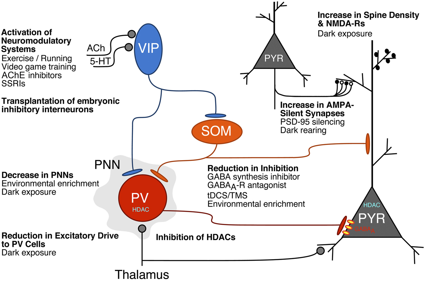
Novel molecular, pharmacological, and behavioral treatments are emerging to harness the brain’s plasticity mechanisms for the recovery from amblyopia (see Fig. 1). Ongoing challenges will be to determine how the various behavioral manipulations engage plasticity factors and how these plasticity factors interact within the cortical networks to promote the desired rewiring. A number of distinct candidate plasticity factors have been identified, and ongoing work will address whether these factors operate independently or as components of a common mechanism. For example, exercise and video games may engage neuromodulatory systems to activate specific types of cortical inhibitory interneurons known to control plasticity. Identifying “hub” circuits, cells, or molecular mechanisms that promote visual plasticity promises to lead to better-targeted treatment strategies that should mitigate side effects and accelerate recovery from amblyopia in human patients.
Cellular and circuit mechanisms underlying molecular, pharmacological, and environmental approaches to increase plasticity in the visual cortex. Release of neuromodulators in the visual cortex, such as acetylcholine (ACh) or serotonin (5-HT = 5-hydroxytryptophan), activate VIP (vasoactive intestinal polypeptide) inhibitory cells that promote cortical activity and plasticity by inhibiting other inhibitory interneuron subtypes, PV (parvalbumin), and SOM (somatostatin) cells (Letzkus et al., Reference Letzkus, Wolff, Meyer, Tovote, Courtin, Herry and Lüthi2011; Pi et al., Reference Pi, Hangya, Kvitsiani, Sanders, Huang and Kepecs2013; Fu et al., Reference Fu, Tucciarone, Espinosa, Sheng, Daracy, Nicoll, Huang and Stryker2014; Kaneko & Stryker, Reference Kaneko and Stryker2014; Fu et al., Reference Fu, Kaneko, Tang, Alvarez-Buylla and Stryker2015). These neuromodulatory systems can be activated by pharmacological treatments such as cholinesterase (AChE) inhibitors (Morishita et al., Reference Morishita, Miwa, Heintz and Hensch2010) or selective serotonin reuptake inhibitors (SSRIs; Maya Vetencourt et al., Reference Maya Vetencourt, Sale, Viegi, Baroncelli, De Pasquale, O’Leary, Castrén and Maffei2008; Thompson et al., Reference Thompson, Lagas, Stinear, Byblow, Russel and Kydd2014) or behavioral therapies such as exercise (Fu et al., Reference Fu, Kaneko, Tang, Alvarez-Buylla and Stryker2015; Lunghi & Sale, Reference Lunghi and Sale2015) and video game training (Bavelier et al., Reference Bavelier, Levi, Li, Dan and Hensch2010). Transplantation of embryonic inhibitory neurons into the postnatal visual cortex induces a second critical period of OD plasticity after the normal one (Southwell et al., Reference Southwell, Froemke, Alvarez-Buylla, Stryker and Gandhi2010; Tang et al., Reference Tang, Stryker, Alvarez-Buylla and Espinosa2014; Isstas et al., Reference Isstas, Teichert, Bolz and Lehmann2017). Plasticity is also enhanced in the adult visual cortex by decreasing perineuronal nets that predominantly enwrap the PV cells by pharmacological (Pizzorusso et al., Reference Pizzorusso, Medini, Berardi, Chierzi, Fawcett and Maffei2002) or behavioral interventions, such as environmental enrichment (Sale et al., Reference Sale, Maya Vetencourt, Medini, Cenni, Baroncelli, De Pasquale and Maffei2007) or dark exposure (Stodieck et al., Reference Stodieck, Greifzu, Goetze, Schmidt and Löwel2014). A reduction in the excitatory drive to PV cells (Huang et al., Reference Huang, Gu, Quinlan and Kirkwood2010; Gu et al., Reference Gu, Tran, Murase, Borrell, Kirkwood and Quinlan2016) and an increase in spine density and NMDA-Rs (Yashiro et al., Reference Yashiro, Corlew and Philpot2005; He et al., Reference He, Hodos and Quinlan2006; Philpot et al., Reference Philpot, Cho and Bear2007; Montey & Quinlan, Reference Montey and Quinlan2011) may also contribute to the enhanced plasticity that occurs with dark exposure. Various manipulations that reduce inhibitory synaptic function have been found to enhance visual cortical plasticity, including drugs that inhibit GABA synthesis or GABAA receptors (Harauzov et al., Reference Harauzov, Spolidoro, Dicristo, Pasquale, Cancedda, Pizzorusso, Viegi, Berardi and Maffei2010), tDCS/TMS (Stagg et al., Reference Stagg, Best, Stephenson, O’Shea, Wylezinska, Kincses, Morris, Matthews and Johansen-Berg2009), and environmental enrichment (Greifzu et al., Reference Greifzu, Pielecka-Fortuna, Kalogeraki, Krempler, Favaro, Schlüter and Löwel2014). Inhibition of HDACs can also reinstate plasticity in the adult visual cortex to allow recovery from amblyopia (Putignano et al., Reference Putignano, Lonetti, Cancedda, Ratto, Costa, Maffei and Pizzorusso2007; Silingardi et al., Reference Silingardi, Scali, Belluomini and Pizzorusso2010). Finally, an increase in AMPA-silent synapses (white synaptic boutons) underlies the heightened plasticity following knock-out or virus-mediated gene silencing of PSD-95 (Huang et al., Reference Huang, Stodieck, Goetze, Cui, Wong, Wenzel, Hosand, Dong, Löwel and Schlüter2015). Silent synapses also persist in the adult visual cortex in dark-reared mice (Funahashi et al., Reference Funahashi, Maruyama, Yoshimura and Komatsu2013). The figure is modified from the one presented by Takao Hensch at the Lasker/IRRF Initiative on Amblyopia workshops, Woods Hole, MA, July/August, 2015.

Recommendations
• The emergence of novel genetic, imaging, and electrophysiological tools to probe circuit and cellular mechanisms is shedding light on new targets to promote plasticity in visual circuits. Pharmacological agents, transcranial direct current or magnetic stimulation (tDCS/TMS), and behavioral therapies may harness these plasticity mechanisms for amblyopia treatment. A focus on understanding how such treatments engage precise circuit, cellular, and molecular mechanisms will provide insights into focused strategies to promote recovery.
• Novel visual training paradigms that exploit our increased understanding of the biological underpinnings of amblyopia recovery are needed. Future work should continue to seek training strategies that are tailored to the individual patient to engage attentional, emotional, and visuomotor circuits for faster and more effective recovery.
• The use of animal models such as mice to study treatments for human amblyopia presents numerous challenges because of differences in both visual function and visual cortical circuits between species. Furthermore, mouse research has been largely confined to studying deprivation amblyopia, the least common form of human amblyopia. New experimental procedures mimicking the more common forms of human amblyopia in mice and other animal models would enhance progress for the treatment of amblyopia in humans. Nevertheless, the knowledge gained over the past 50 years of animal research, with mice as the premier model of the last decade, should continue to inform our understanding of critical periods and their underlying cellular and molecular mechanisms.
• Grating acuity is a reliable measure of amblyopia that can be readily assessed in both animal models and humans. However, developing novel methods of measuring trajectories for recovery of other visual functions, including binocular vision and visuomotor ability, is likely to improve the success of amblyopia treatments in humans. Moreover, future work should be directed at determining whether brain regions outside of the primary visual cortex can additionally be targeted to promote recovery from amblyopia.
Acknowledgments
The authors would like to thank Anne Takesian,* who served as scribe for this discussion section. We would also like to thank all discussion group participants: Yuzo Chino, Nigel Daw, Kevin Duffy, Simon Grant, Paul Harris, Takao Hensch, Suzanne McKee, Ewa Niechwiej-Szwedo, Elizabeth Quinlan, Anu Sharma, Paul Sieving.
*FM Kirby Neurobiology Center, Boston Children’s Hospital, Harvard Medical School, Boston, MA, USA.