Implications
Research has shown that there is a link between the reticulo-rumen (RR) size of sheep and the amount of methane produced from the same amount of feed eaten. However, it is not known if the shape of the rumen differs between individuals. This study describes the application of computational techniques for the analysis and comparison of complex geometries in conjunction with images from computed tomography (CT) to determine morphological (shape) characteristics of the rumen. In contrast to previous studies of rumen morphology, this approach provides information on both size-dependent and size-independent characteristics.
Introduction
The RR is the first compartment in the digestive system of ruminants. Although it performs a similar function in a range of species, breeds and individuals, its morphological features – for example, the total size/capacity of the organ, the relative volume of its compartments, papillation patterns and motility vary widely, for example between concentrate selectors and roughage eaters (Hofmann, Reference Hofmann1989; Clauss et al., Reference Clauss, Hofmann, Fickel, Streich and Hummel2009). These differences are thought to be evolutionary adaptations to differences in the physical characteristics and chemical composition of feed, affecting functions such as retention times, stratification of RR contents (separation into solid, fluid and gas fractions) and fluid passage rates (Clauss et al., Reference Clauss, Hofmann, Streich, Fickel and Hummel2010a and Reference Clauss, Hume and Hummel2010b). Variations in morphology have also been linked to variation in function, for example, in relation to susceptibility to legume bloat (Waghorn and Reid, Reference Waghorn and Reid1984).
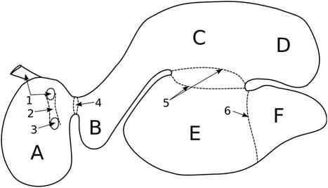
The RR is a complex multi-compartmental organ that consists of six sacs separated by tissue folds and muscular pillars (Figure 1) that help to hold the shape and enable contractions (Wyburn, 1980). The dorsal and ventral sacs are the largest compartments and contribute most of the RR volume. Contractions of the caudo-dorsal and caudo-ventral blind sacs drive digesta towards the cranial sac and the reticulum, whereas contractions of the reticulum (which can be considered as a separate organ) and the cranial sac drive digesta flow caudally (Waghorn and Reid, Reference Waghorn and Reid1977). Digesta is a heterogeneous mixture of plant fragments, saliva and an anaerobic microflora, and its behaviour is dominated by mixing, floatation, sedimentation (Sutherland, 1988). Contents can exit the RR through the reticulo-omasal orifice (ROO) to the omasum (Sellers and Stevens, Reference Sellers and Stevens1966; Wyburn, 1980).
Illustration of the reticulo-rumen internal geometry for sheep. A, reticulum; B, cranial sac/atrium; C, dorsal sac; D, caudo-dorsal blind sac; E, ventral sac; F, caudo-ventral blind sac. 1, Oesophagus; 2, reticular groove; 3, reticulo-omasal orifice; 4, reticulo-rumen orifice; 5, left and right longitudinal pillars; 6, caudo-ventral pillar.

Shape analysis is a technique that is often used to quantify morphological and evolutionary variation of species (Wiley et al., Reference Wiley, Amenta, Alcantara, Ghosh, Kil, Delson, Harcourt- Smith, Rohlf, St John and Hamann2005; Webster and Sheets, Reference Webster and Sheets2010). It has been used to investigate the effect of variables such as gender, age and disease on shape variations, such as for bones in humans (Zhang et al., Reference Zhang, Hislop-Jambrich and Besier2013; Schneider et al., Reference Schneider, Zhang, Crisco, Weiss, Ladd and Nielsen2015) and can be used with non-morphometric variables in parsimony-based cladistics studies to test evolutionary relationships between taxa in population models (Bogdanowicz et al., Reference Bogdanowicz, Juste, Owen and Sztencel2005; Rosenberger, Reference Rosenberger2011). Shape analysis of RR compartments could identify differences between individuals that may affect digesta flow patterns and mixing, and thus influence passage of fluid (with small particulate and suspended/dissolved dry matter (DM)) from the RR, affecting digesta kinetics and methanogenesis.
The procedures developed here were motivated by measurements showing a link between RR physiology and variation in methane (CH4) yield (g CH4/kg DM) between sheep fed the same diet. Such variation was largely explained by the organic matter fill and fractional outflow rate of particulate matter from the RR (Pinares-Patiño et al., Reference Pinares-Patiño, Ulyatt, Lassey, Barry and Holmes2003). More recently, Hammond et al. (Reference Hammond, Pacheco, Burke, Koolaard, Muetzel and Waghorn2014) and Pacheco et al. (Reference Pacheco, Waghorn and Janssen2014) have associated increased intakes with reduced CH4 yields and a faster passage through the RR (reduced residence time), sometimes in association with reduced digestibility (Pinares-Patiño et al., Reference Pinares-Patiño, McEwan, Dodds, Cárdenas, Hegarty, Koolaard and Clark2011a). Selection of sheep having divergent CH4 yield have established a heritability of 0.13±0.03 (Pinares-Patiño et al., Reference Pinares-Patiño, Hickey, Young, Dodds, MacLean, Molano, Sandoval, Kjestrup, Harland, Hunt, Pickering and McEwan2013) and preliminary measurements have shown differences in the surface area, volume, and composition of digestive contents in the RR of sheep with divergent CH4 yields (Goopy et al., Reference Goopy, Donaldson, Hegarty, Vercoe, Haynes, Barnett and Oddy2013; Bain et al., Reference Bain, Pinares-Pantino and McEwan2014). However, there has been no corresponding study of shape variations between high and low CH4 yield animals. Shape variations may affect CH4 yield by influencing outflow rate and mixing. For example, the depth of the reticulum relative to its width and height may influence the size of digesta particles that are transferred through the ROO, due to the density sorting mechanics described by Clauss et al. (Reference Clauss, Hofmann, Streich, Fickel and Hummel2010a), effecting residence time. It has also been suggested that low concentrations of hydrogen in the RR can lead to increased CH4 yield due to thermodynamic effects on fermentation reactions (Janssen, Reference Janssen2010). Differences in the shape of RR compartments may alter the degree of mixing during RR contraction and lead to variations in hydrogen concentrations in the digesta. In this work, we developed methods to quantitatively assess RR shape and analysed RR imaging data from high and low CH4 yield sheep to determine if shape differences contribute to variation in CH4 yield.
Shape is defined as the geometric information that remains when location, scale and rotational effects are removed (Kendall, Reference Kendall1977). Traditionally, shape analysis uses points or other elements (landmarks) that correspond to the same biological feature on different individuals or samples to characterise shape (Bookstein et al., Reference Bookstein, Chernoff, Elder, Humphires, Smith and Strauss1985). The complex curvature of the RR surface and its deformable nature make it difficult to identify biological landmarks consistently across individuals. Therefore, an alternative approach was employed from statistical shape modelling (Heimann and Meinzer, Reference Heimann and Meinzer2009), where a source shape is computationally deformed to match each RR in order to identify corresponding points on each individual. This approach minimises subjective selection by the user and maximises repeatability allowing for the evaluation of shape variation between selection lines. The application of this method to the RR is novel and provides a consistent and quantitative approach for morphological analysis. The methods are developed and used to investigate if shape differences in sheep RR geometry are significant for animals known to differ in CH4 yield.
Material and methods
The data set used in this research was obtained from 45 female sheep, aged ~2 years 10 months, located at AgResearch Invermay (Mosgiel, New Zealand). The study was carried out in strict accordance of the guidelines of the 1999 New Zealand Animal Welfare Act and was approved by the AgResearch’s Invermay Animal Ethics Committee (applications AE12206 and AE12963). The animals had been identified for divergence in CH4 yield (Bain et al., Reference Bain, Pinares-Pantino and McEwan2014) and Pinares-Patiño et al. (Reference Pinares-Patiño, Hickey, Young, Dodds, MacLean, Molano, Sandoval, Kjestrup, Harland, Hunt, Pickering and McEwan2013) has described the overall selection process and estimation of heritability, based upon 1250 ewes aged 8–10 months fed measured amounts of pelleted lucerne (Medicago sativa L.) and screened in respiration chambers over several years. In all, 45 animals that had the highest CH4 yields (18.68±1.04 g/kg dry matter intake (DMI)) and lowest yields (17.23±1.21 g/kg DMI) when fed similar intakes of pellets, were retained and used for measurements of RR morphology. Furthermore, Pinares-Patiño et al. (Reference Pinares-Patiño, Ebrahimi, McEwan, Dodds, Clack and Luo2011b) had shown that divergence in CH4 yield from sheep fed pellets was retained when they were fed fresh pasture. A preliminary analysis of the RR volume and digesta weights was presented by Bain et al. (Reference Bain, Pinares-Pantino and McEwan2014).
Computed tomography measurements (Siemens Somatom ARC; Siemens Medical System, Erlangen, Germany) were undertaken on the selected animals after grazing pasture ad libitum. Measurements were made on the 45 animals (21 with low CH4 yields and 24 with high CH4 yields) over 4 days and their BW (71.61±5.88 and 75.25±7.74 kg (mean±SD) for the respective groups) recorded when removed from grazing at 0900 h. The CT procedure involved sedation with Xylase20 (20 mg/ml), as an intra-muscular injection at 0.01 to 0.02 mg active ingredient (xylazine)/kg BW, and placement in a modified polyvinyl chloride cradle (400 mm diameter pipe) for scanning (Bain et al., Reference Bain, Pinares-Pantino and McEwan2014). Animals were placed in a prone position with the hind legs extended caudally and forelegs folded under the chest. The RR scans were performed starting at the sixth and seventh thoracic vertebrae, through to the sixth lumbar vertebrae. Images were taken at 15 mm intervals with a 450 mm field of view. The process took ~20 min per animal and the time elapsed between removal from pasture and scanning (time removed from pasture (TRP); 4.29±1.51 and 4.95±1.78 h for low and high yielding, respectively) was recorded.
Surface reconstruction
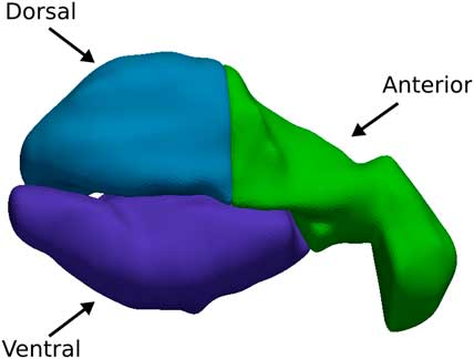
Animal data existed for measurements over 2 years, 2012 and 2013. The 2013 scanning data was selected as the animal position was more consistent between scans. This group contained 39 of the original 45 animals. Computed tomography images consisted of a series of 2D transverse planes which were manually segmented using the software package Stradwin v5.3 (http://mi.eng.cam.ac.uk/-rwp/stradwin) and interpolated using marching tetrahedra (Treece et al., Reference Treece, Prager and Gee1999). Slices that contained significant contractive motion of the RR were removed before surface interpolation. Animals that exhibited body motion during scanning were removed from the study (n=2), resulting in 20 reconstructed high CH4 yielding animals, and 17 low yield. The RR surface consists of a series of tissue folds that separate the organ into various regions and compartments. These folds make it difficult to enforce point correspondence of the RR between different animals. Therefore, the RR was broken into three sections shown in Figure 2, comprising of the reticulum and cranial sac (anterior), dorsal sac and caudal dorsal blind sac (dorsal), and the ventral and caudal ventral blind sac (ventral). This method also helps to remove the effects of local twisting that occurs along the length of the RR due to the positioning of the animal during scanning.
(colour online) Sectioning of the reticulo-rumen in sheep. Blue: the dorsal and caudal dorsal blind sac (dorsal). Green: the reticulum and cranial sac (anterior). Purple: the ventral and caudal ventral blind sac (ventral).

Landmarking
To create a series of meshes (connected points that form a geometric surface) with corresponding points or landmarks for shape analysis, an iterative scheme was used combining radial basis functions (RBF) and principal component (PC) fitting. The RBF method involves the deformation of a source mesh to a target mesh via nearest neighbour searching, in which the closed target point for a given source point is found, and the mesh deformed to minimise this distance (Deng et al., Reference Deng, Zhou and Wu2010). Principal component fitting involves the deformation of a mean shape along a select number of PCs or shape axes to again minimise the nearest neighbour distance. Principal component fitting offers a more constrained fit than RBFs, but with less accuracy in matching topology.
In the first step, a mesh was selected at random from the sample data set as the source, and the points (roughly 20 000 from the surface reconstruction step) were fitted to all other meshes using RBF. The mean shape from this initial fit was then calculated and the process was repeated once with the mean shape as the source to improve the quality of fit.
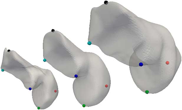
The second step involved an iterative combination of PC and RBF fitting. Principal component analysis (PCA) was carried out on the output from the previous step, and the first five PCs were used to deform the mean shape to each of the sample meshes to provide an initial approximation. This was followed by an RBF fit to improve topological accuracy. A new mean shape and PCs were then calculated and the procedure repeated until a sufficient level of convergence was obtained. An example of the resulting point correspondence for the anterior region is shown in Figure 3. Procedures were performed using the open source python library Geometry Image-Analysis Statistics (GIAS2) (Zhang et al., Reference Zhang, Malcolm, Hislop-Jambrich, Thomas and Nielsen2016). The quality of fit was determined from maximum, and root mean squared (RMS) Hausdorff distance between the fitted and target surface.
(colour online) Example of point correspondence after iterative radial basis function and principal component fitting for the anterior section in the reticulum rumen in sheep. The middle subject is the mean shape that has been deformed to fit the two example subjects on its left and right. Physiological points have been manually identified as nodes on the mean shape and highlighted as spheres, corresponding by colour between meshes.

Statistical analysis
Principal component analysis is a technique that can be used to reduce the dimensionality of data sets that depend on a large number of possibly correlated variables by identifying a smaller set of uncorrelated variables (PCs) that capture the essential features of the data (Zelditch et al., Reference Zelditch, Donald, Swiderski, Sheets and Fink2004). Here, PCA was used to describe the RR geometry information contained in a large number of landmarks with a smaller number of PCs or shape modes. General Procrustes alignment (GPA) was used to remove rotations, translations and scaling (normalising the vertex coordinates by centroid size) from the geometries. General Procrustes alignment and PCA operations were performed using the GIAS2 package. The sample geometries have a score associated with each PC, which can be used to reconstruct the shape as a weighted, linear combination of the components added to the mean.
Principal component analysis produces as many PCs as the n in the data set, not all of which hold meaningful shape information. Dimension reduction was performed using a broken stick method (Jackson, Reference Jackson1993; Cangelosi and Goriely, Reference Cangelosi and Goriely2007) to select the important PCs to be retained for statistical analysis in R. Univariate Welch two-sample t-tests were used to test significance between the retained PCs and for high and low CH4 yield. Logistic regression was then performed to investigate the effect of incorporating covariates (BW, TRP and day of scanning). Only shape modes that are significant under both univariate and logistic regression (P<0.05) were deemed significant to the hypothesis. The physical meaning of significant shape modes is interpreted by deforming the mean RR shape along the PC axis by two standard deviations for visualisation.
Results
Anterior region
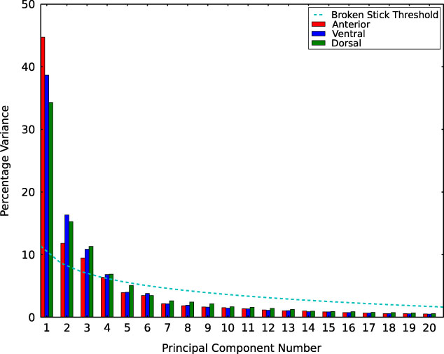
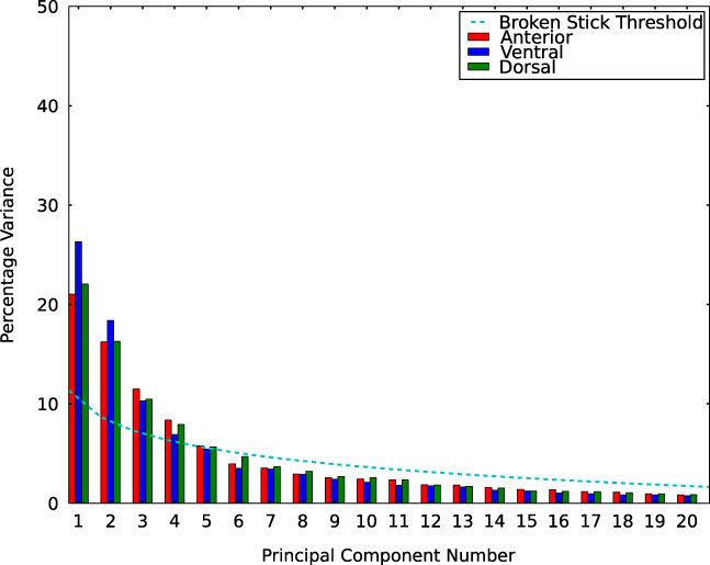
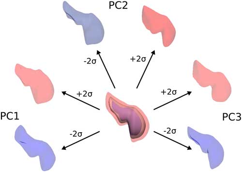
The mean shape of the anterior region of the RR was found to converge after three fitting iterations, with an average maximum and RMS fitting error of 2.4±1.9 and 0.22±0.17 mm, respectively. When size was included in the PCA, the first component contributed 44.7% of the total variation (Figure 4). When this component was inspected, it was found to correspond to a size scaling of the anterior region. A univariate t test indicated no significant difference in PC score value between high and low CH4 yielding animals. When logistic regression was performed to include covariates, the anterior region size was still found to be insignificant. When size was removed from the data set, the first five PCs were deemed important for statistical analysis and were responsible for 63% of the shape variation shown in Figure 5. These shape modes consisted of changes in the relative sizes of the cranial sac and reticulum, as well as changes in anterior length and narrowing of the connection to the dorsal sac. A visual representation of the first three shape components can be seen in Figure 6. No significant difference was detected between any of the shape PCs between high and low CH4 yielding animals.
(colour online) Percentage variance when size is included in the principal component analysis of each segmented region; the anterior (red), ventral (blue), and dorsal (green) reticulo-rumen compartments in sheep.

(colour online) Percentage variance when size is removed from the principal component analysis of each segmented region; the anterior (red), ventral (blue), and dorsal (green) reticulo-rumen compartments in sheep.

(colour online) The size component (centre) of the anterior reticulo-rumen region in sheep overlaid on the mean shape (sold grey), and the first three shape components PC1 (left), PC2 (top) and PC3 (right), which have been deformed by ±2 standard deviations (σ).

Dorsal region
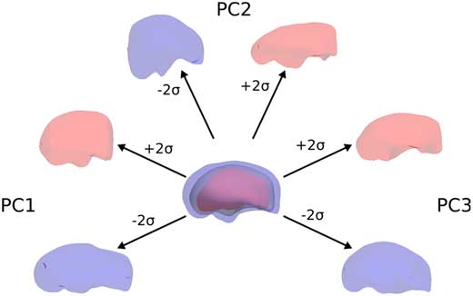
The mean shape of the dorsal region converged after four fitting iterations, with an average maximum and RMS fitting error of 1.8±1.6 and 0.27±0.25 mm, respectively. The first PC, corresponding to the size of the dorsal region, contributed 38.6% of the total sample variation shown in Figure 4. A univariate t test indicated a significant difference (P=0.009) for this component between the low and high emitting groups. When logistic regression was performed to include covariates, the dorsal size was still deemed significant (P=0.012, R 2=0.19). Interpreting the physical meaning of the size component, high emitters are found to have a negative mean score, which corresponds to increased size from the mean shape. Conversely, the low yielding group have a positive mean score corresponding to decreased size from the mean shape. An example of the size component can be seen in Figure 7. When size was removed from the dorsal samples the first four shape components were deemed important for statistical analysis and were responsible for 57% of the shape variation, as shown in Figure 5. Shape modes included elongation of the whole dorsal region, as well as relative size differences between the dorsal sac and the caudal dorsal blind sac. A visual representation of the first three shape components can be seen in Figure 7. No significant difference was detected for any of the mean PC scores for high and low CH4 yield.
(colour online) The size component (centre) of the dorsal reticulo-rumen region in sheep overlaid on the mean shape (sold grey), and the first three shape components PC1 (left), PC2 (top) and PC3 (right), which have been deformed by ±2 standard deviations (σ).

Ventral region
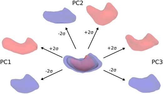
The mean shape of the ventral region converged after four fitting iterations, with an average maximum and RMS fitting error of 2.4±1.5 and 0.36±0.32 mm, respectively. The size component of the ventral region contributed 34.3% of the total sample variation shown in Figure 4. A univariate t test indicated a significant difference (P=0.0002) between the mean PC score for low and high emitting groups. When logistic regression was performed to include covariates, the ventral size was still deemed significant (P=0.002, R 2=0.32). Interpreting the physical meaning of the component scores, high emitters were found to have a negative mean score corresponding to an increase in size from the mean shape, whereas low emitters have a positive mean score, a decrease in size from the mean shape, as shown in Figure 8. When size was removed from the samples and thresh-holding applied, the first four shape components were deemed important for statistical analysis and were responsible for 73% of the shape variation as shown in Figure 5. Shape modes included elongations of the caudal ventral blind sac, an increase in ventral sac depth/height, as well as the relative angle between the two compartments. A visual representation of the first three shape components can be seen in Figure 8. No significant difference was detected for any of shape scores for high and low CH4 yield.
(colour online) The size component (centre) of the ventral reticulo-rumen region in sheep overlaid on the mean shape (sold grey), and the first three shape components PC1 (left), PC2 (top) and PC3 (right), which have been deformed by ±2 standard deviations (σ).

Discussion
The PCA was first performed on the sample groups with size/volume included in the geometric information to determine if the method could detect size as a significant mode of variation and if a difference could be detected between high and low CH4 yield groups, as found by Bain et al. (Reference Bain, Pinares-Pantino and McEwan2014) and Goopy et al. (Reference Goopy, Donaldson, Hegarty, Vercoe, Haynes, Barnett and Oddy2013). Results showed that the mode with the largest contribution (PC1) for all three regions was indeed related to overall size. A univariate comparison indicated that the high CH4 group has a larger dorsal and ventral region than the low CH4 group (P=0.009 and 0.0002, respectively), despite similar BW which may be related to the link between digesta residence times and CH4 emissions for a range of feeds previously reported (Pinares-Patiño et al., Reference Pinares-Patiño, Ulyatt, Lassey, Barry and Holmes2003; Hammond et al., Reference Hammond, Pacheco, Burke, Koolaard, Muetzel and Waghorn2014; Pacheco et al., Reference Pacheco, Waghorn and Janssen2014).
Although the size of the anterior region, corresponding to the reticulum and cranial sacs accounted for 45% of the total sample variation, no significant difference was detected between group means for high and low yielding animals.
When the analysis was repeated after scaling the RR geometries to remove size, the first four to five shape components were found to contribute more than 50% of the total sample variation. Although these modes represented physiologically meaningful shape differences, no significant difference could be detected between high and low CH4 yielding animals for any of the three regions of interest. From these results, we conclude that RR size is correlated with CH4 yield, but we find no significant correlation between the shape of the RR and CH4 yield. Although shape differences account for a significant percentage of variation within the tested animals, we could not detect any systematic differences between the high and low CH4 yielding groups. It may be the case that true shape differences are difficult to detect due to the nature of the RR and confounding factors discussed below.
Challenges in this procedure
Determining the true shape of the RR is complicated by the highly deformable nature of the organ and its domination of the viscera in the abdominal cavity. As a consequence, it has considerable interactions with other internal structures, such as the spleen, omasum, abomasum, intestines and abdominal wall. This means that the orientation and positioning of the animal in the CT scanner can cause significant distortions to the RR shape as well as local twisting of the organ, between the reticulum, rumen and blind sacs. In addition, internal gas pressure and digestive fill could have a significant influence on RR shape. These factors add noise to the PCA and may mask true differences in shape between high and low CH4 yield animals. Although the methods used in this study attempted to address the issue of relative twisting between compartments of the RR, future work could include more pre-processing of the imaging data to remove identifiable RR distortions.
Other possible sources of error include scan quality and alignment of the geometries before PCA. The CT scans were performed with a 15-mm slice plane interval; this can cause a loss of information when significant curvature occurs between slice planes, such as at the end of a compartment at the reticulum or caudal blind sacs. The distance between these slice planes, as well as the contrast quality used to define the interface between the tissue surface and the RR interior, can create reconstruction artefacts that are difficult to remove.
Alignment operations performed on the reconstructed geometries (translation and rotation performed using GPA) also contribute to the overall noise. Each shape is scaled by centroid size and translated to share the same reference centroid position. Rigid body rotations are then performed to minimise the Procrustes distance between the different geometries. However, the shapes cannot be perfectly matched to each other, and so the alignment of shapes is sensitive to the initial orientation, giving rise to an apparent shape difference.
Summary
We have adopted the techniques of statistical shape modelling to investigate RR morphology in sheep. The approach offers a method for comparing geometries that are complex in shape, and lack common physiological features for applying traditional landmarks via an automatic fitting process. This allows us to perform a quantitative analysis of shape differences for the RR of sheep. Using these techniques, we have detected significant differences in size for the dorsal and ventral regions of the RR between high and low CH4 yielding sheep. When size was removed by scaling the geometries, our analysis detected no significant shape difference between high and low CH4 yield animals. This suggests that observed differences in outflow rates and residence times are independent of shape variations within the RR.
Acknowledgements
This study was funded by the Agricultural Marketing and Research Development Trust (AGMARDT) and DairyNZ through scholarship support to Stephen James Waite. The experimental work was funded by the Pastoral Greenhouse Gas Research Consortium, Sustainable Land Management and Climate Change and the New Zealand Agricultural Greenhouse Gas Research Centre. The animals themselves were part of the Ovita partnership and the related Central Progeny Test: both funded in part or whole by Beef+Lamb New Zealand. Thanks also to the Central Progeny Test collaborating organisations: AgResearch Woodlands especially Kevin Knowler, On Farm Research especially Paul Muir, Lincoln University especially Chris Logan and AbacusBio Ltd especially Neville Jopson.
Declaration of interest
This study was performed with no conflicts of interest.
Ethics statement
The study was carried out in strict accordance of the guidelines of the 1999 New Zealand Animal Welfare Act and was approved by the AgResearch’s Invermay Animal Ethics Committee (applications AE12206 and AE12963).
Software and data repository resources
None of the data were deposited in an official repository.