





Non-technical Summary
Because turtles have a bony shell that can preserve evidence of interactions with predators and parasites, they are important hosts of trace fossils. We systematically searched for traces in the shells of freshwater turtles from Tiupampa, Bolivia, from the early Paleocene (ca. 64 million years ago). We found evidence of bite traces consistent with feeding by crocodyliforms, including crushed shells, holes, and linear traces. These could be from either predation or scavenging and might have been produced by either sebecids or dyrosaurids, both of which are known from Tiupampa. We also found very common invertebrate parasite traces in the form of small holes in the shells. Based on comparisons with modern turtles, these were possibly made by leeches or trematodes while the turtles were alive. Rarely, we found evidence of bacterial or fungal infection in small patches on the shells. All these trace fossils offer insight into paleoenvironments and paleoecology, with the distribution of parasites suggesting that the turtles were not experiencing extreme stress and were likely spending most of their time in the water. The trace fossil evidence suggests that complex ecological interactions were present only two million years after the Cretaceous–Paleogene extinction.
Introduction
Turtles, defined here to include all members of the clade Testudines, whether terrestrial, marine, or freshwater (Ernst and Lovich, Reference Ernst and Lovich2009), occupy a vital ecological role at the intersection of habitats (Lovich et al., Reference Lovich, Ennen, Agha and Gibbons2018). Freshwater turtles provide a link between aquatic and terrestrial ecosystems through their ability to occupy both environments. In Amazonia, for example, nesting of the Arrau turtle, Podocnemis expansa Schweigger, Reference Schweigger1812, can introduce more than 16,000 kg of eggs per year to a single beach (Alho and Pádua, Reference Alho and Pádua1982). Roughly half the nutrients contained within these eggs will remain on land and enter terrestrial food chains (Miranda, Reference Miranda2017). In addition to their amphibious lifestyle, key to the ecological significance of many freshwater turtles is the high population densities they can reach, producing extremely large biomasses (Iverson, Reference Iverson1982). For example, the pond slider, Trachemys scripta Thunberg in Schoepff, Reference Thunberg and Schoepff1792, has a biomass of 877 kg/ha in some habitats (Congdon et al., Reference Congdon, Greene and Gibbons1986), whereas the Llanos side-necked turtle, Podocnemis vogli Müller, Reference Müller1935, can reach a population biomass of 1540 kg/ha during dry periods, when populations are geographically restricted, 5.5× the biomass of any terrestrial large herbivorous mammal population (Iverson, Reference Iverson1982).
High densities of freshwater turtles can produce important trophic effects in a range of directions. Of the 228 freshwater turtle species for which diet has been described, 79 have been classified as carnivorous and 125 as omnivorous or opportunistic (Wang et al., Reference Wang, Xu, Zhu, Chen, Zhao and Wang2025). Faunivorous turtles can have significant effects on prey populations and their surrounding ecosystems (Gomez-Mestre and Keller, Reference Gomez-Mestre and Keller2003; Lindsay et al., Reference Lindsay, Zhang, Forstner and Hahn2013; Aresco et al., Reference Aresco, Travis and MacRae2015; Garig et al., Reference Garig, Ennen and Davenport2020; Shoptaugh, Reference Shoptaugh2022). Turtles are important prey items for terrestrial mammals, crocodylians, snakes, birds, and other turtles (e.g., Emmons, Reference Emmons1989; Elsey, Reference Elsey2006; Salera Junior et al., Reference Salera Junior, Malvasio and Portelinha2009; de la Ossa-Velásquez et al., Reference de la Ossa-Velásquez, Vogt and Ferrara2010; Platt and Rainwater, Reference Platt and Rainwater2011; Thomas and Allain, Reference Thomas and Allain2021; Coleman, Reference Coleman2023; Avenant and Gee, Reference Avenant and Gee2025), and are important host taxa for a range of parasites, including platyhelminths, acanthocephalans, leeches, nematodes, and trematodes (e.g., Esch and Gibbons, Reference Esch and Gibbons1967; Stunkard, Reference Stunkard1970; Readel et al., Reference Readel, Phillips and Wetzel2008; Santana et al., Reference Santana, Teixeira, Araujo-Filho, Teles, Pereira, Brito, Almeida and Mesquita2023; Koch and Bolek, Reference Koch and Bolek2025). Therefore, extant freshwater turtles are highly connected to other organisms within ecosystems and are involved in numerous ecological interactions.
Turtles are a valuable, but often overlooked, paleontological resource. Presumably because of their hard shell and their high population density (Iverson, Reference Iverson1982), they are commonly abundant in fossil localities in which they are present, to the extent that they have been used as a taphonomic control for other taxa (Markwick, Reference Markwick1998). Partly given this abundance, they offer a potential opportunity for ecometric reconstructions of paleotemperature and paleolake depth, through measurement of body size (Cadena et al., Reference Cadena, Scheyer, Carrillo-Briceño, Sánchez, Aguilera-Socorro, Vanegas, Pardo, Hansen and Sánchez-Villagra2020; Parker, Reference Parker2021; Parker et al., Reference Parker, Müller, Boisserie and Head2023, Reference Parker, Boisserie, Müller, Brochu and Head2026). Additionally, because they are surrounded by dermal bone (in the form of their shell), turtles (and other taxa with dermal armor such as cingulate xenarthrans) have a greater potential for preserving a record of premortem bioerosion than most other vertebrates (Zonneveld et al., Reference Zonneveld, Bartels, Gunnell and McHugh2015, Reference Zonneveld, AbdelGawad and Miller2022a). Broadly, bioerosive traces may be those emplaced by vertebrates (particularly during predation and scavenging, e.g., bite traces), or by invertebrates (through either scavenging or parasitism, e.g., grooves, pits, borings, and lesions in the shell surface; Zonneveld et al., Reference Zonneveld, AbdelGawad and Miller2022a; Zonneveld and Bartels, Reference Zonneveld and Bartels2022).
The record of vertebrate-induced bioerosion in turtles is extensive (e.g., Mikuláš et al., Reference Mikuláš, Kadlecová, Fejfar and Dvořák2006; Schwimmer, Reference Schwimmer2010; Milàn et al., Reference Milàn, Lindow and Lauridsen2011; Noto et al., Reference Noto, Main and Drumheller2012; Botfalvai et al., Reference Botfalvai, Prondvai and Ösi2014; Hastings et al., Reference Hastings, Bloch and Jaramillo2015; Kahlke et al., Reference Kahlke, Fritz and Kierdorf2015; Shimada and Hooks, Reference Shimada and Hooks2016; Gônet et al., Reference Gônet, Rozada, Bourgeais and Allain2018; Peters et al., Reference Peters, Bosselaers, Post and Reumer2019; Cadena et al., Reference Cadena, Scheyer, Carrillo-Briceño, Sánchez, Aguilera-Socorro, Vanegas, Pardo, Hansen and Sánchez-Villagra2020; Zonneveld et al., Reference Zonneveld, AbdelGawad and Miller2022a; El Hedeny et al., Reference El Hedeny, Mohesn, Tantawy, El-Sabbagh, AbdelGawad and El-Kheir2023), with a range of suspected agents, including terrestrial mammals, sharks, cetaceans, and crocodylians. The record of invertebrate bioerosion in turtles extends to the Triassic, with evidence of ichnofossils associated with ectoparasitism in Proterochersis from the Löwenstein Formation, Germany (Szczygielski et al., Reference Szczygielski, Marchetti and Dróżdż2024). Although recognition of invertebrate-induced bone modifications in fossil turtles is relatively recent (Zonneveld et al., Reference Zonneveld, Bartels, Gunnell and McHugh2015, Reference Zonneveld, AbdelGawad and Miller2022a), there is now widespread recognition of the occurrence of these features in a range of paleohabitats and turtle taxa around the world, reflecting a range of invertebrate agents (e.g., Hutchison and Frye, Reference Hutchison and Frye2001; Scheyer and Sánchez-Villagra, Reference Scheyer and Sánchez-Villagra2007; Zonneveld et al., Reference Zonneveld, Bartels, Gunnell and McHugh2015, Reference Zonneveld, AbdelGawad and Miller2022a; Donovan and Simpson, Reference Donovan and Simpson2020; Jagt et al., Reference Jagt, Deckers, De Leebeeck, Donovan and Nieuwenhuis2020; Sato and Jenkins, Reference Sato and Jenkins2020; Adrian, Reference Adrian2022; Collareta and Bosselaers, Reference Collareta and Bosselaers2022; Collareta et al., Reference Collareta, Merella, Bosselaers, Casati, Di Cencio and Bianucci2022, Reference Collareta, Varas-Malca, Bosio, Urbina and Coletti2023; Zonneveld and Bartels, Reference Zonneveld and Bartels2022, Reference Zonneveld and Bartels2023; Bogado et al., Reference Bogado, Pereira, de la Fuente and Bergqvist2023; Davies, Reference Davies2023; El Hedeny et al., Reference El Hedeny, Mohesn, Tantawy, El-Sabbagh, AbdelGawad and El-Kheir2023; Ascarrunz and Joyce, Reference Ascarrunz and Joyce2024).
Because their dermal armor readily records predator–prey and host–parasite interactions, turtles can provide insight into biotic interactions and food webs in deep time. They therefore provide an opportunity to observe these interactions in post-recovery ecosystems following extinction events. Turtles were persistent across the Cretaceous–Paleogene (K–Pg) boundary 66 million years ago (e.g., Vlachos et al., Reference Vlachos, Randolfe, Sterli and Leardi2018; Cleary et al., Reference Cleary, Benson, Holroyd and Barrett2020; Vlachos, Reference Vlachos2024), defined by a mass extinction event that eliminated ~75% of terrestrial species (Jablonski, Reference Jablonski1994). Labandeira et al. (Reference Labandeira, Rodríguez-Tovar, Uchman, Mángano and Buatois2016) highlighted examples of changes in biotic interactions across the K–Pg boundary. Case studies of insect herbivory show that plant consumption declined immediately after the K–Pg due to a decline in specialist taxa such as leaf miners (Labandeira et al., Reference Labandeira, Johnson and Wilf2002a, Reference Labandeira, Johnson and Langb, Reference Labandeira, Rodríguez-Tovar, Uchman, Mángano and Buatois2016; Wilf et al., Reference Wilf, Labandeira, Johnson and Ellis2006). Wilf et al. (Reference Wilf, Labandeira, Johnson and Ellis2006) suggested that these food webs remained unbalanced until ca. 2 My after the extinction event. Other potential specialist taxa suggested to have gone extinct at the K–Pg boundary include blood-feeding midges, particularly those feeding on large dinosaurs (Borkent, Reference Borkent1995). It is possible that turtle parasites could show the same pattern of decline across the boundary.
The lower Paleocene locality of Tiupampa, Bolivia, has produced the earliest diverse Cenozoic vertebrate assemblage from South America, with mammals, turtles, amphibians, squamates, and crocodyliforms (e.g., de Broin, Reference de Broin1971, Reference de Broin1991; Marshall et al., Reference Marshall, de Muizon and Sigé1983; de Muizon et al., Reference de Muizon, Marshall and Sigé1984, Reference de Muizon, Billet, Argot, Ladevèze and Goussard2015, Reference de Muizon, Ladevèze, Selva, Vignaud and Goussard2018, Reference de Muizon, Billet and Ladevèze2019; Marshall and de Muizon, Reference Marshall and de Muizon1988; Báez, Reference Báez1991; Buffetaut, Reference Buffetaut1991; Buffetaut and Marshall, Reference Buffetaut and Marshall1991; Rage, Reference Rage1991a, Reference Rageb; Marshall et al., Reference Marshall, de Muizon and Sigogneau-Russell1995; de Muizon, Reference de Muizon1998; de Muizon and Cifelli, Reference de Muizon and Cifelli2000, Reference de Muizon and Cifelli2001; Gelfo et al., Reference Gelfo, Goin, Woodburne and de Muizon2009; Ladevèze et al., Reference Ladevèze, de Muizon, Beck, Germain and Cespedes-Paz2011; Scanferla et al., Reference Scanferla, Zaher, Novas, de Muizon and Céspedes2013; de Muizon and Ladevèze, Reference de Muizon and Ladevèze2020, Reference de Muizon and Ladevèze2022; Jouve et al., Reference Jouve, de Muizon, Cespedes-Paz, Sossa-Soruco and Knoll2021; de Muizon and Billet, Reference de Muizon and Billet2022; Pochat-Cottilloux et al., Reference Pochat-Cottilloux, Martin, Jouve, Perrichon, Adrien, Salaviale, de Muizon, Cespedes and Amiot2022). The exceptional preservation of the fossils from Tiupampa offers an opportunity for higher-resolution study of ecological interactions than is possible for many other Paleogene South American assemblages. In this study, we investigate the population-level size distributions in Tiupampan turtles and recognize bioerosive ichnofossils to identify possible predator–prey and host–parasite relationships in the earliest Paleocene of Bolivia, reconstructing biological interactions.
Materials and methods
Fossil material
The fossil turtles from Tiupampa studied here are housed in the Museo de Historia Natural Alcide d’Orbigny (MHNC), Cochabamba, Bolivia, and in the Muséum National d’Histoire Naturelle (MNHN), Paris, France. Most specimens have been assigned to the podocnemidid Lapparentemys vilavilensis (de Broin, Reference de Broin1971) (previously Roxochelys vilavilensis; de Broin, Reference de Broin1971, Reference de Broin1991; Gaffney et al., Reference Gaffney, Meylan, Wood, Simons and Campos2011). Most specimens currently housed in MHNC are considered to represent cf. Lap. vilavilensis, pending a comprehensive taxonomic assessment. A single specimen, MHNC-P-16411, clearly differs from the other specimens based on its higher, more hemispherical carapace, and we propose that it may represent a terrestrial tortoise (considered here as cf. Testudinoidea indet.), previously unknown from Tiupampa and worthy of future taxonomic attention. No ichnofossils were recognized on MHNC-P-16411. Gaffney et al. (Reference Gaffney, Meylan, Wood, Simons and Campos2011) noted that several Tiupampan specimens (particularly MNHN.F.VIL6 and MHNC-P-6904) differed from Lap. vilavilensis and likely represent different taxa. We did not study MNHN.F.VIL6 here but did recognize bioerosive traces on MHNC-P-6904. We do not disagree with Gaffney et al. (Reference Gaffney, Meylan, Wood, Simons and Campos2011) that it may represent a new species and refer to it here by its specimen number unlike for the other individuals examined. In total, we studied 76 specimens across the two collections, ranging from largely complete articulated carapaces and plastra to unidentifiable shell fragments.
Geological and geographical context
All fossil turtles studied originate from the lower Paleocene Santa Lucía Formation, and were collected during fieldwork in the Tiupampa area, Cochabamba Department, Bolivia (de Broin, Reference de Broin1991; Fig. 1). The turtles from Tiupampa were collected from several smaller localities, with particularly abundant remains from Villa Viscarra, roughly 2 km south of the town of Vilavila, and from the Turtle Cemetery of Tiupampa (de Broin, Reference de Broin1991). Given the short distance between these sub-localities, and to aid in comparisons considering the limited numbers of specimens, we combined all turtles when analyzing population-level trends in bioerosive trace fossils or body sizes.
Illustration of the (1) geography and (2) stratigraphic context for the turtles analyzed in this study. The satellite map of Bolivia originates from GISGeography (2025) and the general location of Tiupampa from Gayet et al. (Reference Gayet, Sempere, Cappetta, Jaillard and Lévy1993). The stratigraphy is adapted from Pochat-Cottilloux et al. (Reference Pochat-Cottilloux, Martin, Faure-Brac, Jouve, de Muizon, Cubo, Lécuyer, Fourel and Amiot2023). The Lapparentemys vilavilensis silhouette is based on MNHN.F.VIV1.

Figure 1. Long description
Panel 1 shows a satellite map of Bolivia. A small inset map of South America on the right shows Bolivia highlighted in green. The main map shows Tiupampa marked with a black silhouette of Lapparentemys vilavilensis.Other cities marked include Cochabamba, La Paz, Santa Cruz de la Sierra, Oruro, Sucre, Potosí, and Tarija. Panel 2 is a stratigraphic chart. From left to right:
* An Age column labeled Danian, spanning from 66.0 to 61.6 million years ago.
* A Magnetic polarity column with alternating black and white blocks labeled C 28 n, C 28 r, C 29 n, C 29 r, and C 30 n.
* A Tiupampa column showing a black turtle silhouette of Lapparentemys vilavilensis positioned between 64.886 and 65.065 million years ago.
* A Formation column showing the Santa Lucia formation at the top and the El Molino formation at the bottom, separated by a dotted line near the 65.065 mark. Diagonal lines correlate the Danian age boundaries to the magnetic polarity scale.
The most fossiliferous site within the Tiupampa locality represents an oxbow lake, suddenly flooded by the main river, bringing a major influx of sand (Gelfo et al., Reference Gelfo, Goin, Woodburne and de Muizon2009). The age inferred for the Tiupampa fossils, and the Tiupampan South American Land Mammal Age (SALMA) has changed since this locality’s original description. Lapparentemys was originally described (de Broin, Reference de Broin1971) as Late Cretaceous in age, but the Santa Lucía Formation at Tiupampa has since been considered to represent early Paleocene deposition (Marshall, Reference Marshall1989; Gayet et al., Reference Gayet, Marshall and Sempere1991). The age of Tiupampa was reviewed in detail by de Muizon and Ladevèze (Reference de Muizon and Ladevèze2020). These authors favored an early Danian age for the Tiupampa fauna, which is correlated with Chron 28r, with an absolute age close to 65 Ma, based on taxonomic comparisons across the mammal faunas of Tiupampa, the Patagonian Paleocene assemblage from Punta Peligro, and North American Cretaceous and Paleocene localities. Unfortunately, no radiometric dating is available yet for Tiupampa (de Muizon and Ladevèze, Reference de Muizon and Ladevèze2020).
Terminology and measurements
Anatomical terminology for the turtles follows Gaffney et al. (Reference Gaffney, Meylan, Wood, Simons and Campos2011). Although Lapparentemys vilavilensis shows some variability in shell morphology (e.g., some specimens have six neural plates and some seven, and two different epiplastral morphotypes have been recognized; Gaffney et al., Reference Gaffney, Meylan, Wood, Simons and Campos2011), these differences do not influence describing the position of the bioerosion marks. The external surface refers to the outward-facing surfaces of the carapace and plastron, which were covered with epidermal scutes in life. We did not observe any bioerosive traces on the visceral surface, which refers to the internal parts of the carapace and plastron, contacting the internal organs. The internal surface refers to the inward-facing parts of the carapace or plastron, in the outlying regions, that were covered with epidermal scutes and did not contact the viscera. The epidermal scutes were not preserved, but their boundaries are visible on the surface of the dermal bone as epidermal sulci (grooves). These sulci do not overlap with the sutures between dermal bones (Zonneveld et al., Reference Zonneveld, Bartels, Gunnell and McHugh2015; Fig. 2).
Summary of osteological terminology applied to Tiupampan turtle specimens. The outline is Lapparentemys vilavilensis (MNHN.F.VIL1). Line art adapted from Weems and Knight (Reference Weems, Knight, Brinkman, Holroyd and Gardner2013) and de Broin (Reference de Broin1971). Black lines show sutures between dermal bone. Orange lines show positions of epidermal sulci. Gray-shaded region shows a broken region, where no sulci are illustrated. Abbreviations: l = left; r = right; Nu = nuchal; C = costal; N = neural; P = peripheral; SP = suprapygal; Py = pygal; Epi = epiplastron; Ent = entoplastron; Hyo = hyoplastron; Mes = mesoplastron; Hyp = hypoplastron; Xip = xiphiplastron. Scale bar is 50 mm.

Figure 2. Long description
The diagram consists of two panels showing the anatomy of Lapparentemys vilavilensis. The left panel shows a dorsal view of the carapace. From top to bottom, the labelled bones along the midline of the carapace are: nuchal (Nu), the first to seventh neurals (N1-7), the suprapygal (SP) and the pygal (Py). The costals (C1-8) surround the midline on the left (l) and right (r) sides, and on the lateral edge of the carapace are the peripherals (P1-11). The right panel shows the anataomy of the plastron. At the front, in the centre, the entoplastron (Ent) is labelled, with the left and right epiplastra (Epi) anterolateral to this. Posterior to the entoplastron are the hyoplastra (Hyo), then successively, the hypoplastra (Hyp) and xiphiplastra (Xip). In the lateral region of the plastron, at the midline between the hyoplastra and hypoplastra, there are left and right mesoplastra (Mes). In both the carapace and the plastron, black lines show dermal bone boundaries, and orange lines the positions of sulci. The scale bar for both is 50 mm.
All ichnofossils described here, by definition, result from the actions of an external agent, rather than from a response of the host organism to pathology (Bertling et al., Reference Bertling, Braddy, Bromley, Demathieu and Genise2006, Reference Bertling, Buatois, Knaust, Laing and Mángano2022; Zonneveld et al., Reference Zonneveld, Bartels, Gunnell and McHugh2015, Reference Zonneveld, Fiorillo, Hasiotis and Gingras2022b Reference Zonneveld, Wilson and Holroyd2024; Zonneveld and Bartels, Reference Zonneveld and Bartels2019). We follow Vallon et al. (Reference Vallon, Rindsberg and Martin2015) in using “trace” rather than “mark” when describing a biogenic structure (e.g., “bite trace”). Use of the terms “pit” and “hole” follows Zonneveld et al. (Reference Zonneveld, Bartels, Gunnell and McHugh2015), with the former defined as a trace terminating within the bone and the latter as a perforation through the bone of the shell. In this way, holes can be considered “penetrative” and pits (and other traces that do not pass through the bone) as “non-penetrative”. We use the word “score” to mean a groove along the surface of the bone (Drumheller et al., Reference Drumheller, D’Amore, Njau, Woodward and Farlow2023). In the bite-trace literature (e.g., Binford, Reference Binford1981; Drumheller et al., Reference Drumheller, D’Amore, Njau, Woodward and Farlow2023; LaBarge and Njau, Reference LaBarge and Njau2024), distinction is often drawn between pits and “punctures” (rather than between pits and holes), where punctures are defined as penetrative bite traces. For consistency with other studies of traces emplaced by vertebrates, we apply this definition, so that a puncture is considered a hole produced through biting by a vertebrate agent.
Where possible, we measured three dimensions for the emplaced ichnofossils. For MNHN specimens, length (defined as the maximum diameter) and width (defined as the minimum diameter) were measured using digital calipers to the nearest 0.01 mm. Depth was measured using the insertion of a metal probe into the bioerosive trace, with the depth of penetration measured using the digital calipers. This depth-measurement approach was used for specimens in both collections, but for logistical reasons, depth measurements of MHNC specimens used analogue calipers to the nearest 0.1 mm, and their length and width were measured from photos using Fiji for ImageJ (Schindelin et al., Reference Schindelin, Arganda-Carreras, Frise, Kaynig and Longair2012).
Turtle body size
We estimated body mass (BM) for all specimens from carapace length (CL), using regressions specific for Testudines families from Parker (Reference Parker2021) and Wilson and Parker (Reference Wilson and Parker2023). For MHNC-P-16411, we used the Testudinidae equation:

For all other specimens, we used the Podocnemididae equation:

As for the ichnofossils, measurements of shell elements were taken from photographs due to equipment availability. Lengths of individual elements were measured using Fiji for ImageJ (Schindelin et al., Reference Schindelin, Arganda-Carreras, Frise, Kaynig and Longair2012). To reduce challenges with perspective when using these photos, all measured CL values were adjusted using a correction factor (CF), given differences in vertical scale bar position based on measured values, to produce mean (CF = 1.09), maximum (CF = 1.20), and minimum (CF = 1) CL values. This means that in some cases, mean calculated body size is likely to differ slightly from the true value, but the treatment of all specimens is consistent for comparison. For specimens without a completely preserved carapace, we constructed linear regression equations for mean CL value against plastron length (PL) and PL against other measurements (entoplastron length, xiphiplastron length, and xiphiplastron width). The measurements and equations used for the body size calculations are available in Supplementary Material 1 and 2.
We compared the maximum and minimum body masses for Lapparentemys to recorded maximum and hatchling masses of modern freshwater turtles from the CheloniansTraits database (Wang et al., Reference Wang, Xu, Zhu, Chen, Zhao and Wang2025), using all freshwater turtle species for which data were available. We used these modern freshwater turtle masses to construct a linear regression for maximum mass against hatchling mass (both log10 transformed). This was then used to predict the expected hatchling mass for Lap. vilavilensis, given the maximum recorded size, and vice versa. We used the body size distributions (recorded through CL) of the Tiupampan turtles as a proxy for individual age, with the assumption that older turtles would be more likely to have a larger body size (Zweifel, Reference Zweifel1989; Browne and Hecnar, Reference Browne and Hecnar2007). We tested for normality of the body size distribution using a Shapiro–Wilk normality test (Shapiro and Wilk, Reference Shapiro and Wilk1965) and qualitatively compared this distribution to those of modern turtle populations (McKnight et al., Reference McKnight, Bower, Ariel, Beatty and Clulow2025).
Minimum bite force estimates
To estimate the minimum bite force required to produce the ichnofossils observed in the carapaces of Lapparentemys specimens, we used the area of the bite trace, following a similar procedure to Wilson et al. (Reference Wilson, Moreno-Bernal, Suarez, Cardozo Tovar, Vanegas, Vanegas, Shirley, Holroyd and Zonneveld2026). The bite trace area reflects the region that underwent compressive failure as a result of contact with the crocodyliform teeth, which have a conical shape and a finite tip radius. We used the equation:

where F is the minimum required bite force in newtons (N), σ is the compressive strength of the bone, and A is the area of the bite trace. Such area-based approaches do not necessarily account for differences in the material properties of different layers of the carapace and plastron (e.g., epidermal scutes, external and internal cortical bone, trabecular bone; Achrai and Wagner, Reference Achrai and Wagner2013) and therefore differ from bite-force estimation approaches applied by other authors (e.g., Erickson et al., Reference Erickson, Van Kirk, Su, Levenston, Caler and Carter1996; Gignac et al., Reference Gignac, Makovicky, Erickson and Walsh2010; Gignac and Erickson, Reference Gignac and Erickson2017; Peterson et al., Reference Peterson, Tseng and Brink2021). However, given the uncertainty in tracemaker identity, we think that bite trace area can provide an approximation for bite force, which can be used for identification of etiological agent, in combination with other factors such as shape. Area was directly measured from polygon traces of the ichnofossils using the Fiji platform for ImageJ (Schindelin et al., Reference Schindelin, Arganda-Carreras, Frise, Kaynig and Longair2012). We used a compressive strength (σ) of 151.3 ± 42.4 MPa for turtle shell (Magwene and Socha, Reference Magwene and Socha2013).
These minimum bite forces were compared to estimated bite forces from early Paleocene Bolivian crocodyliforms. Estimates come from length measurements taken from photos using the Fiji platform for ImageJ (Schindelin et al., Reference Schindelin, Arganda-Carreras, Frise, Kaynig and Longair2012) of the sebecids Zulmasuchus querejazus Buffetaut and Marshall, Reference Buffetaut and Marshall1991 (MHNC-P 3701; Pochat-Cottilloux et al., Reference Pochat-Cottilloux, Martin, Jouve, Perrichon, Adrien, Salaviale, de Muizon, Cespedes and Amiot2022) and Sebecidae indet. (MHNC-P 6671; Buffetaut, Reference Buffetaut1991) and the dyrosaurids Rodeosuchus machukiru Jouve et al., Reference Jouve, de Muizon, Cespedes-Paz, Sossa-Soruco and Knoll2021 (MHNC-P 6070, MHNC-P 6670, and MHNC-P 8126; Buffetaut, Reference Buffetaut1991), Luciasuchus lurusinqa Jouve et al., Reference Jouve, de Muizon, Cespedes-Paz, Sossa-Soruco and Knoll2021 (MHNC-P 14061), and Dorbignysuchus niatu Jouve et al., Reference Jouve, de Muizon, Cespedes-Paz, Sossa-Soruco and Knoll2021 (MHNC-P 13500). We used the equations of Erickson et al. (Reference Erickson, Gignac, Steppan, Lappin, Vliet, Brueggen, Inouye, Kledzik and Webb2012) to calculate bite force (BF) to body mass (BM). Because modern gharials have a lower BF than expected given their BM (Erickson et al., Reference Erickson, Gignac, Steppan, Lappin, Vliet, Brueggen, Inouye, Kledzik and Webb2012), we also used a separate regression for longirostrine dyrosaurids calculated using the measured BF and BM values for the two individual gharials of Erickson et al. (Reference Erickson, Gignac, Steppan, Lappin, Vliet, Brueggen, Inouye, Kledzik and Webb2012). To estimate BM, we used the general Crocodylia equation of Slavenko et al. (Reference Slavenko, Tallowin, Itescu, Raia and Meiri2016), which used total body length (TL). Total body mass was estimated from dorsal cranial length (DCL) using the equations of Sereno et al. (Reference Sereno, Larsson, Sidor and Gado2001), with the “intermediate” (Crocodylus porosus Schneider, Reference Schneider1801) equation for sebecids and the “longirostrine” (Gavialis gangeticus Gmelin, Reference Gmelin1789), equation for dyrosaurids, although we acknowledge that using equations based on modern crocodylians are unlikely to produce highly accurate body masses for these clades, particularly for sebecids, given differences in body proportions (Paiva et al., Reference Paiva, Godoy, dos Santos, Langer and Mannion2026). For MHNC-P 3701, we used the frontal length (FL) equation of Bravo et al. (Reference Bravo, Pol, Leardi, Krause, Nicholl, Rougier and Mannion2025) to estimate DCL. For MHNC-P 6671, we constructed an equation relating DCL to mandibular symphysis length (MSL) for sebecids, using values either reported in the literature or measured from photos of published sebecid specimens (N = 4, R 2 = 0.88, p = 0.04): Bretesuchus bonapartei Gasparini, Fernandez, and Powell, Reference Gasparini, Fernandez and Powell1993 (PVL 4735), Barinasuchus arveloi Paolillo and Linares, Reference Paolillo and Linares2007 (MAAT-0260), Lorosuchus nodosus Pol and Powell, Reference Pol and Powell2011 (PVL 6219), and Sahitisuchus fluminensis Kellner, Pinheiro, and Campos, Reference Kellner, Pinheiro and Campos2014 (MCT.R.1730). For the incomplete Bolivian dyrosaurids, we estimated DCL from average mandibular interalveolar length (AMIL), using an equation built using other longirostrine South American dyrosaurids where DCL and AMIL could be associated (N = 6, R 2 = 0.99, p < 0.0005): Lu. lurusinqa (MHNC-P 14061; Jouve et al., Reference Jouve, de Muizon, Cespedes-Paz, Sossa-Soruco and Knoll2021), Do. niatu (MHNC-P 13500; Jouve et al., Reference Jouve, de Muizon, Cespedes-Paz, Sossa-Soruco and Knoll2021), Cerrejonisuchus improcerus Hastings et al., Reference Hastings, Bloch, Cadena and Jaramillo2010 (UF/IGM 30), Acherontisuchus guajiraensis Hastings, Bloch, and Jaramillo, Reference Hastings, Bloch and Jaramillo2011 (UF/IGM 35 and UF/IGM 34), and Guarinisuchus munizi Barbosa, Kellner, and Viana, Reference Barbosa, Kellner and Viana2008 (DG-CTG-UFPE 5723). A full list of these equations is shown in Table 1.
Equations used for estimating bite force in early Paleocene Bolivian crocodyliforms. All length measurements are in mm, masses are in kg and forces are in N. BF = bite force, BM = body mass, TL = total length, DCL = dorsal cranial length, FL = frontal length, MSL = mandibular symphysis length, AMIL = average mandibular interalveolar length

Table 1. Long description
The table is organized into three columns: Taxon, Equation, and Source.
* Row 1: All crocodyliforms. BF = (29.632 × BM) + 569.35. Source: Erickson et al., 2012.
* Row 2: Gavialis gangeticus. BF = 12.33 × B M + 513.67. Source: This study, from Erickson et al., 2012.
* Row 3: All crocodyliforms. log10 (B M) equals 2.79 × log10 (T L) - 4.67. Source: Slavenko et al., 2016.
* Row 4: Longirostrine crocodyliforms. TL = 0.74 × DCL - 69.369. Source: Sereno et al., 2001.
* Row 5: Intermediate rostrum crocodyliforms. TL = 0.7717 × DCL - 20.224. Source: Sereno et al., 2001.
* Row 6: Sebecidae. DCL = 4.75 × FL - 31.8. Source: Bravo et al., 2025.
* Row 7: Sebecidae. DCL = 2.134 × MSL + 104.309. Source: This study.
* Row 8: Longirostrine South American dyrosaurids. DCL = 26.303 × AMIL - 9.225. Source: This study.
Initialisms used: BF (bite force), BM (body mass), TL (total length), DCL (dorsal cranial length), FL (frontal length), MSL (mandibular symphysis length), and AMIL (average mandibular interalveolar length).
Bite trace circularity
To aid identification of the agent that emplaced the puncture traces, we measured the circularity of these traces. Trace fossil circularity (fcirc) was calculated following the equation of Zonneveld et al. (Reference Zonneveld, Bartels, Gunnell and McHugh2015):

where A is the area of the puncture traces and p is the perimeter. Area and perimeter were measured from the traces using Fiji for ImageJ, alongside maximum and minimum diameters (Schindelin et al., Reference Schindelin, Arganda-Carreras, Frise, Kaynig and Longair2012).
Repositories and institutional abbreviations
Ah SGDB = Collection “Ahníkov” of the Doly Bílina Company, Doly Bílina, Czechia; DG-CTG-UFPE = Departamento de Geologia, Centro de Tecnologia e Geociências, Universidade Federal de Pernambuco, Recife, Brazil; DPC = Division of Fossil Primates, Duke Lemur Center, Duke University, Durham, USA; IGM = Museo Geológico Nacional, Servicio Geológico Colombiano, Bogotá, Colombia; IUPC = Indiana University Paleontology Collection, Bloomington, USA; LPP = Laboratório de Paleoecologia e Paleoicnologia, Federal University of São Carlos, São Carlos, São Paulo, Brazil; MAAT = Museo Alberto Arvelo Torrealba, Barinas, Venezuela; MCT.R = Reptile Collection, Museu de Ciências da Terra, Serviço Geológico do Brasil, Rio de Janeiro, Brazil; MHNC = Museo de Historia Natural Alcide d’Orbigny, Cochabamba, Bolivia; MNHN = Muséum National d’Histoire Naturelle, Paris, France; PS/C = Center for Paleontological Interpretation, Viator, Spain; PVL = Colección de Paleontología de Vertebrados Lillo, Tucumán, Argentina; RTMP = Royal Tyrrell Museum of Paleontology, Drumheller, Canada; UA-TF = University of Alberta Trace Fossil collections, Edmonton, Canada; UF = Florida Museum of Natural History, University of Florida, Gainesville, USA.
Systematic ichnology
Ichnogenus Nihilichnus Mikuláš et al., Reference Mikuláš, Kadlecová, Fejfar and Dvořák2006
Type ichnospecies
Nihilichnus nihilicus Mikuláš et al., Reference Mikuláš, Kadlecová, Fejfar and Dvořák2006.
Diagnosis
“Roughly triangular, circular, elliptical, or lenticular holes, or external pits, found on skeletal material, occurring solitarily or in groups, which may show recurring patterns. Outer margin of the concavity is either smooth and continuous with surrounding skeletal material or shows minute irregular jags. Lacks organized boring patterns” (LaBarge and Njau, Reference LaBarge and Njau2024, p. 44).
Nihilichnus clavus LaBarge and Njau, Reference LaBarge and Njau2024
Examples of non-bisected Nihilichnus clavus ichnofossils. (1) Nihilichnus clavus in the carapace of MHNC-P-15412; (2) Nihilichnus clavus in the carapace of MHNC-P-18408 associated with Machichnus bohemicus; (3) Nihilichnus clavus in the plastron of MHNC-P-18408. Outlines of the ichnotaxa are shown to highlight the absence of vertices. Scale bars are 10 mm.

Figure 3. Long description
The photo consists of three columns, each with a top and bottom panel. All panels include a black scale bar in the bottom right representing 10 mm.
Column 1: The top panel shows the fossil surface of carapace MHNC-P-1 54 12. The bottom panel is shaded, with a black outline circling a small, circular pit labelled as Nihilichnus clavus..
Column 2: The top panel shows the surface of MHNC-P-18408. The bottom panel features a black outline around a circular pit labeled Nihilichnus clavus. To the left, several straight black lines highlight linear grooves labeled Machichnus bohemicus.
Column 3: The top panel shows the plastron surface of MHNC-P-18408. The bottom panel uses a black outline to highlight a circular depression, with a line pointing to it from the label Nihilichnus clavus.
Holotype
IUPC 101303.
Referred material
MHNC-P-15412, MHNC-P-18408, MNHN.F.VIL3, MNHN.F.VIL4, MNHN.F.VIL48, MNHN.F.VIL84.
Diagnosis
“Circular to elliptical pits on the surface of vertebrate bone, not passing through cortical bone. Interior of pit evenly slopes to singular point with no obvious facets. Concavity may either be conical or hemispherical” (LaBarge and Njau, Reference LaBarge and Njau2024, p. 47).
Remarks
Distinguishing between N. nihilicus and N. clavus in dense bone (e.g., in turtles) can be challenging, but we do not recognize any obvious cases of N. nihilicus, in which the ichnofossil passes through cortical bone without any opposing vertices indicating the presence of carination. We therefore identify all Nihilichnus without such vertices as N. clavus (LaBarge and Njau, Reference LaBarge and Njau2024). The N. clavus observed here are variable in size, ranging from 3.79 to 81.27 mm2 in area (mean = 25.18 mm2, standard deviation = 31.81 mm2). They are broadly circular, with a mean circularity of 0.83 (standard deviation = 0.08). As observed by LaBarge and Njau (Reference LaBarge and Njau2024), the internal morphology of N. clavus is also variable. In some cases (e.g., MHNC-P-18408), the internal structure consists of compacted cortical bone (Fig. 3.1, 3.3), whereas in other cases (e.g., MNHN.F.VIL84), the internal structure is fractured cortical bone (Fig. 4.3). Nihilichnus clavus were observed in four carapaces and four plastra.
Bisected Nihilichnus ichnofossils. (1) Nihilichnus hastarius in MHNC-P-16409; (2) Nihilichnus hastarius in MHNC-P-13827; (3) Nihilichnus sicarius in MNHN.F.VIL84, with arrows showing the N. sicarius (black arrowheads) and Nihilichnus clavus (white arrowheads) identified; (4) zoomed area showing the detail of two of these ichnofossils (outlines of these two ichnotaxa are used to highlight the presence of opposing vertices). Scale bars are 10 mm (1–3) and 5 mm (4).

Figure 4. Long description
A four-panel figure displays bite marks in the surface of turtle shells.
Panel 1 at the top-left shows a close-up of a deep, irregular pit. A white scale bar representing 10 mm is in the bottom-right. Below this, a duplicate image has a black outline around the pit, which is labeled Nihilichnus hastarius.
Panel 2 in the top-center shows a dark trace on a lighter speckled surface. A black scale bar representing 10 mm is in the bottom-right. Below it, a duplicate image outlines the feature and labels it Nihilichnus hastarius.
Panel 3 at the top-right shows a larger area of shell. Two black arrowheads point to dark traces identified as Nihilichnus sicarius, and two white arrowheads point to lighter traces identified as N dot clavus. A black square box outlines a central region. A black scale bar representing 10 mm is at the bottom-left.
Panel 4 at the bottom-right is a zoomed-in view of the boxed area from panel 3. It shows two distinct, irregular shapes outlined in black, labeled Nihilichnus sicarius. These outlines highlight opposing vertices within the ichnofossils. A black scale bar representing 5 mm is in the bottom-right.
Nihilichnus hastarius LaBarge and Njau, Reference LaBarge and Njau2024
Holotype
IUPC 101303.
Referred material
MHNC-P-16409, MHNC-P-13827.
Diagnosis
“Lenticular holes in vertebrate bone, passing through the cortical bone. Outline presents two vertices on opposing sides of trace” (LaBarge and Njau, Reference LaBarge and Njau2024, p. 46).
Remarks
Bisected punctures penetrating the cortical bone are present in the left anterior portion of the peripheral carapace of two specimens, MHNC-P-16409 and MHNC-P-13827, which are identified as N. hastarius. In both cases, only a single N. hastarius is present. The puncture in MHNC-P-16409 has a maximum diameter of 17.28 mm and a minimum diameter of 10.19 mm. The same dimensions in MHNC-P-13827 are 12.87 mm and 10.53 mm. The opposing vertices in each case are at the lateral extremities of the puncture traces.
Nihilichnus sicarius LaBarge and Njau, Reference LaBarge and Njau2024
Holotype
IUPC 101303.
Referred material
MNHN.F.VIL84.
Diagnosis
“Lenticular pits on the surface of vertebrate bone, not passing through the cortical bone. Width to length ratio is greater than 0.25, and sides slope to an internal vertex dividing pit, resulting in a ‘V-shaped’ cross section” (LaBarge and Njau, Reference LaBarge and Njau2024, p. 49).
Remarks
A single plastron fragment is identified as having evidence of three bisected pits assigned to the ichnotaxon N. sicarius. The surface of the bone is somewhat weathered making identification of the edges of the pits challenging. The bone surface is oligoichnotaxic (featuring two or three ichnotaxa; Lages et al., Reference Lages, Carneiro, Andrade, Silva and Perini2025), with two pits identified as N. clavus. The N. sicarius traces are variable in size and shape, with maximum length ranging from 3.02 to 4.21 mm. The weakly visible vertices are orientated anteroposteriorly in each case, and the pits do not show penetration through the cortical bone, precluding assignment to N. hastarius.
Ichnogenus Brutalichnus Mikuláš et al., Reference Mikuláš, Kadlecová, Fejfar and Dvořák2006
Type ichnospecies
Brutalichnus brutalis Mikuláš et al., Reference Mikuláš, Kadlecová, Fejfar and Dvořák2006.
Diagnosis
“Straight to arcuate lines on bones composed of cortical bone and cancellous bone, characterized by breaking the cortical bone into internal part of the bone. The breaking inside the line shows in some places a radial arrangement” (Mikulás et al., Reference Mikuláš, Kadlecová, Fejfar and Dvořák2006, p. 123).
Brutalichnus brutalis Mikuláš et al., Reference Mikuláš, Kadlecová, Fejfar and Dvořák2006
Brutalichnus brutalis in MHNC-P-18408. (1) Dorsal view of carapace of MHNC-P-18408; dashed white line indicates boundary of the crushed region of the carapace assigned to the ichnotaxon Brutalichnus brutalis; (2) lateral view of close-up of Brutalichnus brutalis highlighting the associated semicircular punctures; the approximate positions of the punctures in dorsal view are shown by asterisks in (1). Scale bars are 10 mm.

Holotype
Ah-1444 SGDB.
Referred material
MHNC-P-18408.
Diagnosis
“Straight to arcuate lines on bones composed of cortical bone and cancellous bone made by breaking the cortical bone into its internal part. Ratio (width of the line)/(length of the line) ranges from 0.2 to 0.4.” (Mikulás et al., Reference Mikuláš, Kadlecová, Fejfar and Dvořák2006, p. 125).
Remarks
In a single specimen, MHNC-P-18408, a large portion of the left part of the lateral and dorsal carapace has been crushed and removed, with the cortical bone broken into the internal part of the bone. Within the crushed area, at the lateral edge of the bone, there are at least three semicircular features resembling puncture traces (Fig. 5B). These range from a maximum diameter of 8.31 mm to 9.94 mm. Wisshak et al. (Reference Wisshak, Knaust and Bertling2019) suggested that Brutalichnus may be a nomen dubium because deformation in the type material described by Mikuláš et al. (Reference Mikuláš, Kadlecová, Fejfar and Dvořák2006) could not be confirmed to be of biogenic origin. We disagree with this interpretation. As noted by Wilson et al. (Reference Wilson, Moreno-Bernal, Suarez, Cardozo Tovar, Vanegas, Vanegas, Shirley, Holroyd and Zonneveld2026), most of the material from the assemblage described by Mikulás et al. (2006) that featured the holotype of Brutalichnus lacks significant evidence of compaction due to burial. Mikuláš et al. (Reference Mikuláš, Kadlecová, Fejfar and Dvořák2006) further argued that Brutalichnus ichnofossils feature regions of increased deformation more consistent with biting than burial. We note that the asymmetry of the deformation in the carapace MHNC-P-18408, and particularly the association of the crushed region assigned to the ichnogenus Brutalichnus with puncture traces, supports the idea that the breakage of the shell was biogenic in origin and offers further support to the validity of Brutalichnus as an ichnotaxon. Further evidence comes from the association of other ichnofossils with faunivory (here, the puncture traces, as well as observations of N. clavus and linear traces assigned to Machichnus). So far, only a single Brutalichnus ichnospecies has been described, B. brutalis Mikuláš et al., Reference Mikuláš, Kadlecová, Fejfar and Dvořák2006, which was defined as having a ratio of width to length between 0.2 and 0.4. The measured ratio in MHNC-P-18408 is 0.23, supporting assignment to this ichnospecies.
Ichnogenus Machichnus Mikuláš et al., Reference Mikuláš, Kadlecová, Fejfar and Dvořák2006
Type ichnospecies
Machichnus regularis Mikuláš et al., Reference Mikuláš, Kadlecová, Fejfar and Dvořák2006.
Diagnosis
“Groups of shallow, linear or regularly arcuate, parallel to subparallel grooves on firm or hard lithic or biogenic substrates (e.g., bones, teeth, coprolites), with flat-bottomed to U-shaped transverse sections. Trace-parallel striae within the grooves may be present” (Lindholm et al., Reference Lindholm, Godfrey, Ward and Collareta2022, p. 187).
Machichnus bohemicus Mikuláš et al., Reference Mikuláš, Kadlecová, Fejfar and Dvořák2006
Linear scores assigned to the ichnotaxa Machichnus bohemicus in MHNC-P-18408 (1, 2) and Linichnus bromleyi in MNHN.F.VIL.48 (3); black arrows show the positions and orientations of the scores. (1) Narrow, laterally oriented M. bohemicus on dorsal surface of carapace; (2) narrow, anteroposteriorly oriented M. bohemicus on dorsal surface of carapace; (3) shallow, wide Li. bromleyi on peripheral fragment; the drag-snag type score (Nihilichnus in association with Li. bromleyi) is shown with an asterisk; black dashed lines show the approximate boundaries of individual Li. bromleyi traces. Scale bars are 10 mm.

Figure 6. Long description
The three panels are arranged horizontally. Each contains a 10 mm scale bar in the bottom right corner.
* Panel 1 shows a close-up of a dorsal carapace surface with two black arrows pointing to narrow, laterally oriented Machichnus bohemicus scores. One arrow is in the upper right quadrant and the other is in the lower right quadrant.
* Panel 2 displays a similar carapace surface where four black arrows, stacked vertically on the right side, point to narrow, anteroposteriorly oriented Machichnus bohemicus scores.
* Panel 3 shows a peripheral fragment with shallow, wide Linichnus bromleyi traces. Three black arrows point to different traces. One arrow in the lower center is accompanied by an asterisk, indicating a drag-snag type score known as Nihilichnus. Black dashed lines outline the approximate boundaries of these individual traces.
Holotype
Ah-1435 SGDB.
Referred specimen
MHNC-P-18408.
Diagnosis
“Shallow, thin, discrete, parallel to subparallel, smooth-bottomed scratches. The scratches occur in small groups or series of corresponding width and length” (Mikuláš et al., Reference Mikuláš, Kadlecová, Fejfar and Dvořák2006, p. 123).
Remarks
In MHNC-P-18408, there are two regions of the carapace with concentrated narrow scores, with a smooth base. In one region, there are two laterally orientated scores (Fig. 6.1), with a maximum length of 21.75 mm and a maximum width of 0.93 mm. These are separated by 2.37 mm. In another part of the same carapace, there are four parallel similar anteroposteriorly orientated grooves (Fig. 6.2), although they are shorter and narrower, with a maximum length of 5.91 mm and a maximum width of 0.35 mm. The average spacing between these scores is 1.34 mm. The fact that these are roughly perpendicularly oriented to one another suggests that they represent two separate bioerosive events (e.g., separate biting events by the same or different individuals).
The ichnotaxonomy of linear traces is not straightforward. Various ichnogenera have been used to describe linear traces of the type observed here, particularly Machichnus (Mikuláš et al., Reference Mikuláš, Kadlecová, Fejfar and Dvořák2006), Knethichnus (Jacobsen and Bromley, Reference Jacobsen and Bromley2009), and Linichnus (Jacobsen and Bromley, Reference Jacobsen and Bromley2009). The ichnospecies Li. serratus Jacobsen and Bromley, Reference Jacobsen and Bromley2009, can be easily ruled out because it is defined by its serrated morphology (Jacobsen and Bromley, Reference Jacobsen and Bromley2009), which is absent in the scores observed here. Nine ichnospecies of Machichnus are currently recognized: Mac. bohemicus; Mac. fatimae de Araújo-Júnior, de Souza Barbosa, and da Silva, Reference de Araújo-Júnior, Barbosa and Silva2017; Mac. harlandi Chumakov, Dronov, and Mikuláš, Reference Chumakov, Dronov and Mikuláš2013; Mac. inrosus Yanenko and Kovalchuk, Reference Yanenko and Kovalchuk2023; Mac. jeansi Chumakov, Dronov, and Mikuláš, Reference Chumakov, Dronov and Mikuláš2013; Mac. multilinearis Mikuláš et al., Reference Mikuláš, Kadlecová, Fejfar and Dvořák2006; Mac. normani Chumakov, Dronov, and Mikuláš, Reference Chumakov, Dronov and Mikuláš2013; Mac. regularis Mikuláš et al., Reference Mikuláš, Kadlecová, Fejfar and Dvořák2006; and Mac. rodicus Andrade et al., Reference Andrade, Carneiro, Trifilio, da Silva, Peters, de Araújo-Júnior and Oliveira2025. Machichnus dimorphodon, originally described by Lindholm et al., Reference Lindholm, Godfrey, Ward and Collareta2022, was reassigned to Piscidenticulus dimorphodon (Lindholm et al., Reference Lindholm, Godfrey, Ward and Collareta2022) by Pimentel et al. (Reference Pimentel, Hunt, Barroso-Barcenilla and Berrocal-Casero2025) based on its coprolitic substrate.
The narrow scores in MHNC-P-18408 can be clearly differentiated from Mac. regularis, Mac. multilineatus, and Mac. normani by the number of grooves, from Mac. harlandi by the cross-section of the groove (absence of longitudinal striae), from Mac. jeansi by the absence of longitudinal tapering, from Mac. fatimae by the orientation of the grooves, from Mac. inrosus in the absence of the double striation, and from Mac. rodicus in the shape of the grooves, with straight rather than fan-like morphology.
There are similarities between Mac. bohemicus and other ichnotaxa, particularly Linichnus bromleyi Muñiz et al., Reference Muñiz, Belaústegui, Toscano, Ramirez-Cruzado and Gámez Vintaned2020 (Zonneveld, Reference Zonneveld2025a) and Knethichnus parallelum Jacobsen and Bromley, Reference Jacobsen and Bromley2009. We consider that this is an area of ichnotaxonomy in need of further consideration, given that narrow parallel grooves can currently be reasonably considered part of each of these ichnotaxa. Here, we aim to apply a pragmatic, though admittedly contestable approach. Knethichnus is defined as including traces made by serrated teeth on bone (Jacobsen and Bromley, Reference Jacobsen and Bromley2009). It is possible that there are as-yet-undescribed taxa from Tiupampa with serrated teeth, but to date, none has been described, and in the Tiupampan sebecids, a potential Knethichnus-producing taxon elsewhere (Carneiro et al., Reference Carneiro, Andrade and Silva2025; Wilson et al., Reference Wilson, Moreno-Bernal, Suarez, Cardozo Tovar, Vanegas, Vanegas, Shirley, Holroyd and Zonneveld2026), there is no serration in the dentition. The parallel linear grooves in MHNC-P-18408 are close together and narrow and are morphologically similar to those identified as Mac. bohemicus by Mikuláš and Dvořák (Reference Mikuláš and Dvořák2010) and de Araújo-Junior et al. (Reference de Araújo-Júnior, Barbosa and Silva2017).
Ichnogenus Linichnus Jacobsen and Bromley, Reference Jacobsen and Bromley2009
Type ichnospecies
Linichnus serratus Jacobsen and Bromley, Reference Jacobsen and Bromley2009.
Diagnosis
“Single, elongated, straight, or curved groove of biogenic origin on skeletal material (e.g., bones, teeth). The groove may be U- or V-shaped in transverse section and occurs in vertebrate bone. The groove has a serrated or non-serrated morphology” (Wilson et al., Reference Wilson, Moreno-Bernal, Suarez, Cardozo Tovar, Vanegas, Vanegas, Shirley, Holroyd and Zonneveld2026, p. 12, from Muñiz et al., Reference Muñiz, Belaústegui, Toscano, Ramirez-Cruzado and Gámez Vintaned2020).
Linichnus bromleyi Muñiz et al., Reference Muñiz, Belaústegui, Toscano, Ramirez-Cruzado and Gámez Vintaned2020
Holotype
PS/C/001.
Referred material
MNHN.F.VIL48.
Diagnosis
“Linichnus with a non-serrated edge” (Muñiz et al., Reference Muñiz, Belaústegui, Toscano, Ramirez-Cruzado and Gámez Vintaned2020, p. 347).
Remarks
In MNHN.F.VIL48, the scores are shallower and wider than those identified as Mac. bohemicus in MHNC-P-18408 and are laterally oriented on the peripheral fragment (Fig. 6.3). These are clearly visible by the different color on the bone surface but do not pass through the cortical bone. There are subparallel scores in two slightly different orientations, with a maximum length of 11.71 mm and a maximum width of 2.22 mm. The mean spacing between the grooves is 9.97 mm. In the region of the Li. bromleyi, there is also one Nihilichnus cf. N. clavus, which is in association with the linear features. This seems to represent ‘drag-snag’, a behavior associated with modern crocodilian predation (Drumheller et al., Reference Drumheller, D’Amore, Njau, Woodward and Farlow2023). We therefore draw a distinction between these thick, shallow, widely spaced predation traces and the narrow, closely spaced gnawing traces in MHNC-P-18408 and assign them to Li. bromleyi, given that there is no serration in the scores.
Ichnogenus Karethraichnus Zonneveld et al., Reference Zonneveld, Bartels, Gunnell and McHugh2015
Type ichnospecies
Karethraichnus lakkos Zonneveld et al., Reference Zonneveld, Bartels, Gunnell and McHugh2015.
Diagnosis
Circular to subcircular and oval pits and holes bored into bony substrates. The trace may penetrate fully through the substrate or terminate within the substrate as a shallow, bowl-shaped pit or as a deeper shaft with a rounded, blunt, or pointed terminus (adapted from Zonneveld et al., Reference Zonneveld, AbdelGawad and Miller2022a, p. 314).
Karethraichnus lakkos Zonneveld et al., Reference Zonneveld, Bartels, Gunnell and McHugh2015
Detail of bioerosive ichnofossils in freshwater turtles of Tiupampa, identified as Karethraichnus lakkos (black arrows) and Karethraichnus kulindros (white arrows). (1) MHNC-P-9629; (2) MHNC-P-15407; (3) MHNC-P-13827; (4) MHNC-P-2704; (5) MNHN.F.VIL49; (6) MNHN.F.VIL2; (7) MHNC-P-6905; (8) MHNC-P-6911; (9) MNHN.F.VIL1; (10) MHNC-P-7716; (11) MNHN.F.VIL3; (12) MHNC-P-6904. Scale bars are 10 mm.

Figure 7. Long description
A collection of 12 close-up photos of fossilized turtle shell surfaces, each containing a black scale bar representing 10 mm in the bottom right corner. Black arrows show the position of Karethraichnus lakkos traces, while white arrows show the position of Karethraichnus kulindros traces.
* Panel 1: A single black arrow points to a small, circular pit.
* Panel 2: A black arrow points to a dark, shallow pit.
* Panel 3: Multiple black arrows point to a cluster of small, distinct circular pits scattered across the shell surface.
* Panel 4: Four black arrows point to individual circular pits located near the sutures of the shell plates.
* Panel 5: A black arrow points to a faint, circular pit.
* Panel 6: A black arrow points to a deep, well-defined circular pit.
* Panel 7: Two black arrows point to irregular circular pits.
* Panel 8: A black arrow points to a subtle circular pit.
* Panel 9: Two black arrows point to separate circular pits, near a prominent boundary in the shell.
* Panel 10: A white arrow points to a deep, cylindrical hole (Karethraichnus kulindros) while a black arrow points to a shallower pit (Karethraichnus lakkos).
* Panel 11: A white arrow points to a small, deep hole and a black arrow points to a shallower, wider pit to its left.
* Panel 12: Two white arrows point to an elongated, figure-eight shaped boring representing a compound Karethraichnus kulindros trace, while a black arrow points to a nearby shallow pit.
Bioerosive traces identified in MHNC-P-6904. (1) Dorsal view of carapace showing the three regions where Violinichnus punctatus ichnofossils are clustered; (2) right anterior region with detail of V. punctatus; (3) dorsal central region with detail of V. punctatus and Karethraichnus lakkos; (4) left central region with detail of V. punctatus and cf. Thatchtelithichnus holmani. Black and white arrowheads illustrate V. punctatus and Ka. lakkos, respectively; the asterisk shows the position of cf. Th. holmani. Detail of the compound Ka. kulindros in MHNC-P-6904 is visible in Figure 7.12. Scale bars are 10 mm (1) and 5 mm (2–4).

Figure 8. Long description
Panel 1 is a large dorsal view of a fossilized carapace with a 10 mm scale bar at the bottom right. Three black square frames labeled 2, 3, and 4 indicate regions of interest. Region 2 is at the top right (anterior), region 3 is in the center, and region 4 is on the left side.
Panel 2 is a close-up of the right anterior region with a 5 mm scale bar. Six black arrowheads point to small, circular pits identified as Violinichnus punctatus clustered around a large fracture in the shell surface.
Panel 3 shows the dorsal central region with a 5 mm scale bar. Ten white arrowheads point to a cluster of small Karethraichnus lakkos pits. A single black arrowhead on the right side of the panel points to a larger, oval-shaped depression identified as Violinichnus punctatus.
Panel 4 displays the left central region with a 5 mm scale bar. A black arrowhead on the left points to a Violinichnus punctatus trace. On the right side of the panel, an asterisk marks the position of a shallow, elongated trace identified as cf. Thatchtelithichnus.
Holotype
UA-TF140-1
Referred material
MHNC-P-2704, MHNC-P-6903, MHNC-P-6904, MHNC-P-6905, MHNC-P-6909, MHNC-P-6911, MHNC-P-7716, MHNC-P-15407, MHNC-P-9629, MHNC-P-13827, MNHN.F.VIL1, MNHN.F.VIL2, MNHN.F.VIL3, MNHN.F.VIL33, MNHN.F.VIL36, MNHN.F.VIL37, MNHN.F.VIL40, MNHN.F.VIL41, MNHN.F.VIL44, MNHN.F.VIL45, MNHN.F.VIL47, MNHN.F.VIL49, MNHN.F.VIL66, MNHN.F.VIL68, MNHN.F.VIL73, MNHN.F.VIL77, MNHN.F.VIL81, MNHN.F.VIL87.
Diagnosis
“Nonpenetrative Karethraichnus with a simple profile and a rounded to flattened, hemispherical terminus” (Zonneveld et al., Reference Zonneveld, AbdelGawad and Miller2022a, p. 314).
Remarks
By far the most common bone modification traces in the freshwater turtles from Tiupampa are smooth, circular to subcircular shallow pits in the surface of the bone, which we identify as Karethraichnus lakkos. We identified at least 109 of these pits from 28 individuals. For well-preserved specimens including a complete carapace and/or plastron, the presence of these pits was the norm, with only a few individuals without identifiable Ka. lakkos. These were MHNC-P-18408 (although the discontinuous surface of the bone made identification of pits challenging), MHNC-P-9628, MHNC-P-16409, MHNC-P-15410, MHNC-P-13829, and MNHN.F.VIL5.
The pits identifiable as Ka. lakkos are variable in their morphology, ranging from ~1 mm to a maximum observed length of 15.90 mm. Some Ka. lakkos exhibited a rounded base, whereas others had a flattened base. An extreme case is the very large Ka. lakkos, observed on the midline of the entoplastron–hyoplastra boundary of MNHN.F.VIL1, the holotype of Lap. vilavilensis (Fig. 7.9), where other sizeable Ka. lakkos are also present along this midline. Karethraichnus lakkos is present on both the carapace and the plastron in roughly similar proportions, being found in 38% of carapaces or carapace fragments, and in 34% of plastra or plastron fragments.
The ichnogenus Karethraichnus currently contains five ichnospecies: Ka. lakkos, Ka. kulindros Zonneveld et al., Reference Zonneveld, Bartels, Gunnell and McHugh2015, Ka. fiale Zonneveld et al., Reference Zonneveld, Bartels, Gunnell and McHugh2015, Ka. minimum Zonneveld et al., Reference Zonneveld, Bartels, Gunnell and McHugh2015, and Ka. zaratan Sato and Jenkins, Reference Sato and Jenkins2020 (Moura et al., Reference Moura, Nascimento, Peixoto, Barros, Robbi and Fernandes2021). Karethraichnus lakkos can be differentiated from Ka. kulindros and Ka. fiale by their depth (with Ka. lakkos representing only relatively shallow bioerosive traces) and from Ka. minimum and Ka. zaratan by their shape, with Ka. minimum having a conical to ogive profile (Moura et al., Reference Moura, Nascimento, Peixoto, Barros, Robbi and Fernandes2021) and Ka. zaratan having a flask-shaped profile (Sato and Jenkins, Reference Sato and Jenkins2020). The non-penetrative Karethraichnus have a simple profile which is neither conical nor flask-shaped, and we therefore support assignment to Ka. lakkos for almost all cases identified here.
Karethraichnus kulindros Zonneveld et al., Reference Zonneveld, Bartels, Gunnell and McHugh2015
Holotype
UA-TF140-8.
Referred material
MHNC-P-6904, MHNC-P-7716, MNHN.F.VIL3.
Diagnosis
Deep, nonpenetrative Karethraichnus with a cylindrical profile, an axis approximately perpendicular to the substrate surface and a rounded to flattened, hemispherical terminus (adapted from Zonneveld et al., Reference Zonneveld, Bartels, Gunnell and McHugh2015).
Remarks
Only three individuals were identified as having bone modification features attributable to Ka. kulindros. In all cases, rounded holes in the bone surface were present, which were much deeper than those attributed to Ka. lakkos but the holes did not penetrate through the entire bone (as for Ka. fiale; Zonneveld et al., Reference Zonneveld, Bartels, Gunnell and McHugh2015). In both MHNC-P-7716 and MNHN.F.VIL3, Ka. kulindros are located on the plastron (right xiphiplastron in MNHN.F.VIL3 and left xiphiplastron in MHNC-P-7716). Only a single hole is identified in each case. In MHNC-P-7716, it has a length of 2.45 mm, a width of 2.26 mm, and a depth of 4.20 mm (Fig. 7.10). In MNHN.F.VIL3, the equivalent dimensions are 1.30 mm, 1.27 mm, and 4.46 mm (Fig. 7.11). In MHNC-P-6904, we interpret a deep feature on the posterior left costal region (?LC7) with a lemniscate shape as likely consisting of two conjoined Ka. kulindros (Fig. 7.12). The larger lobe has a length of 6.05 mm, a width of 4.60 mm and a depth of 4.08 mm, whereas the smaller lobe has a length of 4.36 mm, a width of 3.34 mm, and a depth of 3.32 mm. None of the recognized Ka. kulindros lie on any sutures or sulci.
Ichnogenus Violinichnus Moura et al., Reference Moura, Nascimento, Peixoto, Barros, Robbi and Fernandes2021
Type ichnospecies
Violinichnus punctatus Moura et al., Reference Moura, Nascimento, Peixoto, Barros, Robbi and Fernandes2021.
Diagnosis
Consecutive grouped pits always on the external surface of the bone, forming a curved or wavy perimeter line in overall elliptical shape. It can show constriction in the perimeter region of the ellipse’s smaller axis, forming a violin-like shape. The consecutive pits can merge, or the bone between the consecutive pits can be missing (adapted from Moura et al., Reference Moura, Nascimento, Peixoto, Barros, Robbi and Fernandes2021).
Violinichnus punctatus Moura et al., Reference Moura, Nascimento, Peixoto, Barros, Robbi and Fernandes2021
Holotype
LPP-PV-002-0201.
Referred material
MHNC-P-6904.
Diagnosis
As for ichnogenus by monotypy.
Remarks
The carapace of MHNC-P-6904, which is not considered to represent Lap. vilavilensis (Gaffney et al., Reference Gaffney, Meylan, Wood, Simons and Campos2011), contains evidence of several small, ring-like traces consisting of a shallow groove linking circular microtraces, here considered to represent the ichnotaxon V. punctatus (Fig. 8). These are morphologically similar to those identified in cf. Roxochelys from the Late Cretaceous Presidente Prudente Formation, Brazil (Bogado et al., Reference Bogado, Pereira, de la Fuente and Bergqvist2023). At least eight V. punctatus are present on the surface of the carapace, four of which show at least some constriction in the smaller axis, producing the characteristic violin-like shape of this ichnotaxon. The V. punctatus occur in three separate zones (Table 2, Fig. 8): one on the right anterior portion of the carapace, one on the dorsal central portion, and one on the left central-posterior portion. They range in maximum length from 1.91 mm to 4.11 mm (mean = 3.12 mm) and in maximum width from 1.36 to 2.49 mm (mean = 1.93 mm).
Measurements of the ichnofossils assigned to Violinichnus and cf. Thatchtelithichnus holmani in MHNC-P-6904. The numbers in brackets in the width column are the width of the constricted region. Regions correspond to those in Figure 8, where 2 is the right anterior portion of the carapace, 3 is the dorsal central portion, 4 is the left central-posterior portion

Table 2. Long description
The table contains nine rows of data organized into five columns. The regions correspond to the panels in Figure 8.
Row 1: Region 2, Violinichnus punctatus, Constriction No, Length 1.91 mm, Width 1.79 mm.
Row 2: Region 2, Violinichnus punctatus, Constriction No, Length 2.51 mm, Width 1.81 mm.
Row 3: Region 2, Violinichnus punctatus, Constriction Yes, Length 3.09 mm, Width 1.92 mm with a constricted width of 1.45 mm.
Row 4: Region 2, Violinichnus punctatus, Constriction Yes, Length 3.96 mm, Width 2.49 mm with a constricted width of 2.08 mm.
Row 5: Region 2, Violinichnus punctatus, Constriction No, Length 1.97 mm, Width 1.36 mm.
Row 6: Region 2, Violinichnus punctatus, Constriction No, Length 3.69 mm, Width 2.30 mm.
Row 7: Region 3, Violinichnus punctatus, Constriction Yes, Length 3.71 mm, Width 2.02 mm with a constricted width of 1.56 mm.
Row 8: Region 4, Violinichnus punctatus, Constriction Yes, Length 4.11 mm, Width 1.78 mm with a constricted width of 1.51 mm.
Row 9: Region 4, cf. Thatchtelithichnus holmani, Constriction No, Length 4.54 mm, Width 2.50 mm.
Ichnogenus Thatchtelithichnus Zonneveld et al., Reference Zonneveld, Bartels, Gunnell and McHugh2015
Type ichnospecies
Thatchtelithichnus holmani Zonneveld et al., Reference Zonneveld, Bartels, Gunnell and McHugh2015.
Diagnosis
“Circular to subcircular grooves with a central boss or platform, bored into a bone substrate. Grooves penetrate into the cortical layer of the bone. The central platform may consist of an unmodified platform or a rounded nubbin. Burrow walls rounded, convex. or sharp and perpendicular to the external substrate surface” (Zonneveld et al., Reference Zonneveld, Bartels, Gunnell and McHugh2015, p. 809).
cf. Thatchtelithichnus holmani Zonneveld et al., Reference Zonneveld, Bartels, Gunnell and McHugh2015
Holotype
UA-TF149-1.
Referred material
MHNC-P-6904.
Diagnosis
As for ichnogenus by monotypy.
Remarks
A single bioerosive trace, here assigned to cf. Th. holmani is identified in MHNC-P-6904, in its left, central-posterior region (Fig. 8). It is slightly larger than the ring-like traces assigned to V. punctatus, with a maximum length of 4.54 mm and a maximum width of 2.50 mm. It differs from the V. punctatus in having a more excavated ring, particularly in the lateral portion, and in showing a central depression within the raised pedestal. In these aspects, it also differs from other previously identified Thatchtelithichnus (Zonneveld et al., Reference Zonneveld, Bartels, Gunnell and McHugh2015, Reference Zonneveld, AbdelGawad and Miller2022a; Zonneveld and Bartels, Reference Zonneveld and Bartels2019; Collareta et al., Reference Collareta, Tsai, Coletti and Bosselaers2021; Bogado et al., Reference Bogado, Pereira, de la Fuente and Bergqvist2023; Mulè et al., Reference Mulè, Casati, Bonfigli, Di Cencio and Collareta2025). We tentatively assign this trace to cf. Th. holmani, given the presence of a ring-shaped groove and the raised central pedestal element, although acknowledge the further modification of this pedestal.
Ichnogenus Gunnellichnus Zonneveld, AbdelGawad, and Miller, Reference Zonneveld, AbdelGawad and Miller2022a
Type ichnospecies
Gunnellichnus moghraensis Zonneveld, AbdelGawad, and Miller, Reference Zonneveld, AbdelGawad and Miller2022a.
Diagnosis
“Shallow, wide depressions on the external surface of cortical bone; typically, many times wider than deep; outer margin edge smoothly scalloped to gently undulose, rarely straight; marginal troughs deeper than center of trace variably present; width-to-depth ratio >10:1, commonly exceeds 100:1” (Zonneveld et al., Reference Zonneveld, AbdelGawad and Miller2022a, p. 315)
Gunnellichnus moghraensis Zonneveld, AbdelGawad, and Miller, Reference Zonneveld, AbdelGawad and Miller2022a
Bioerosive features identified as Gunnellichnus moghraensis in two Lapparentemys vilavilensis from Tiupampa. (1–3) MNHN.F.VIV3; (1) ventral view of plastron; (2) detail of Gun. moghraensis on right hyoplastron; (3) detail of Gun. moghraensis on left hypoplastron. (4–6) MHNC-P-2704; (4) dorsal view of carapace; (5) detail of Gun. moghraensis on the right third costal; (6) detail of corroded bone on sixth(?) neural. Scale bars are 50 mm (1, 4) and 10 mm (2, 3, 5, 6).

Figure 9. Long description
A multi-panel display of fossilized Lapparentemys vilavilensis remains.
Panel 1 shows a ventral view of a plastron with a 50 mm scale bar. Numbers 2 and 3 indicate specific regions on the right hyoplastron and left hypoplastron.
Panel 2 is a close-up of the right hyoplastron with a 10 mm scale bar. A black line outlines an irregular, elongated pit labeled Gunnellichnus moghraensis.
Panel 3 is a close-up of the left hypoplastron with a 10 mm scale bar. It shows a complex, branching bioerosive trace outlined in black and labeled Gunnellichnus moghraensis.
Panel 4 displays a dorsal view of a carapace with a 50 mm scale bar. Numbers 5 and 6 point to the right third costal and the sixth neural respectively.
Panel 5 provides a detail of the right third costal with a 10 mm scale bar. Two distinct Gunnellichnus moghraensis traces are outlined: a smaller circular one at the top and a larger, hourglass-shaped depression at the bottom.
Panel 6 shows a detail of the sixth neural bone with a 10 mm scale bar. A large, roughly circular area of corroded bone with a pitted texture is outlined and labeled Gunnellichnus moghraensis.
Holotype
DPC 7401-01.
Referred material
MHNC-P-2704, MNHN.F.VIL3.
Diagnosis
“Shallow, wide, millimeter to submillimeter depth, nonpenetrative borings into the surface of cortical bone, much wider than deep; outer margin edge smoothly scalloped to gently undulose, width-to-depth ratio >25:1, commonly exceeds 100:1” (Zonneveld et al., Reference Zonneveld, AbdelGawad and Miller2022a, p. 316).
Remarks
In MNHN.F.VIL3, two clear Gun. moghraensis are identified, but there might be further smaller cases, obscured by bone weathering. A subcircular Gun. moghraensis is present on the right hyoplastron (Fig. 9.2). Its area is 89.73 mm2, with a maximum length of 13.02 mm and a maximum width of 11.33 mm. In the right hypoplastron, a much more irregular feature, with an area of 117.06 mm2, maximum length of 18.56 mm and maximum width of 95.50 mm, is also identified as Gun. moghraensis (Fig. 9.3). In both cases, the internal surface of the ichnofossil has a rougher texture than the surrounding bone. The margin walls are gently undulose and the depth in both cases is shallow (<1 mm). The Gun. moghraensis specimen on the hypoplastron does branch slightly but it is not regularly slender, therefore being more consistent with Gun. moghraensis than Gun. akolouthiste Zonneveld, AbdelGawad, and Miller, Reference Zonneveld, AbdelGawad and Miller2022a (Zonneveld et al., Reference Zonneveld, AbdelGawad and Miller2022a).
In MHNC-P-2704, three features are identified as Gun. moghraensis. A single small depression is identified on the third right costal plate (Fig. 9.5). As for the features observed in MNHN.F.VIL3, the internal surface is rugose, and the depth is shallow. Here, the maximum length is 9.24 mm, the maximum width is 5.38 mm, and the area is 31.34 mm2. It differs from those in MNHN.F.VIL3 in the form of the margin. Here, it is punctate resembling Violinichnus in the presence of small holes defining the edge of the bioerosive feature. In MHNC-P-2704, there are also two areas of bone with a highly rugose texture, resembling those of corroded bone identified in modern Trachemys by Zonneveld and Bartels (Reference Zonneveld and Bartels2023). Following this study, we refer to these as Gun. moghraensis although there is no depression in these regions. These irregular regions roughly follow the boundaries of the bones and sutures. The larger patch has an hourglass shape and lies roughly level with the fifth costal. The area of this region is 497.59 mm. The smaller corroded region is subcircular and lies on the midline, at the sixth(?) neural plate (Fig. 9.6). The area of this Gun. moghraensis is 266.19 mm2.
Discussion
Etiological agent: vertebrate-induced bioerosion
The occurrence of the ichnogenera Nihilichnus, Linichnus, and Brutalichnus is consistent with consumption of Lapparentemys vilavilensis by carnivores, either through predation or scavenging. The carnivore fauna of Tiupampa includes the metatherians Allqokirus australis Marshall and de Muizon, Reference Marshall and de Muizon1988 (de Muizon et al., Reference de Muizon, Ladevèze, Selva, Vignaud and Goussard2018) and Mayulestes ferox de Muizon, Reference de Muizon1994 (de Muizon, Reference de Muizon1998), various squamate taxa, including boid, tropiophiid, and possible madtsoid snakes (Rage, Reference Rage1991a), the sebecid Zulmasuchus querejazus (Buffetaut and Marshall, Reference Buffetaut and Marshall1991; Paolillo and Linares, Reference Paolillo and Linares2007), an unnamed large sebecid that may represent a different taxon (Buffetaut, Reference Buffetaut1991), the dyrosaurid Rodeosuchus machukiru (Buffetaut, Reference Buffetaut1991; Jouve et al., Reference Jouve, de Muizon, Cespedes-Paz, Sossa-Soruco and Knoll2021) and possibly another dyrosaurid species (Jouve et al., Reference Jouve, de Muizon, Cespedes-Paz, Sossa-Soruco and Knoll2021). Two more dyrosaurids, Dorbignysuchus niatu and Luciasuchus lurusinqa, have been found at nearby Blanco Rancho (Jouve et al., Reference Jouve, de Muizon, Cespedes-Paz, Sossa-Soruco and Knoll2021), and may represent animals of the same age (Gayet et al., Reference Gayet, Marshall and Sempere1991).
Of these, we consider the crocodyliform taxa to be the most likely to have produced the ichnofossils observed in Lap. vilavilensis (Fig. 10). The dimensions of the upper molar teeth (given that the vertebrate-emplaced ichnofossils largely occur on the carapace) of Al. australis (de Muizon et al., Reference de Muizon, Ladevèze, Selva, Vignaud and Goussard2018) and Ma. ferox (de Muizon, Reference de Muizon1998) overlap slightly with some of the Nihilichnus observed (e.g., on MHNC-P-15412), but the body masses of Allqokirus and Mayulestes are estimated as 247.7–445.9 g and 181.3–355.5 g, respectively (Beck, Reference Beck2015). It seems implausible either taxon would predate adult turtles. In particular, the estimated bite forces required to break the carapace would have been far beyond those of these metatherian predators, given that even large carnivorous South American metatherians such as Borhyaena tuberata Ameghino, Reference Ameghino1887, and Prothylacynus patagonicus Ameghino, Reference Ameghino1891, only produced bite forces of 538 N and 489 N, respectively (Blanco et al., Reference Blanco, Jones and Grinspan2011). Although the estimates of minimum required bite forces (Table 3) are indirect proxies to aid in tracemaker identification, we suggest that they support a larger required force than possible for these metatherians. The ichnofossils also have a largely circular form (Table 3), and a greater area than any cusp of the upper molars of these metatherians, so are most consistent with crocodyliform feeding. The combination of the crushed regions (Brutalichnus), linear scores (Linichnus, Machichnus), and circular and subcircular pits and punctures (Nihilichnus) are consistent with actualistic studies of chelonivory by crocodylians (Milàn et al., Reference Milàn, Kofoed and Bromley2010).
Paleoartistic representation of a possible predation event at Tiupampa involving Lapparentemys vilavilensis (based on MHNC-P-18408), a sebecid, and a dyrosaurid. Reconstruction by Miguel Hernandez.

Figure 10. Long description
In the immediate foreground, several Lapparentemys vilavilensis turtles swim through murky, shallow water. One turtle is positioned at the bottom left, while two others are on the right, with their shells partially submerged. Leeches can be seen on the carapaces of the turtles. In the midground, two large predatory reptiles wade through the water from left to right. On the left is a dyrosaurid with a long, slender snout and dark, pebbled skin. To its right and slightly behind it is a sebecid with a deep, robust skull and tan, skin. The sebecid has its jaws open, clutching a Lapparentemys vilavilensis turtle, with water droplets splashing from its mouth. The background features a calm body of water under a bright, cloudy sky with distant low-lying hills on the horizon.
Summary statistics for the dimensions of ichnofossils assigned to Nihilichnus, including predicted minimum bite force to produce each of the puncture traces. Where multiple Nihilichnus are recorded for a single specimen, the minimum bite force required to produce the traces emplaced on that specimen is a sum of the calculated minimum bite force for each trace

Table 3. Long description
The table contains eight columns: Specimen, Carapace length in millimeters, Ichnospecies, Area in millimeters-squared, Circularity, Minimum length in millimeters, Maximum length in millimeters, and Minimum bite force in Newtons.
* MHNC-P-13827: Carapace 347.4, Nihilichnus hastarius, Area 93.76, Circularity 0.82, Min length 10.53, Max length 12.87, Bite force 14185 plus or minus 3975.
* MHNC-P-15412: Carapace 272.6, three entries for Nihilichnus clavus with areas of 10.66, 4.30, and 7.42. Bite forces are 1612, 650, and 1122 respectively.
* MHNC-P-16409: Carapace 203.2, Nihilichnus hastarius, Area 114.27, Circularity 0.79, Min length 10.19, Max length 17.28, Bite force 17288 plus or minus 4845.
* MHNC-P-18408: Carapace 192.6, three entries for Nihilichnus clavus with areas of 7.54, 28.02, and 15.76. Bite forces are 1141, 4240, and 2385 respectively.
* MNHN.F.VIL3: Carapace 331.4, two entries for Nihilichnus clavus with areas of 81.27 and 68.03. Bite forces are 12296 and 10292 respectively.
* MNHN.F.VIL4: Carapace 383.2, Nihilichnus clavus, Area 21.03, Circularity 0.86, Bite force 3181 plus or minus 892.
* MNHN.F.VIL48: Carapace 428.3, two entries for Nihilichnus clavus with areas of 3.79 and 13.27. Bite forces are 573 and 2008 respectively.
* MNHN.F.VIL84: Carapace length not provided. Five entries including Nihilichnus sicarius (areas 7.65, 10.61, 6.66) and Nihilichnus clavus (areas 4.87, 22.84). Bite forces range from 737 to 3456.
Both N. hastarius and N. sicarius are defined by the presence of two vertices on opposing sides of the puncture or pit (Njau and Blumenschine, Reference Njau and Blumenschine2006; LaBarge and Njau, Reference LaBarge and Njau2024). They are associated with crocodylian feeding (Njau and Blumenschine, Reference Njau and Blumenschine2006), in particular by those with carinated teeth (LaBarge and Njau, Reference LaBarge and Njau2024). Although the Tiupampan sebecids do not have the serrated teeth of later sebecids (Buffetaut, Reference Buffetaut1991; Buffetaut and Marshall, Reference Buffetaut and Marshall1991), both the anterior and posterior teeth are carinated (Prasad and de Lapparent de Broin, Reference Prasad and de Lapparent de Broin2002). We therefore propose that the two N. hastarius traces (MHNC-P-13827 and MHNC-P-16409) and the N. sicarius trace (MNHN.F.84) were most likely emplaced by sebecids, either by predation or scavenging. In both Z. querejazus and MHNC-P 6671, the anterior teeth are more circular in cross section, whereas posterior teeth are more mesiodistally elongated (Buffetaut, Reference Buffetaut1991; Buffetaut and Marshall, Reference Buffetaut and Marshall1991). Given the general circularity of the Nihilichnus identified here, they are likely to have been produced by anterior teeth. Based on shape and size, the two N. hastarius traces, the N. clavus trace on MNHN.F.VIL3, and the Brutalichnus isp. trace on MHNC-P-18408 would have been produced by an animal with large, round teeth that could have produced a powerful bite (>10,000 N; Tables 3 and 4). We therefore suggest that the large Tiupampan sebecid was the most likely predator of Lap. vilavilensis in these cases, given the calculated bite forces and tooth sizes for this taxon (Table 4; Buffetaut, Reference Buffetaut1991). We note that for the series of N. sicarius on MNHN.F.84, the mean distance between the centers of the pits is 8.37 mm, compared to 15.80 mm and 26.99 mm between alveoli for MHNC-P 3701 (Z. querejazus) and MHNC-P 6671 (Sebecidae indet.) respectively. This suggests that further discoveries at Tiupampa may reveal other predators, or other ontogenetic stages of the currently recognized sebecids with dentition that could more closely match the spacing of the emplaced ichnofossils.
Estimated body masses and bite forces for early Paleocene Bolivian crocodyliforms. Bite forces are estimated using the equation of Erickson et al. (Reference Erickson, Gignac, Steppan, Lappin, Vliet, Brueggen, Inouye, Kledzik and Webb2012). The number in brackets for the dyrosaurids is the lower bite force estimate using the equation for Gavialis gangeticus. DCL = dorsal cranial length, FL = frontal length, MSL = mandibular symphysis length, AMIL = average mandibular interalveolar length

Table 4. Long description
The table contains nine entries organized by locality.AMIL = Average mandibular interalveolar length, FL = Frontal length, MSL = Mandibular symphysis length, DCL = Dorsal cranium length.
At Tiupampa:
* Zulmasuchus querejazus (Sebecidae, MHNC-P 3701): 9 kg body mass, 833 N bite force, based on FL of 40.40 mm.
* Sebecidae indet. (MHNC-P 6671): 384 kg body mass, 11938 N bite force, based on MSL of 205.14 mm.
* Rodeosuchus machukiru (Dyrosauridae, MHNC-P 6070): 195 kg body mass, 6354 N bite force (2921 N lower estimate), based on AMIL of 19.97 mm.
* Rodeosuchus machukiru (Dyrosauridae, MHNC-P 8126): 157 kg body mass, 5211 N bite force (2445 N lower estimate), based on AMIL of 18.75 mm.
* cf. Rodeosuchus machukiru (Dyrosauridae, MHNC-P 6670): 161 kg body mass, 5331 N bite force (2495 N lower estimate), based on AMIL of 18.89 mm.
At Molle Cancha (Torotoro):
* Rodeosuchus machukiru (Dyrosauridae, MHNC-P 14067): 154 kg body mass, 5145 N bite force (2417 N lower estimate), based on DCL of 481.95 mm.
* Rodeosuchus machukiru (Dyrosauridae, MHNC-P 13829): 82 kg body mass, 3003 N bite force (1526 N lower estimate), based on DCL of 403.36 mm.
At Blanco Rancho:
* Luciasuchus lurusinqa (Dyrosauridae, MHNC-P 14061): 820 kg body mass, 24865 N bite force (10623 N lower estimate), based on DCL of 800.00 mm.
* Dorbignysuchus niatu (Dyrosauridae, MHNC-P 13500): 13 kg body mass, 955.29 N bite force (674 N lower estimate), based on DCL of 253.76 mm.
A central tenet of ichnotaxonomy is that the same ichnofossil can be produced by multiple agents (Buatois and Mángano, Reference Buatois and Mángano2011). However, in many cases, ichnospecies can be used to identify the most likely candidate. For example, bisected bite traces (e.g., N. hastarius or N. sicarius) can be considered diagnostic of tracemakers with carinated dentition (LaBarge and Njau, Reference LaBarge and Njau2024) and are used here to propose sebecids as a likely tracemaker because these are the only taxa with such dentition currently known from Tiupampa.
Several bite traces attributed to sebecids have now been reported in the South American fossil record using similarly diagnostic ichnotaxa. Carneiro et al. (Reference Carneiro, Andrade and Silva2025) recognized the ichnotaxa Li. serratus and Kn. parallelum on the mandibles of the metatherian Epidolops ameghinoi Paula Couto, Reference Paula Couto1952 (~400 g; Zimicz, Reference Zimicz2014) from the Itaboraí Basin, Brazil (MCT-1 following Bergqvist et al., Reference Bergqvist, Carneiro, Zanesco, Castro and Oliveira2024) and suggested that the sebecids Sahitisuchus fluminensis or Sebecus sp. were the most likely tracemakers. Carneiro et al. (Reference Carneiro, Andrade, Arêas and da Silva2026) came to the same conclusion as to the likely tracemaker for Kn. parallelum, Li. serratus, and Mac. bohemicus traces emplaced on a Didelphopsis mandible (prey size between 1.8 and 2.25 kg), also from the MCT-1 fissure. Wilson et al. (Reference Wilson, Moreno-Bernal, Suarez, Cardozo Tovar, Vanegas, Vanegas, Shirley, Holroyd and Zonneveld2026) observed Kn. parallelum emplaced in the mandible of a Xenastrapotherium kraglievichi Cabrera, Reference Cabrera1929 individual weighing 1687 kg, previously killed and dismembered by a Purussaurus. These traces were provisionally attributed to Langstonia huilensis Langston, Reference Langston1965.
The ichnofossils observed in Lap. vilavilensis do not include the two characteristic sebecid ichnotaxa (Kn. parallelum and Li. serratus), although this is presumably related to the absence of serration on the dentition of the Tiupampan sebecids. Recognition of the inclusion of turtles in the diet of Tiupampan sebecids further increases the size of the Sebecidae dietary niche, with evidence available for scavenging of large mammals (Wilson et al., Reference Wilson, Moreno-Bernal, Suarez, Cardozo Tovar, Vanegas, Vanegas, Shirley, Holroyd and Zonneveld2026), predation of small ones (Carneiro et al., Reference Carneiro, Andrade and Silva2025, Reference Carneiro, Andrade, Arêas and da Silva2026), potential hunting of juvenile sauropod dinosaurs (Sellés et al., Reference Sellés, Blanco, Vila, Marmi, López-Soriano, Llácer, Frigola, Canals and Galobart2020), and now chelonivory. A reasonable interpretation for this diversity might be that sebecids were relatively flexible and opportunistic carnivores with variable diets according to body size, similar to modern large varanids (e.g., Auffenberg, Reference Auffenberg1981; Shine, Reference Shine1986; Mayes et al., Reference Mayes, Thompson and Withers2005; Shine and Somaweera, Reference Shine and Somaweera2019; Purwandana et al., Reference Purwandana, Ciofi, Imansyah, Ariefiandy, Rudiharto and Jessop2021), a group that has previously been considered a plausible modern analogue for sebecids (Busbey, Reference Busbey1986).
The other vertebrate-emplaced ichnofossils (Li. bromleyi, Mac. bohemicus, N. clavus) are less diagnostic and we do not propose a specific tracemaker, preferring to outline the quantitative evidence provided by indirect proxies (e.g., estimated bite forces and tooth spacing) from the traces and from the crocodyliform taxa known from Tiupampa. Based on the reconstructed minimum required bite forces and estimated bite forces for each early Paleocene crocodyliform, it seems unlikely (although not impossible) that Z. querejazus (at least based on MHNC-P 3701) and Do. niatu were the tracemakers. The Li. bromleyi emplaced on MNHN.F.VIL48 has a mean intertrace spacing of 9.97 mm, again smaller than the distance between teeth of the known sebecid specimens. This spacing is similar to the mean interalveolar distance in Do. niatu, but smaller than that of other dyrosaurids, which would have been capable of eating a turtle based on size and bite force estimates. The ichnofossils emplaced in MHNC-P-18408 are very narrow and narrowly spaced (1.37–2.34 mm apart). Their uniformity and association with other ichnofossils demonstrating a strong bite show that they resemble the Mac. bohemicus described as produced by crocodylians in bones from the Miocene Ahníkov Site, Czech Republic, including the holotype of the ichnospecies (Mikuláš and Dvořák, Reference Mikuláš and Dvořák2010). It is also possible that they reflect a scavenging event by an unknown mammalian carnivore, given their overall morphological similarity to mammalian-emplaced Mac. bohemicus elsewhere (e.g., de Araújo-Junior et al., Reference de Muizon, Ladevèze, Selva, Vignaud and Goussard2018).
Dyrosaurids, like modern longirostrine crocodylians, may have been generalist feeders (Schwarz-Wings, Reference Schwarz-Wings2014). In modern longirostrines—Gavialis gangeticus, Tomistoma schlegelii Müller, Reference Müller1838, Mecistops cataphractus Cuvier, Reference Cuvier1824, and Crocodylus johnstoni Krefft, Reference Krefft1873—the long snout has been suggested to relate to the abundance of fish within the environments in which they evolved, rather than because they have a particular preference for piscivory (Grigg and Kirshner, Reference Grigg, Kirshner, Grigg and Kirshner2015). Predation on turtles has been recorded in Ga. gangeticus (Shortt, Reference Shortt1921), To. schlegelii (Selvaraj, Reference Selvaraj2012), and Cr. johnstoni (Tucker et al., Reference Tucker, Limpus, McCallum and McDonald1996). The case of To. schlegelii may be particularly instructive, given that the two individuals observed eating turtles ranged from 2.8–3.8 m in TL (Selvaraj, Reference Selvaraj2012), comparable with the reconstructed TL of Rod. machukiru (2.3–3.1 m). The turtle observed in this case was estimated to have a carapace length of 250 mm, close to the estimated sizes of Lap. vilavilensis. Bite traces attributed to the brevirostrine dyrosaurid Anthracosuchus balrogus Hastings, Bloch, and Jaramillo, Reference Hastings, Bloch and Jaramillo2015, have been observed in the carapace of a pelomedusoid turtle from the middle Paleocene Cerrejón Formation in Colombia, suggesting that at least some dyrosaurids consumed turtles (Hastings et al., Reference Hastings, Bloch and Jaramillo2015). In particular, the robust dentition of Rod. machukiru (Jouve et al., Reference Jouve, de Muizon, Cespedes-Paz, Sossa-Soruco and Knoll2021) might have supported this opportunistic chelonivory.
Etiological agent: invertebrate-induced bioerosion
The most commonly identified ichnofossil within the Tiupampan turtles was Karethraichnus, particularly Ka. lakkos. Following its recognition by Zonneveld et al. (Reference Zonneveld, Bartels, Gunnell and McHugh2015), this ichnotaxon has been commonly observed in turtles (Jagt et al., Reference Jagt, Deckers, De Leebeeck, Donovan and Nieuwenhuis2020; Adrian, Reference Adrian2022; Collareta et al., Reference Collareta, Merella, Bosselaers, Casati, Di Cencio and Bianucci2022, Reference Collareta, Varas-Malca, Bosio, Urbina and Coletti2023; Zonneveld et al., Reference Zonneveld, AbdelGawad and Miller2022a; Bogado et al., Reference Bogado, Pereira, de la Fuente and Bergqvist2023; Davies, Reference Davies2023; Szczygielski et al., Reference Szczygielski, Marchetti and Dróżdż2024; Mulè et al., Reference Mulè, Casati, Bonfigli, Di Cencio and Collareta2025). Similar Ka. lakkos borings have also been identified in terrestrial cingulate xenarthrans and have been considered most likely the result of the action of hectopsyllid fleas (Ezquiaga et al., Reference Ezquiaga, Linardi, de Avelar and Lareschi2015; Zonneveld et al., Reference Zonneveld, Wilson and Holroyd2024; Lima et al., Reference Lima, Porpino and Ribeiro2025; Zonneveld, Reference Zonneveld2025a; Wilson and Zonneveld, Reference Wilson and Zonneveld2025). Instead, in terrestrial tortoises, such pits may have been produced by ixodid ticks (Fairchild, Reference Fairchild1943; Ernst and Ernst, Reference Ernst and Ernst1977; Zonneveld et al., Reference Zonneveld, AbdelGawad and Miller2022a), suggesting that the etiological agent responsible for Ka. lakkos is variable. In freshwater turtles, leeches have been posited as the potential most likely etiological agent, particularly using comparative observations between modern and fossil turtles (Zonneveld and Bartels, Reference Zonneveld and Bartels2022). During feeding, leeches produce salivary secretions from their proboscis, which are capable of bone decalcification (Siddall and Gaffney, Reference Siddall and Gaffney2004). We support the conclusion of other authors that leeches are the most likely agent responsible for the Ka. lakkos observed here. However, we note that alternative agents are also possible (e.g., spirorchid liver flukes, trematodes, and cestodes), all of which have been recognized as ectoparasites in modern turtles (Johnson et al., Reference Johnson, Griffith, Tenorio, Hytrek and Lang1998; Harris et al., Reference Harris, Shinn, Cable and Bakke2004; Mitchell, Reference Mitchell and Baker2007).
Some authors (Siddall and Gaffney Reference Siddall and Gaffney2004; Fediras et al., Reference Fediras, Roang, Ziane, Olivier, Béchet and Benyacoub2017; Zonneveld and Bartels, Reference Zonneveld and Bartels2022; Mulè et al., Reference Mulè, Casati, Bonfigli, Di Cencio and Collareta2025) have identified that in both modern and fossil turtles, leech parasitism-associated pits commonly occur at the sulci between the keratinous scutes. Interestingly, although we did find many cases of Ka. lakkos at these sulci, our results do not follow this pattern, with many instances of pits on the bone surface, outside of the sulci (Fig. 7). However, we observed particularly large pits, assigned to Ka. lakkos, at the midline sulcus in MNHN.F.VIL1, which ranged from 8.22–15.90 mm in length (Fig. 7.9). Zonneveld and Bartels (Reference Zonneveld and Bartels2022) suggested that the diameter of Ka. lakkos-like pits in turtles could relate to two factors: (1) anterior sucker diameter of leeches; and (2) attachment time, with larger pits suggesting longer feeding time. These larger Ka. lakkos clearly differ in their dimensions from most others observed here (Fig. 11) and we speculate that it is possible that this represents utilization by multiple leech species, including one that was very large.
Lengths and widths of ichnofossils identified as Karethraichnus in the freshwater turtles from Tiupampa. More circular Karethraichnus are closer to the L = W line; more ovoid Karethraichnus are closer to the L = 2W line.

Figure 11. Long description
A scatter plot on a grid. The horizontal X-axis is labeled Length (mm) and ranges from 0 to 16. The vertical Y-axis is labeled Width (mm) and ranges from 0 to 16. Two dashed diagonal lines originate from the point 0, 0. The upper line is labeled L equals W, representing a 1 to 1 ratio. The lower line is labeled L equals 2W, representing a 2 to 1 length-to-width ratio.
Black circular data points are concentrated in the lower-left quadrant.
* Between 0 and 4 m m on the X-axis, there is a dense cluster of points following the L equals W line closely.
* Between 4 and 10 m m on the X-axis, the points become more dispersed, mostly falling between the two dashed lines, indicating an ovoid shape.
* A single outlier is located at approximately 16 m m length and 13.5 m m width, positioned just below the L equals W line.
The largest modern leech is the tropical South American glossiphoniid Haementeria ghilianii de Filippi, Reference de Filippi1849, which can be up to 50 cm in length (Sawyer et al., Reference Sawyer, Lepont, Stuart and Kramer1981). Other species of Haementeria have been observed parasitizing freshwater turtles (Santana et al., Reference Santana, Pereira, Romão, Ferreira and Lima2020) and Ha. ghilianii will feed on turtle blood in captive settings (Sawyer et al., Reference Sawyer, Lepont, Stuart and Kramer1981). Hence, we consider it plausible that these traces are evidence of similar giant leech activity. This inference is highly speculative, and it is also possible that the larger traces in MNHN.F.VIL1 reflect particularly prolonged attachment time. Further evidence from modern turtle–parasite interactions will help to elucidate the significance of trace size.
The prevalence of Ka. lakkos can also indicate aspects of turtle ecology. For example, turtles in slow-moving or still water have a higher number of ectoparasites (Readel et al., Reference Readel, Phillips and Wetzel2008; Trivalairat et al., Reference Trivalairat, Chiangkul and Purivirojkul2020), whereas higher ectoparasite load is also associated with less subaerial basking time (i.e., spending more time at the bottom of the water column; Ernst, Reference Ernst1971; McAuliffe, Reference McAuliffe1977; Dodd, Reference Dodd1988; Ryan and Lambert Reference Ryan and Lambert2005; McCoy et al., Reference McCoy, Failey, Price and Dorcas2007; Readel et al., Reference Readel, Phillips and Wetzel2008; Santana et al., Reference Santana, Iwama, Teixeira, Moura, Faria and Mesquita2019). The high prevalence of Ka. lakkos in the freshwater turtles of Tiupampa is consistent with the paleoenvironment reconstructed for the deposits corresponding to an oxbow lake with limited water movement (Gelfo et al., Reference Gelfo, Goin, Woodburne and de Muizon2009). Furthermore, it is possible that these turtles were not frequent baskers and spent most of their time in the water.
The high temperatures reconstructed for Tiupampa (Pochat-Cottilloux et al., Reference Pochat-Cottilloux, Martin, Faure-Brac, Jouve, de Muizon, Cubo, Lécuyer, Fourel and Amiot2023) would have promoted desiccation, and we found a relatively equal rate of occurrence of Ka. lakkos on the carapace and plastron (38% and 34% respectively). In areas where the risk of desiccation for ectoparasites is high, they are typically more abundant on the plastron (Vogt, Reference Vogt1979; Ayres and Alvarez, Reference Ayres and Alvarez2008; Fediras et al., Reference Fediras, Roang, Ziane, Olivier, Béchet and Benyacoub2017; Zonneveld and Bartels, Reference Zonneveld and Bartels2022), whereas higher loads are recorded in the carapace when there is greater risk of abrasion with the substrate from plastron attachment (Trivalairat et al., Reference Trivalairat, Chiangkul and Purivirojkul2020; Zonneveld and Bartels, Reference Zonneveld and Bartels2022). An even distribution may suggest that neither of these factors was a significant risk to the ectoparasites, possibly because the Tiupampan turtles spent most of their time in the water.
Finally, we note that the two juvenile specimens observed here, MNHN.F.VIL5 and MHNC-P-13829, do not record any evidence of ectoparasitism. This is consistent with other studies that suggest adults tend to have higher ectoparasite loads (Esch and Gibbons, Reference Esch and Gibbons1967). It is unclear whether this is related to size (McCoy et al., Reference McCoy, Failey, Price and Dorcas2007; Readel et al., Reference Readel, Phillips and Wetzel2008; Zonneveld and Bartels, Reference Zonneveld and Bartels2022), or some other factor.
It is notable that two ichnotaxa, cf. Th. holmani and V. punctatus, were only recognized in one specimen (MHNC-P-6904), which is not included in Lap. vilavilensis (Gaffney et al., Reference Gaffney, Meylan, Wood, Simons and Campos2011). In other contexts, invertebrate ectoparasites, particularly leeches, have been proposed as the etiological agents responsible for Th. holmani in turtles (Zonneveld et al., Reference Zonneveld, Bartels, Gunnell and McHugh2015, Reference Zonneveld, AbdelGawad and Miller2022a; Zonneveld and Bartels, Reference Zonneveld and Bartels2019, Reference Zonneveld and Bartels2022; Collareta et al., Reference Collareta, Casati, Zuffi and Di Cencio2020; Mulè et al., Reference Mulè, Casati, Bonfigli, Di Cencio and Collareta2025) but direct observation of a parasite–trace relationship remains lacking. The feature identified here differs from those previously assigned to Thatchtelithichnus in the modification to the central pedestal, and in that it is present on the carapace. Previous work has only identified similar ring-shaped bioerosive features on turtle plastra (Zonneveld et al., Reference Zonneveld, Bartels, Gunnell and McHugh2015, Reference Zonneveld, AbdelGawad and Miller2022a; Zonneveld and Bartels, Reference Zonneveld and Bartels2019, Reference Zonneveld and Bartels2022; Collareta et al., Reference Collareta, Casati, Zuffi and Di Cencio2020; Mulè et al., Reference Mulè, Casati, Bonfigli, Di Cencio and Collareta2025).
Previous authors have proposed a fungal origin for ichnofossils assigned to Violinichnus (Moura et al., Reference Moura, Nascimento, Peixoto, Barros, Robbi and Fernandes2021; Bogado et al., Reference Bogado, Pereira, de la Fuente and Bergqvist2023), given that their shapes superficially resemble lesions produced during mycoses, including dermatophytosis (Moura et al., Reference Moura, Nascimento, Peixoto, Barros, Robbi and Fernandes2021). We also note that there is apparent rough similarity between Violinichnus and necrotic bone patches identified in modern Trachemys, also considered to relate to microbial, possibly fungal or bacterial, infection (Zonneveld et al., Reference Zonneveld, AbdelGawad and Miller2022a). This uncertainty suggests that identification of a specific etiological attribution is not possible, although a fungal origin is perhaps most likely. Previously, this ichnogenus was known from Holmesina (Pampatheriidae) from the Quaternary of Brazil (Moura et al., Reference Moura, Nascimento, Peixoto, Barros, Robbi and Fernandes2021) and the podocnemidid turtle Roxochelys from the Late Cretaceous Presidente Prudente Formation, Brazil (Bogado et al., Reference Bogado, Pereira, de la Fuente and Bergqvist2023).
We cannot comment here on the specific etiological agent that may be responsible for Violinichnus in MHNC-P-6904 but rather focus on the paleoecological implications of the recognition of this ichnotaxon. Although any interpretation based on a single specimen is highly tentative, we speculate that the absence of Violinichnus and cf. Thatchtelithichnus from the large sample of Lap. vilavilensis at Tiupampa, and their presence in MHNC-P-6904, might suggest some host–parasite specificity. We are not aware of any neontological studies of turtle host species specialization in fungi. In modern turtles, more work is also required to identify the relationships between hosts and ectoparasites like leeches. For example, native turtles have been found to have a higher leech load in Brazil than non-native turtles, which has been used to propose an important role for parasite–host coevolution (Rocha et al., Reference Rocha, Rouco, Louback-Franco, Grou and Takemoto2025). The occurrence of Violinichnus in podocnemidids across the K–Pg boundary possibly indicates survival of the etiological agent during the mass extinction. We hypothesize that the rarity of Violinichnus and cf. Thatchtelithichnus within the sample studied here also suggests that specialized interactions between micro/mesoparasites and turtles might have been present roughly 1 Ma after the K–Pg boundary, earlier than the 2 Ma identified for plant–insect herbivore interactions (Labandeira et al., Reference Labandeira, Rodríguez-Tovar, Uchman, Mángano and Buatois2016).
Gunnellichnus has rarely been systematically identified, but it has been previously described in widely distributed turtles from the Middle Eocene Bridger Basin, U.S.A. (Zonneveld and Bartels, Reference Zonneveld and Bartels2023; Zonneveld, Reference Zonneveld2025b), the Lower Miocene Moghra Formation, Egypt (Zonneveld et al., Reference Zonneveld, AbdelGawad and Miller2022a), the Upper Cretaceous Presidente Prudente Formation, Brazil (Bogado et al., Reference Bogado, Pereira, de la Fuente and Bergqvist2023), the Middle Miocene Berchem Formation, Belgium (Collareta and Bosselaers, Reference Collareta and Bosselaers2022), the Upper Pliocene Valdelsa Basin, Italy (Mulè et al., Reference Mulè, Casati, Bonfigli, Di Cencio and Collareta2025), and the middle Eocene Messel Pit Formation, Germany (Ascarrunz and Joyce, Reference Ascarrunz and Joyce2024). Gunnellichnus has been interpreted to be an ichnological correlate of shell disease caused by fungal, bacterial, or algal infection in freshwater turtles (Zonneveld et al., Reference Zonneveld, AbdelGawad and Miller2022a; Zonneveld and Bartels, Reference Zonneveld and Bartels2023). Shell diseases in turtles are often informally known as ‘shell rot’, and include CSD (carapacial shell disease), NSD (necrotizing scute disease), PTSD (pond turtle shell disease), USD (ulcerative shell disease), and SCUD (septicemic cutaneous ulcerative disease). Bone corrosion, as identified here, occurs in modern aquatic turtles with all four of these diseases (Assan Kasim et al., Reference Assan Kasim, Senthil Kumar and Palanivelrajan2017; Getto, Reference Getto2021; Lambert et al., Reference Lambert, Hernández-Gómez, Krohn, Mutlow, Patterson, Rosenblum, Timmer, Willis and Bushell2021; Zonneveld and Bartels, Reference Zonneveld and Bartels2023; Zonneveld, Reference Zonneveld2025b). The relationships between these diseases are somewhat unclear. The names may be used interchangeably, and there are likely common etiologies (Zonneveld and Bartels, Reference Zonneveld and Bartels2023).
The occurrence of Gunnellichnus as a proxy for shell diseases has paleoecological implications, because these diseases tend to be associated with a low basking efficiency, perhaps related to high cloud cover, high rainfall, or some form of systemic stress, such as changing temperatures or nutrient deficiency (Rose et al., Reference Rose, Koke, Koehn and Smith2001; Paré and Jacobsen, Reference Paré, Jacobson and Jacobson2007; Zonneveld and Bartels, Reference Zonneveld and Bartels2023; Mulè et al., Reference Mulè, Casati, Bonfigli, Di Cencio and Collareta2025; Zonneveld, Reference Zonneveld2025b). Theoretically, this means that Gunnellichnus frequency could be used as a paleoenvironmental proxy, but this has not yet been done in a systematic way. The proportion of individuals with Gunnellichnus was relatively small (2.6%), so we suggest that there is no evidence for systematic stress revealed by this ichnotaxon. Instead, individuals experiencing shell disease likely reflect separate stresses, such as secondary infections (Zonneveld and Bartels, Reference Zonneveld and Bartels2023).
Tiupampan turtle population ecology
Lapparentemys vilavilensis is a polymorphic species (de Broin, Reference de Broin1991) and has been previously recognized to have two epiplastron morphotypes (Gaffney et al., Reference Gaffney, Meylan, Wood, Simons and Campos2011). Here we assigned all specimens with the anterior portion of the plastron preserved to a morphotype based on the morphology of the epiplastra. Morphotype 1 was defined as having a smoothly rounded, convex anterior plastron margin with no elaboration or ornamentation. Morphotype 2 was defined as having a complex anterior plastron margin, with a more angular medial portion and lateral portions with anteriorly directed projections (Fig. 12). As for some other podocnemidids (e.g., Stupendemys geographicus Wood, Reference Wood1976; Cadena et al., Reference Cadena, Scheyer, Carrillo-Briceño, Sánchez, Aguilera-Socorro, Vanegas, Pardo, Hansen and Sánchez-Villagra2020), we did not observe dimorphism in the xiphiplastron. Of the 14 specimens with a sufficiently well-preserved anterior portion of the plastron, only a single specimen (MHNC-P-9629) belongs to Morphotype 2. It is possible that this represents a case of sexual dimorphism, as has been observed for the carapace horns of S. geographicus (Cadena et al., Reference Cadena, Scheyer, Carrillo-Briceño, Sánchez, Aguilera-Socorro, Vanegas, Pardo, Hansen and Sánchez-Villagra2020), although the anteriorly directed projections do not resemble the horns in this taxon.
Morphotypes assigned to cf. Lapparentemys vilavilensis specimens, anterior plastra figured. (1) Morphotype 1 (smoothly convex anterior margin; MNHN.F.VIL3). (2) Morphotype 2 (complex anterior margin; MHNC-P-9629); the left side of the plastron in MHNC-P-9629 is broken. Figures not to scale.

Indeed, we consider it plausible, though in need of further assessment, that the MHNC-P-9629 anterior projections are somewhat analogous to the gular “horns” of some other testudines, which tend to be longer in males, and used for intraspecific combat (Obst, Reference Obst1988). Pending a full taxonomic revision of the Tiupampa turtles, if this interpretation is correct, it would suggest a sex ratio of ~7% males to ~93% females. Turtle population sex ratios can vary, with recorded cases of male-skewed (e.g., Fachín-Terán and Vogt, Reference Fachín-Terán and Vogt2004) and female-skewed (e.g., Mogollones et al., Reference Mogollones, Rodríguez, Hernández and Barreto2010) modern Podocnemis populations. Sex determination in ~29% of modern turtle species (Valenzuela, Reference Valenzuela2021) is temperature dependent, and in Pelomedusoidea (the clade containing podocnemidids), this appears to have resulted from an evolutionary reversal around 144 Mya (Valenzuela, Reference Valenzuela2021). This suggests that Lap. vilavilensis was also likely to have had temperature-dependent sex determination (TSD). In modern Podocnemis, higher incubation temperatures, and particularly the amount of time incubating at a temperature of >31°C produce a more female-skewed population sex ratio (e.g., de Souza and Vogt, Reference de Souza and Vogt1994; Valenzuela et al., Reference Valenzuela, Botero and Martínez1997). Although our interpretation of the sex ratio of Lap. vilavilensis is tentative, a female-biased sex ratio, assuming similar TSD to modern Podocnemis, could be consistent with paleoenvironmental reconstructions indicating high temperatures for Tiupampa, with carbon and oxygen isotopes indicating a dry, tropical environment (Pochat-Cotilloux et al., Reference Pochat-Cottilloux, Martin, Faure-Brac, Jouve, de Muizon, Cubo, Lécuyer, Fourel and Amiot2023). We were able to reconstruct body mass from 23 specimens. One is the possible testudinoid (MHNC-P-16411), estimated at 4.10 (3.12–5.51) kg, and one is MHNC-P-6904, not considered to represent Lap. vilavilensis, estimated at 0.41 (0.31–0.55) kg. The reconstructed body masses of Lap. vilavilensis from Tiupampa range from 0.09 (0.07–0.12) kg (MNHN.F.VIL5) to 11.94 (9.11–16.07) kg (MNHN.F.VIL.48).
In modern freshwater turtles, we found a weak, but statistically significant relationship between hatchling mass and maximum mass (Fig. 13; ANOVA: F1,148 = 57.38, p <0.0001; R 2 = 0.275). Using the maximum measured body mass, we therefore predict the hatchling size of Lap. vilavilensis as 0.06 kg and, using the mass reconstructed for the smallest measured specimen, we predict its maximum body mass as 17.92 kg. Because of the sampling limits, the 21 Lap. vilavilensis specimens measured here are unlikely to truly account for the maximum or minimum size values (Mallon and Hone, Reference Mallon and Hone2024). The mean carapace length for the population at Tiupampa was 277.29 (254.38–305.53) mm, with a standard deviation of 82.82 (75.70–90.92) mm. The mean and standard deviation for body mass are 3.93 (3.01–5.30) kg and 2.99 (2.28–4.01) kg, respectively. In the range of observed carapace lengths and maximum adult body masses (particularly accounting for the predicted maximum value calculated here), the taricaya (Podocnemis unifilis Troschel, Reference Troschel and Schomburgk1848) may represent a reasonable modern analogue of Lap. vilavilensis (Wang et al., Reference Wang, Xu, Zhu, Chen, Zhao and Wang2025).
Relationship between maximum adult body mass and maximum hatchling body mass in modern freshwater turtles, using data from CheloniansTraits (Wang et al., Reference Wang, Xu, Zhu, Chen, Zhao and Wang2025). The black point with error bars shows the measured values for Lapparentemys vilavilensis from Tiupampa; the black line indicates the linear regression log10(maximum mass) = 2.4091 + 1.116log10(hatchling mass).

Figure 13. Long description
A scatter plot on a grid with the x axis labeled log 10 Hatchling mass in kg ranging from -2.5 to negative 1.0 and the y axis labeled log 10 Maximum mass in kg ranging from -1 to 2. A black regression line slopes upward from the bottom-left to the top-right.
Data points are color-coded by family.
* Chelidae in light purple.
* Chelydridae in light green.
* Dermatemydidae in dark blue.
* Emydidae in yellow.
* Geoemydidae in orange-red.
* Kinosternidae in olive brown.
* Pelomedusidae in navy blue.
* Platysternidae in pink.
* Podocnemididae in teal.
* Trionychidae in dark brown.
Most data points are clustered between negative 2.5 and negative 1.5 on the x axis. A single prominent black data point representing Lapparentemys vilavilensis is located at approximately negative 1.05 on the x axis and 1.1 on the y axis, featuring horizontal and vertical error bars. The regression line passes through the main cluster and continues toward the upper right quadrant.
Studies of population structure in modern podocnemidids usually use CL (e.g., Mogollones et al., Reference Mogollones, Rodríguez, Hernández and Barreto2010; Bernhard and Vogt Reference Bernhard and Vogt2012; Bernardes et al., Reference Bernardes, Ferrara, Vogt and Schneider2014; Portelinha et al., Reference Portelinha, Malvasio, Piña and Bertoluci2014; McKnight et al., Reference McKnight, Bower, Ariel, Beatty and Clulow2025), so this is probably a more useful measure for comparison with the Tiupampan turtles than BM. The distribution of CL suggests a normally distributed structure with respect to size (Shapiro–Wilk normality test, W = 0.97915, p = 0.91; Fig. 14) for the Lap. vilavilensis population of Tiupampa. This is consistent with the observed CL distributions in previous studies of modern podocnemidid populations and in populations of modern turtles in general (McKnight et al., Reference McKnight, Bower, Ariel, Beatty and Clulow2025). The absence of a negative skew in these data could suggest that the population of Lap. vilavilensis was relatively stable (Browne and Hecnar, Reference Browne and Hecnar2007).
Body size distributions shown as histograms for Lapparentemys cf. L. vilavilensis from Tiupampa using carapace length (N = 21).

Two individuals, MNHN.F.VIL5 and MHNC-P-13829, represent juveniles; however, there can be a significant overlap in CL between immature and adult individuals in Podocnemis (e.g., Bernhard and Vogt, Reference Bernhard and Vogt2012; Bernardes et al., Reference Bernardes, Ferrara, Vogt and Schneider2014). We do not, therefore, estimate the proportion of immature turtles in the Lap. vilavilensis population at Tiupampa as a measure of recruitment. However, we consider that the size distribution of the Tiupampa turtles is unlikely to have been significantly affected by taphonomic factors. Small vertebrates, including mammals, squamates, and amphibians, have been recovered from the biocenosis at Tiupampa, including some articulated skeletons (e.g., Marshall et al., Reference Marshall, de Muizon and Sigé1983, Reference Marshall, de Muizon and Sigogneau-Russell1995; de Muizon et al., Reference de Muizon, Marshall and Sigé1984, Reference de Muizon, Billet, Argot, Ladevèze and Goussard2015, Reference de Muizon, Ladevèze, Selva, Vignaud and Goussard2018, Reference de Muizon, Billet and Ladevèze2019; Marshall and de Muizon, Reference Marshall and de Muizon1988; Báez, Reference Báez1991; Rage, Reference Rage1991a, Reference Rageb; de Muizon, Reference de Muizon1998; de Muizon and Cifelli, Reference de Muizon and Cifelli2000, Reference de Muizon and Cifelli2001; Gelfo et al., Reference Gelfo, Goin, Woodburne and de Muizon2009; Ladevèze et al., Reference Ladevèze, de Muizon, Beck, Germain and Cespedes-Paz2011; Scanferla et al., Reference Scanferla, Zaher, Novas, de Muizon and Céspedes2013; de Muizon and Ladevèze, Reference de Muizon and Ladevèze2020, Reference de Muizon and Ladevèze2022; de Muizon and Billet, Reference de Muizon and Billet2022). It is, therefore, unlikely that there was any taphonomic bias against small turtles within the available sample, and we consider the fossilized body size distribution to represent roughly the true size distribution of the turtle population. McKnight et al. (Reference McKnight, Bower, Ariel, Beatty and Clulow2025) demonstrated that low numbers of juveniles do not necessarily demonstrate that turtle populations are declining, and our observations of body size distributions are consistent with a reasonably stable turtle population at Tiupampa. However, we acknowledge that our sample size is below that suggested as optimal by these authors (50–100 individuals).
Conclusions
The dermal armor of turtles provides an opportunity for reconstructing biotic interactions in the fossil record, which has been largely previously untapped in South America. We used the early Paleocene Tiupampa fauna to explore this potential in the earliest Cenozoic, through identification of bioerosive bone modification in freshwater turtles. We identified a large number of ichnofossils within the available turtle population, including those associated with predation and/or scavenging by vertebrates, and parasitism. The vertebrate-emplaced trace assemblage included the ichnogenera Nihilichnus, Machichnus, Linichnus, and Brutalichnus, all of which are commonly associated with predation by crocodyliforms. The Tiupampa fauna contains various crocodyliform taxa, including dyrosaurids and sebecids. In some cases, we take sebecids to be the most likely consumer of the Tiupampan turtles, given the presence of diagnostic ichnofossils such as N. sicarius and N. hastarius, but in other cases, it is possible that different crocodyliform taxa from Tiupampa were consuming these turtles. The abundance of bite traces on the Tiupampa turtles suggests that the ecologically significant contribution of turtles to modern terrestrial food webs has a deep-time origin, extending to at least the earliest Cenozoic in South America, although at least some of their potential predators were also semi-aquatic.
Ectoparasites of modern turtles are varied, and invertebrate-emplaced ichnofossils include the ichnogenera Karethraichnus, cf. Thatchtelithichnus, Violinichnus, and Gunnellichnus. The latter two are related to fungal or bacterial infection and are only infrequently observed in the turtles studied here, suggesting that the population did not experience any significant environmental stressor, which is in contrast to previous assessments (Zonneveld and Bartels, Reference Zonneveld and Bartels2023; Zonneveld, Reference Zonneveld2025b), but consistent with body mass distributions suggesting a relatively stable population. We propose that the unique traces Violinichnus and cf. Thatchtelithichnus emplaced only in MHNC-P-6904, likely representing a different species than Lap. vilavilensis, by far the most common Tiupampan turtle, supports the presence of host–parasite specialization in the early Cenozoic, only ca. 1 My after the K–Pg boundary. Karethraichnus traces in the Tiupampan turtles are most consistent with ectoparasites such as leeches, identified as the most likely etiological agents for similar pits in other contexts. Further, the high prevalence of Karethraichnus agrees with the relatively still-water habitat in which these turtles were living and possibly suggests that they spent most of their time underwater with limited subaerial basking.
Acknowledgments
This work was funded by the Research Council of Finland grant NEPA - Non-Analogue Ecosystems of the Past [340775/346292] and by a travel grant from the Nordenskiöld Samfundet. For planning collections visits, we thank G. Billet, C. de Muizon, and F. de Lapparent de Broin. For support in accessing the MHNC and MNHN collections, we are grateful to R. Sahonero Hidalgo and N.-E. Jalil, respectively. We are also thankful to M. Hernandez for the artistic reconstruction of biotic interactions at Tiupampa. For their constructive comments that helped to improve the manuscript, we thank M. Langer, D. Sedorko, and an anonymous reviewer.
Competing interests
The authors declare no competing interests.
Data availability statement
Supplementary data are available from the Mendeley Data Digital Repository: https://doi.org/10.17632/jm82y4pcfp.1.


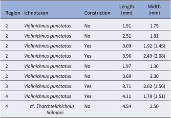


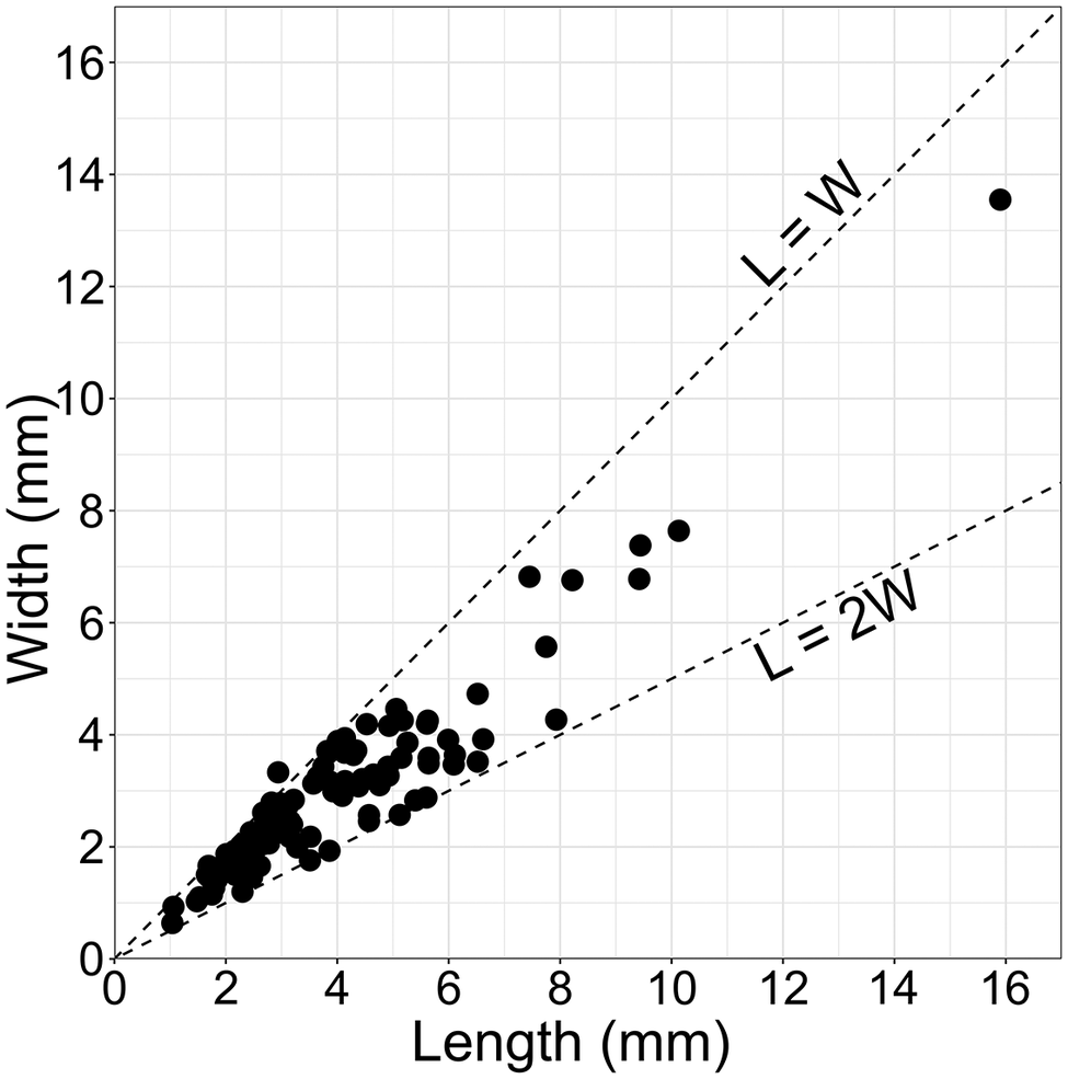


 Open access

