Introduction
Cerebrospinal fluid (CSF) rhinorrhoea is the leakage of CSF from the arachnoid space into the nasal cavity via a defect in the dura, bone and mucosa.
Historically, the most common cause of CSF rhinorrhoea has been traumatic head injury.Reference Park, Strelzow and Friedman1, Reference Ye, Zuo, Zhao, Liu, An and Liu2 Few authorsReference Presutti, Mattioli, Villari, Marchioni and Alicandri-Ciufelli3, Reference Banks, Palmer, Chiu, O'Malley, Woodworth and Kennedy4 have reported iatrogenic injury (i.e. from functional endoscopic sinus surgery and neurosurgical procedures) as a common cause of CSF rhinorrhoea. Spontaneous CSF leakage is even less common; it has been variously reported to account for 25 per cent,Reference Presutti, Mattioli, Villari, Marchioni and Alicandri-Ciufelli3 40 per centReference Banks, Palmer, Chiu, O'Malley, Woodworth and Kennedy4 and 49 per centReference Ye, Zuo, Zhao, Liu, An and Liu2 of CSF rhinorrhoea cases.
In patients with untreated CSF rhinorrhoea, the incidence of developing one or more episodes of meningitis has been variously cited as 24 per cent,Reference Banks, Palmer, Chiu, O'Malley, Woodworth and Kennedy4 40 per centReference Park, Strelzow and Friedman1 and 60 per cent of cases.Reference Ye, Zuo, Zhao, Liu, An and Liu2, Reference Eljamel and Foy5
Wigand first reported endoscopic repair of CSF rhinorrhoea in 1981, and it has now become the preferred treatment.Reference Wigand6
In 2000,Reference Ramsden, Corbridge and Bates7 Ramsden et al. reported bilateral, spontaneous CSF rhinorrhoea in a 47-year-old woman who presented initially with a CSF leak on the left side. Four years after successful endoscopic closure of this leak, she developed CSF rhinorrhoea on the right side; this was also repaired endoscopically, with no further episodes of leakage.
We present the case of a 47-year-old woman with bilateral, spontaneous CSF rhinorrhoea occurring simultaneously. This patient's CSF leaks were closed simultaneously using a uninasal, trans-septal approach, a previously unreported technique.
Case report
A 47-year-old woman presented to the emergency department with a one-day history of fever, vomiting, headache and altered sensorium. She also complained of intermittent, bilateral, watery nasal discharge which increased on bending forwards, for the previous six weeks. She was a known hypertensive and had received medication for this for the past five years. She also gave a history of pneumococcal meningitis five years earlier, treated at our institution; at that time, she did not have any nasal symptoms or evidence of para-meningeal foci of infection.
Clinical examination revealed signs of meningeal irritation. Cerebrospinal fluid analysis was consistent with acute bacterial meningitis.
The patient was commenced on intravenous antibiotics (ceftriaxone) along with a short course of steroids.
Nasal endoscopy showed active leakage medial to the middle turbinate, in both nasal cavities. Fluid collected from both sides was subjected to biochemical analysis for sugar and protein; results were suggestive of CSF.
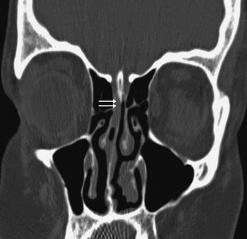
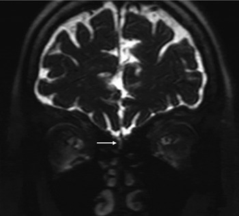
A high resolution computed tomography (CT) scan showed a small defect in the right cribriform plate (Figure 1). Magnetic resonance imaging (MRI) revealed a small meningocoele defect and adjacent CSF leakage via the right cribriform plate (Figure 2). However, no defect or leakage could be radiologically identified on the left side.
Coronal, thin section computed tomography scan through the anterior cranial fossa, showing a small defect in the right cribriform plate, with a small, linear hypodensity extending from the anterior cranial fossa through the defect into the right nasal cavity (double arrows).

Coronal magnetic resonance imaging scan, showing a small meningocoele and adjacent cerebrospinal fluid leak (arrow) through a defect in the right cribriform plate.

The patient was scheduled to undergo endoscopic repair of her CSF fistulae on completion of her anti-meningitic medication regime (i.e. two weeks of intravenous antibiotics as an in-patient, followed by oral antibiotics for one week as an out-patient). During her in-patient admission, she was also given the pneumococcal vaccine.
However, while taking oral antibiotics and awaiting surgery, the patient presented once again to the emergency department with signs of meningitis. She was admitted and treated for meningitis with intravenous ceftriaxone.
On the 14th day of her anti-meningitic regime, the patient underwent uninasal, trans-septal, endoscopic repair of her bilateral CSF leak. Following harvesting of fascia lata and fat from the right thigh, the fistulae were closed using standard endoscopic sinus surgery equipment, without the use of computer-assisted surgical navigation.
Initially, a right anterior ethmoidectomy was performed, followed by removal of the right middle turbinate to expose the roof of the right nasal cavity. Active CSF leakage was noted. The mucosa over the adjoining roof and superior part of the septum was elevated, revealing a large (4–5 mm) defect in the right cribriform plate area. The bony edges of the defect were identified.
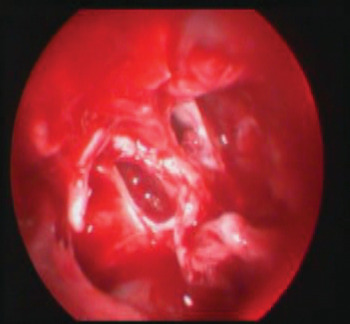
Through the right nasal cavity, the superior part of the perpendicular plate of the ethmoid and adjoining mucosa were removed to expose the roof of the left nasal cavity, where active CSF leakage was noted. The mucosa around the leak was elevated to expose bare bone. A large (approximately 6–7 mm) defect was noted in the left cribriform plate area (Figure 3).
Endoscopic view showing bilateral cribriform plate defects, with intervening superior aspect of the nasal septum, seen through the trans-septal surgical approach.

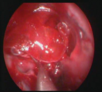
Fat was then tucked in between the dura and the bony defect on both sides, followed by fibrin glue and fascia lata grafting over the defect, including the bare bone on the roof of both nasal cavities (Figure 4). Turbinate mucosa plus fibrin glue was then applied, followed by Surgicel® and Gelfoam®. Both nasal cavities were then packed with bismuth iodoform paraffin paste.
Endoscopic view showing a single fascia lata graft applied over both defects.

Post-operatively, the patient was restricted to complete bed rest, with continuous lumbar subarachnoid drainage, for five days. Intravenous ceftriaxone was continued for a further eight days. The nasal pack was removed on the seventh post-operative day, following which the patient was discharged with a further week's course of oral antibiotics (cefixime).
At eight months' follow up, the patient had no evidence of recurrence of either CSF leakage or meningitis.
Discussion
The first surgical intervention for repair of CSF rhinorrhoea was performed by Dandy in 1926, via an intracranial approach using a bifrontal craniotomy. In 1948, Dohlman was the first to use an extracranial approach via a nasofrontal incision. The first successful endoscopic closure of a CSF leak was performed by Wigand in 1981.Reference Presutti, Mattioli, Villari, Marchioni and Alicandri-Ciufelli3, Reference Wigand6
The aetiology of CSF leaks can be broadly divided into traumatic (which can be subdivided into accidental and surgical) and non-traumatic (causes may be congenital, neoplastic or idiopathic). Traditionally, CSF rhinorrhoea caused by craniomaxillary trauma has been treated conservatively with bed rest, head elevation and strict sinus precautions,Reference Banks, Palmer, Chiu, O'Malley, Woodworth and Kennedy4 as observations show that the majority of closed injuries resolve spontaneously. However, long term follow up of post-traumatic patients has found that 29 per cent subsequently develop meningitis.Reference Eljamel and Foy5, Reference Bernal-Sprekelsen, Bleda-Vazquez and Carrau8 Therefore, all post-traumatic, ongoing CSF leaks need to be repaired. Iatrogenic, post-surgical leaks should be closed as soon as they are identified. When there is no history of trauma (either accidental or surgical) and the CSF leak is spontaneous, the clinician should exclude benign intracranial hypertension (or pseudotumour cerebri), empty sella syndrome, morbid obesity and galactorrhoea.Reference Ye, Zuo, Zhao, Liu, An and Liu2, Reference Banks, Palmer, Chiu, O'Malley, Woodworth and Kennedy4
In a suspected case of CSF rhinorrhoea, clinical examination should be followed by confirmation of the presence of CSF in the nasal fluid. Testing the fluid for the presence of β-2 transferrin remains the ‘gold standard’ for CSF identification.Reference Presutti, Mattioli, Villari, Marchioni and Alicandri-Ciufelli3, Reference Banks, Palmer, Chiu, O'Malley, Woodworth and Kennedy4 Testing for β-trace protein has been performed by some authors, as it is highly sensitive, quicker (taking 20 minutes, versus 120 minutes for β-2 transferrin testing) and less expensive than β-2 transferrin testing. If there is any doubt over the results, β-2 transferrin testing can be performed subsequently.Reference Presutti, Mattioli, Villari, Marchioni and Alicandri-Ciufelli3 Ye et al. Reference Ye, Zuo, Zhao, Liu, An and Liu2 performed a retrospective study estimating the glucose concentration of nasal discharge, to confirm the presence of extracranial CSF. In our patient's case, we estimated the glucose concentration in the nasal fluid, as the other two tests were not available in our centre.
Clinic-based nasal endoscopy is of primary importance when attempting to identify the site of a CSF leak.
Additional pre-operative evaluation of the exact site of the leak is achieved using high resolution CT scanning of the paranasal sinuses and anterior skull base, this being accepted as the imaging modality of choice. Magnetic resonance imaging (with T2-weighted sequences) is indicated, in addition, when parenchymal or meningeal herniation is suspected.Reference Presutti, Mattioli, Villari, Marchioni and Alicandri-Ciufelli3, Reference Banks, Palmer, Chiu, O'Malley, Woodworth and Kennedy4
Many authorsReference Banks, Palmer, Chiu, O'Malley, Woodworth and Kennedy4, Reference Lund, Savy, Lloyd and Howard9 have advised intra-operative localisation of the CSF leak using intrathecal fluorescein (after waiting approximately 30 minutes, a blue light is used to detect green-yellow fluid in the nasal cavity, indicating the leak site). Banks et al. Reference Banks, Palmer, Chiu, O'Malley, Woodworth and Kennedy4 considered this to be the most accurate test to identify the CSF leak site. However, the use of intrathecal fluorescein is controversial due to its multiple reported complications.Reference Keerl, Weber, Draf, Wienke and Schaefer10 Recently, Seth et al. Reference Seth, Rajasekaran, Benninger and Batra11 reported a series of 103 patients in which lack of intra-operative fluorescein visualisation did not exclude the presence of CSF leakage, as evidenced by a false negative rate of 26.2 per cent.
Presutti et al. Reference Presutti, Mattioli, Villari, Marchioni and Alicandri-Ciufelli3 have advocated that the simple and safe Valsalva manoeuvre be performed intra-operatively, by the anaesthestist. This has the dual advantage of identifying the leak site and testing the adequacy of closure.
In our patient, the right leak location was confirmed pre-operatively using diagnostic endoscopy and high resolution CT scan with MRI. The left leak site, although noted on nasal endoscopy, could not be exactly defined using high resolution CT with MRI; however, its location was confirmed intra-operatively using the Valsalva manoeuvre.
Materials utilised for CSF leak closure included fat, fascia (temporalisReference Stankiewicz12 and lataReference Kelly, Stankiewicz, Chow, Origitano and Shea13), free mucoperichondrial graft from the nasal septum,Reference Presutti, Mattioli, Villari, Marchioni and Alicandri-Ciufelli3 and free middle turbinate mucosal graft without bone.Reference Ye, Zuo, Zhao, Liu, An and Liu2 Composite osteomucosal or chondromucosal flaps have also been advocatedReference Stankiewicz12, Reference Kelly, Stankiewicz, Chow, Origitano and Shea13 for the repair of defects larger than 3–4 cm. Although management must be tailored according to the aetiology and site of the leak, precise localisation of the leak and adequate preparation of the defect site is mandatory. A flat surface must be created, to which the graft can be made to conform fully, in order for closure to be successful.Reference Banks, Palmer, Chiu, O'Malley, Woodworth and Kennedy4 The multilayer reconstructive technique has been recommended by Ye et al. Reference Ye, Zuo, Zhao, Liu, An and Liu2
• This is the first reported case of bilateral, spontaneous, simultaneous cerebrospinal fluid (CSF) rhinorrhoea
• History-taking, examination, nasal endoscopy, high resolution computed tomography (CT) and (if possible) biochemical confirmation are required to diagnose and localise the leak site
• In this case, high resolution CT and magnetic resonance imaging did not identify the leak site on one side
• An endoscopic, uninasal, trans-septal approach (a new technique) achieved effective closure
Presutti et al. Reference Presutti, Mattioli, Villari, Marchioni and Alicandri-Ciufelli3 conducted a five-year, retrospective study of 52 patients, with endoscopic closure of CSF leaks using septo-mucoperichondrial grafting, and without lumbar drainage or fluorescein testing; they reported a success rate of 88.5 per cent for the first closure attempt. Banks et al. Reference Banks, Palmer, Chiu, O'Malley, Woodworth and Kennedy4 undertook a 21-year, retrospective study of 193 patients, using endoscopic closure with intrathecal fluorescein localisation of the leak site and lumbar drainage in 73 per cent; they had an initial success rate of 85–90 per cent, and an overall success rate of 98 per cent. Ye et al. Reference Ye, Zuo, Zhao, Liu, An and Liu2 conducted a 10-year, retrospective study of 69 patients, using endoscopic multilayer reconstruction without pre-operative fluorescein injection, and reported a success rate of 89 per cent at the first closure attempt.
Conclusion
Non-traumatic, bilateral, simultaneous CSF rhinorrhoea is indeed a diagnostic and therapeutic challenge. Our patient represents the first reported case. An endoscopic, uninasal, trans-septal approach is effective in achieving simultaneous closure of leaks on both sides.