Introduction
The quality assurance (QA) of complex treatment planning has become an essential part of radiation delivery techniques. For this purpose three-dimensional (3D) dosimetry like PRESAGE® is considered one of the possible best tool. Human tissue-equivalent material is being used as an ideal dosimeter.Reference Babic, Battista and Jordan 1 The properties of dosimeters, like scattering and absorption of the radiation are comparable with water.Reference Sakhalkar, Sterling, Adamovics, Geffry and Mark 2 The PRESAGE® (Heuris Inc., Skillman, NJ, USA) has an effective 7·6 atomic number which is very close to 7·42 atomic number of water.Reference Sakhalkar, Adamovics, Ibbott and Oldham 3 The PRESAGE® dosimeter is composed of polyurethane and radiochromic components, which can measure dose in three dimensions using an optical-computer tomography (CT) scanner.Reference Doran 4 – Reference Oldham, Sakhalkar, Guo and Adamovics 7 The dose distribution is being compared with measurements taken with thermoluminescent detectors (TLDs), Gafchromic® EBT film and PRESAGE® which are enclosed in the Imaging and Radiation Oncology Core (IROC) head and neck (H&N) phantom.Reference Molineu, Followill and Balter 8
It was observed that if TLD and film were used for QA then many treatment plans were failed.Reference Geoffrey, David, Molineu, Jessica, Paola and Joye 9 It was a turning point for the discovery of 3D dosimetry. The PRESAGE® dosimeter has been recognised to maintain the standard of QA of complex treatment planning techniques like intensity-modulated radiation therapy (IMRT).Reference McJury, Oldham and Cosgrove 10 It is proved by many researchers that the PRESAGE® dosimeter has shown acceptable agreement between calculated and measured dose.Reference Newton, Oldham, Li, Adamovics, Kirsch and Das 11 – Reference Sakhalkar and Oldham 13 It is also found that the dose profile has good agreement with EBT film, PRESAGE® and Eclipse dose distribution. A report has already been published regarding the feasibility of PRESAGE® as 3D dosimetry in the IMRT H&N phantom.Reference Devic, Seuntjens and Sham 14
The aim of this research is to evaluate the performance of PRESAGE® dosimeter for use in patient-specific for volumetric-modulated arc therapy (VMAT) QA. VMAT is the one of most precise and accurate delivery technique for the treatment of H&N cancer. Before the start of any VMAT plan, patient-specific QC treatment plan is necessary, however.Reference Arumugam, Xing and Young 15
Materials and Methods
The IROC H&N phantom and insert
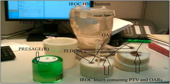
The IROC phantom, previously named the Radiological Physics Center (RPC) head & neck phantom, can accommodate either an IROC insert or a PRESAGE® insert. Owing to the different electron densities of the insert materials, three different structures appear on a CT scan. These structure were contoured and labelled as the planning target volume (PTV) and two organs at risk (OARs) mimicking the oesophagus and brain. The insert also contains slots for three pieces of film and eight TLDs. Two pieces are combining to form a sagittal slice and one central axial slice. Four TLDs are present in the PTV at superior–anterior, superior–posterior, inferior–anterior and inferior–posterior positions relative to the axial EBT film plane. Additionally two TLDs were placed in the brain and oesophagus at superior and inferior positions for a total of four, as shown in Figure 1.
Image Radiation Oncology Core (IROC) head and neck (HN) anthropomorphic Phantom with IROC insert and PRESAGE® insert. Abbreviations: PTV, planning target volume; TLD, thermoluminescent detectors; OAR, organs at risk.

VMAT treatment planning using CT scan of IROC H&N phantom
IROC comes with an instruction sheet for using the H&N phantom for treatment planning. According to the instructions, the H&N phantom was filled with water to maintain the dose distribution homogeneity. There should be no air bubbles present in the water of the phantom. An X-ray CT scan was taken using a Philips CT scanner (Philips Healthcare, Andover, MA, USA) with a slice thickness of 1·5 mm to ensure that the TLDs could be seen. The CT scan was imported into the Pinnacle (9.4 version) TPS for treatment planning, where the structures (PTV, OARs) and TLDs were manually contoured for VMAT plans. Treatment planning was done using VMAT (dual arcs of 182°–178° and 178°–182°). The treatment plan was designed such that PTV coverage was V90=100% at least of the prescription dose and got best plan to spare OARs.Reference Yavas, Yavas, Gul, Acar and Atal 16 True beam linear accelerator was used for dose delivery with a photon energy of 6 MV and collapsed cone convolution algorithm was used for dose calculation.Reference Mesbahi and Dadgar 17 The adaptive convolution algorithm was used for optimisation.Reference ur Rehman, Tailor, Isa, Muhammad, James and Ibbot 18 The VMAT plan was delivered three times for reproducibility and reliability of results.
QA, TLDs and EBT film
Before delivery, plan specific QC using arc check was performed to check consistency of planned fluence with delivered fluence for individual beams. The treatment plan was delivered to the phantom using a TrueBeam® linear accelerator (Varian Medical System, Palo Alto, CA, USA). The treatment plans and dosimeters such as TLDs and EBT film were sent to IROC for analysis and dose measurement. The mean calculated dose from the TPS for TLD contours were compared with measured TLD dosimeter doses. The standard IROC procedure was used for dose measurements with the help of a technical expert available at IROC to reduce uncertainty. The films were digitised using a 48-bit transmission/reflection flatbed photo scanner (Flat-Bed Epson-10000XL, Epson America Inc, Long Beach, CA, USA). Each film was scanned with an IROC film scanner in transmission mode using three colour channels: red, green and blue. Only the red channel was extracted for analysis because EBT has a maximum response to red light at 633 nm.Reference Ibbott 19 We did gamma analysis using the IROC film software by scaling the film dose to the TLD measurements.
3D dose measurement using PRESAGE® dosimeter
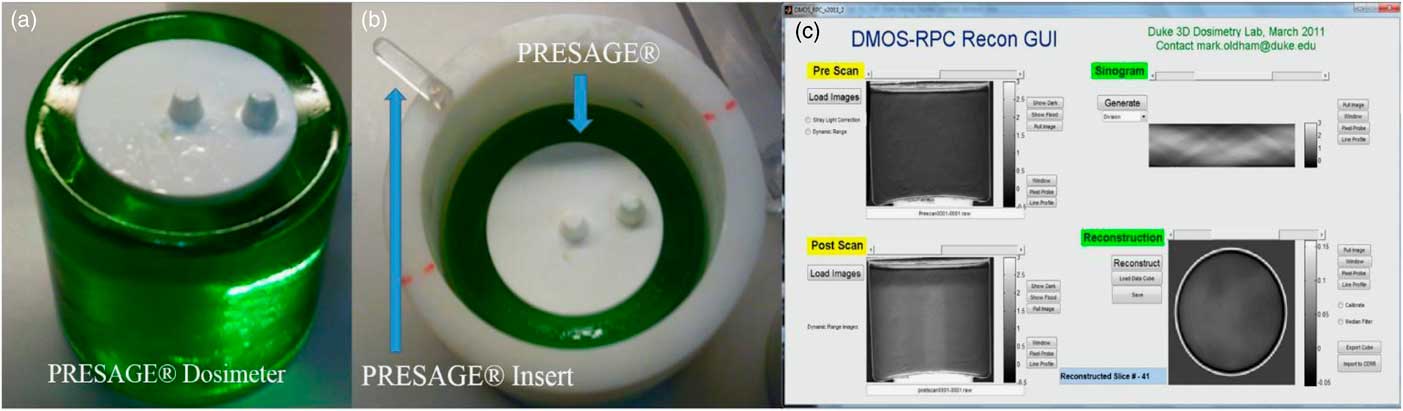
The PRESAGE® dosimeter was placed into the modified insert and put into the IROC H&N phantom before radiation delivery. The Duke Midsized Optical-CT Scanner (DMOS – Duke University, Durham, NC, USA) was used to acquire a 3D dose readout from the irradiated PRESAGE® dosimeter. A solid, radiochromic leuco dye-doped polyurethane plastic PRESAGE® dosimeter was moulded to fit inside a plastic sleeve that was compatible with the IROC H&N phantom as shown in Figure 2b.
(a) PRESAGE® dosimeter and (b) PRESAGE® insert containing plastic insert and PRESAGE®. (c) Duke Midsized Optical-CT Scanner (DMOS) reconstruction graphical user interfaces.

The radiochromic response was determined spectrophotometrically using cuvette irradiations and was found to be linear with a slope of 0·046 optical density/cm/Gy. The CT scan of the phantom with the PRESAGE® insert was imported into the Pinnacle TPS and registered with the CT scan of the phantom with the IROC insert using the auto-register tool, with the only difference that the prescription dose was reduced from 6·6 to 4 Gy/fraction.
After the treatment, the PRESAGE® dosimeter was kept refrigerated at 4°C and away from room light for 12 hours. The 3D dose distribution was read out using the DMOS which is dedicated for the IROC phantom (DMOS-IROC) using 1° per step to produce 360 projection images. The PRESAGE® dosimeters were scanned before and after irradiation. The scanner was configured for a voxel size of 1 mm3 and using a linear projection geometry.Reference Rehman, Iqbal and Tailor 20
Data registration and dose analysis
The dose data from DMOS and Pinnacle TPS were exported to CERR (Washington University, St Louis, MO, USA), a MATLAB based software. Three types of data were available for analyses: (1) Pinnacle calculated dose distributions, (2) two-dimensional dose distribution of EBT film and TLDs absolute dose distribution, (3) PRESAGE® dosimeter dose distribution. The calculated dose distribution from Pinnacle were compared with the measured dose distribution from the PRESAGE® dosimeter and EBT film for the VMAT plan. EBT film doses were measured using IROC images analysis software. Gamma analysis was performed on the line profiles in order to quantify the agreement between the calculated and measured dose distributions. Whereas a 3D gamma map was used between the PRESAGE® dose distribution and Pinnacle dose distribution in specific regions of interest (ROI) – that is, PTV, oesophagus and brain. The gamma map criteria of 5%/3 mm dose difference/distance to agreement, which was more stringent than currently used for IROC H&N credentialing protocols (7%/4 mm).
Results
Before direct comparison of EBT film with the 3D dosimeter called PRESAGE®, it was our responsibility to check that the relative dose distribution was similar in the IROC insert which contains both EBT film and the PRESAGE® insert. The differences could come from the non-linear scaling of the dose-response between 6·6 and 4 Gy plans for EBT film and PRESAGE®, respectively. The Arc CHECK™ VMAT QA device observed that the relative fluences were similar between these two plans, with a greater than 98% gamma pass rate which was achieved for all fields with a 3%/3 mm criteria.
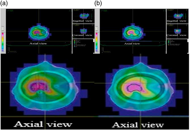
The relative dose distributions from these two treatment plans compared with the dose calculated by Pinnacle is shown in Figure 3. Dosimetrically, both plans when normalised to the prescription dose agreed to within 1%, signifying minimal non-linearities in the sensitivity of the PRESAGE dosimeter when prescription dose change from 6 to 4 Gy.
Volumetric-modulated arc therapy dose distribution from Pinnacle® version 9.4 axial views (a) dose prescription 6·6 Gy with IROC standard insert. (b) Dose prescription 4 Gy with PRESAGE® insert scan.

Absolute point dose analysis using TLDs
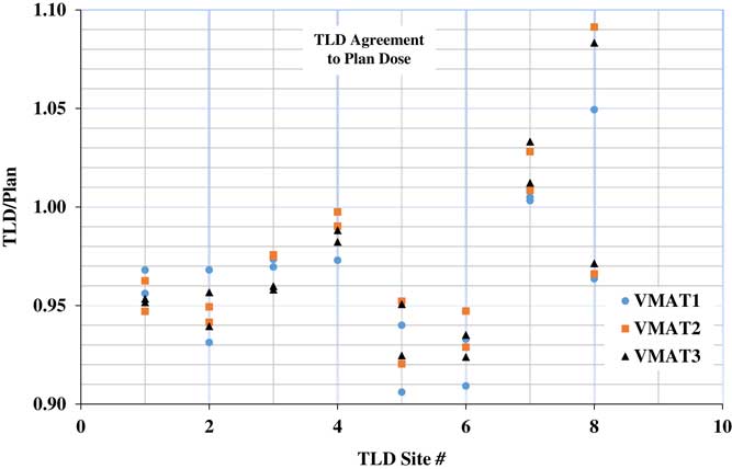
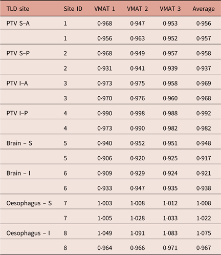
The mean doses for each TLD contour were calculated by the Pinnacle treatment planning system. The Table 1 contains the mean ratio of TLD doses between what was measured to what was calculated for the VMAT plan over three deliveries. These results are also shown graphically in Figure 4. The eight points of measurement in the PTV and OARs were evaluated to determine the agreement between TLD measured dose and that calculated by Pinnacle. It was observed that the results were within the IROC absolute dose difference criteria of ±7% dose. The doses to the OARs are also included in this analysis since VMAT H&N plans generally contain steep dose gradients and thus small errors in position may cause larger errors in dose. The ratio of TLD measured dose to Pinnacle calculated dose at the four PTV locations in the H&N phantom were within 0·95 to 0·99 for all three VMAT deliveries, thus passing the IROC criteria of a ±7% dose difference.
The ratio of thermoluminescent detectors (TLD) measured and calculated doses in head and neck phantom for three deliveries of the volumetric-modulated arc therapy (VMAT) plan.

The ratio of thermoluminescent detectors (TLD) measured and calculated doses in head and neck phantom for three deliveries of the volumetric-modulated arc therapy (VMAT) plan

Abbreviations: S, Superior; I, inferior; A, anterior; P, posterior.
Dose profile comparison between PRESAGE® and Pinnacle
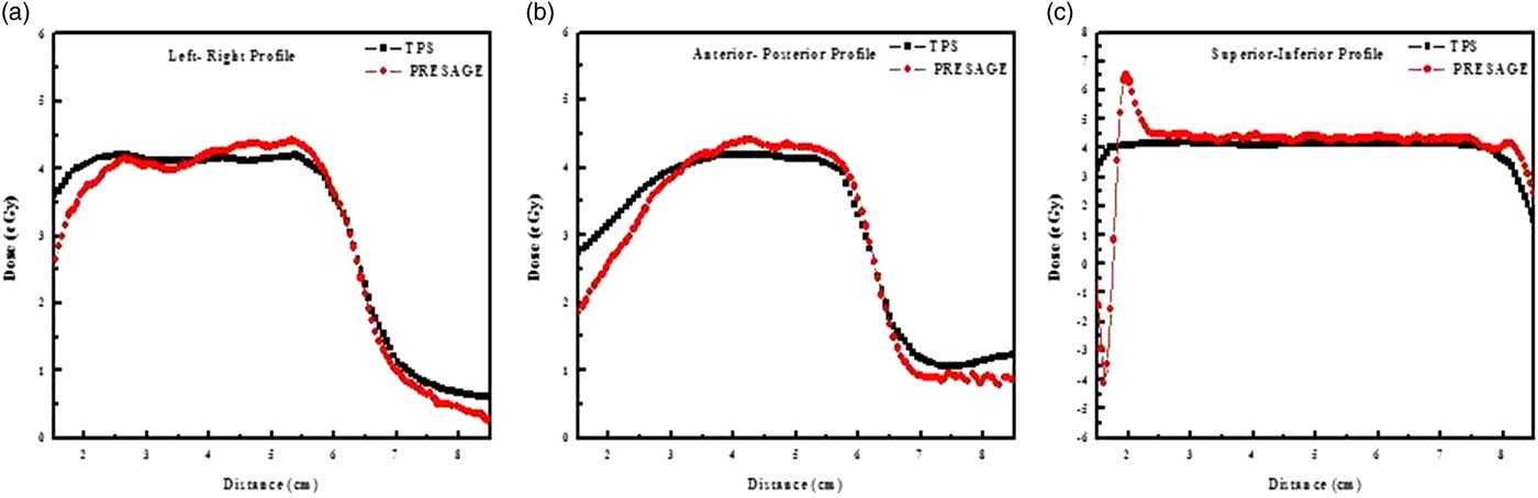
Dose profiles were taken along the left–right, anterior–posterior and superior–inferior axis from the Pinnacle (TPS) and PRESAGE® dosimeter read out as shown in Figure 5. Subplots A, B and C display dose profiles along the left–right, anterior–posterior and superior–inferior directions, respectively. Many comparatively minor differences can be discerned between the distributions due to setup error but trends are not readily apparent and it is not possible to state whether the Pinnacle dose distribution agrees more closely with one or the other of the measured distributions.
Dose profiles for Pinnacle (TPS) and PRESAGE® of volumetric-modulated arc therapy plan along left–right profile (a), anterior–posterior profiles (b) and superior–inferior profile (c).

Figure 5 shows agreement between the PRESAGE® and the Pinnacle® calculated dose on the order of ±10% in high-dose regions such as the PTV while larger disparities appear to exist in the high gradient regions between the PTV and OARs as seen at the peripherals of Figure 5a–5c.
Gamma results analysis
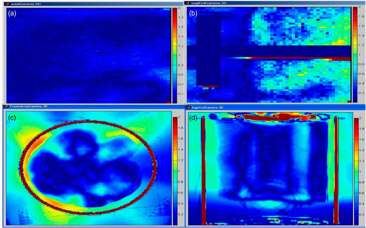
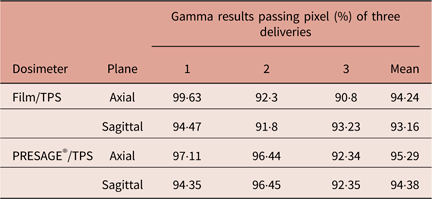
Gamma analysis was performed between the film dose distribution, the PRESAGE® dose distribution and the dose calculated by the Pinnacle TPS in the axial and sagittal planes. The gamma indexes are shown in Figures 6a, 6b and 6c, 6d for film versus the TPS, and PRESAGE® versus the TPS, respectively, using a criteria of ±5%/3 mm. During the gamma analysis masking of the dose distributions was performed in order to avoid the pricked region and edge of the film area. The gamma index results given in terms of percentage of passing pixels is summarised in Table 2 for all three deliveries.
Gamma maps using a ±5%/3 mm criteria comparing Pinnacle versus EBT film in the axial (a) and sagittal (b) planes and Pinnacle versus PRESAGE® in the axial (c) and sagittal (d) planes for the first volumetric-modulated arc therapy delivery.

Gamma analysis results of a volumetric-modulated arc therapy plan with ±5% dose difference and 3 mm DTA in axial and sagittal planes

The 3D gamma results of criteria ±5%/3 mm between PRESAGE® dose distribution and Pinnacle dose distribution in the specific ROI – that is, PTV, oesophagus and brain were 99·5%, 99·9% and 100% passing pixel, respectively. It is observed that overall plan passing the pixel more than 90%, it means treatment plan pass the QA test.
Discussion
The TLDs results show that calculated dose is greater than measured dose with TLDs as shown in Table 1. The ratio of measured and calculated TLD doses in Table 2 shows and average ratio between TLD measured dose and calculated dose that varies from 0·95 to 0·97 for all three VMAT deliveries. This lies within the IROC credentialing criteria of ±7% dose difference. In the IROC credentialing criteria, OARs are excluded but results of OARs were also within IROC tolerance limits.
Dose profiles were drawn across the centre of the axial, sagittal and coronal planes and close agreement was found even in the dose gradient region. The mean displacement between the measured dose gradient and calculated dose gradient from Pinnacle was <2 mm between PTV and OARs, which was less than IROC standard of 4 mm. In general, the Pinnacle calculated and PRESAGE® measured profiles agreed well except at the edges due to edge artefacts in PRESAGE® and film dose distributions.Reference Vieillevigne, Molinier, Brun and Ferrand 21
Gamma analysis was used for further verification of the agreement between Pinnacle, PRESAGE® and EBT film in axial, sagittal and coronal planes. The gamma results of sagittal and axial planes were presented in text and where similar to results in the coronal plane. A gamma criteria of 3% dose difference and 3 mm distance to agreement (DTA) was used, which was less than the 7%/4 mm criteria used in the IROC credentialing test. Gamma analysis showed that accuracy of the PRESAGE® dose distribution in the sagittal and central axial plane, supports its use as a reliable 3D standard for patient-specific QA. Some areas of the PRESAGE® and Pinnacle dose distributions disagreed due to high-dose gradient regions and appear as yellow to red in Figure 6. This is attributable to edge artefacts at the peripheral regions of the PRESAGE® insert.Reference Oldham and Kim 22 The pass rate between the PRESAGE® dosimeter and pinnacle (TPS) for the axial and sagittal planes was 95·29 and 94·38%, respectively, when excluding edge artefacts.
Conclusions
3D dosimetry tools have the potential to improve QA of complex VMAT treatment plans for H&N cancers. This research shows the application and feasibility of the 3D PRESAGE® dosimetry system for relative dosimetry in an IROC H&N phantom. The PRESAGE® dose measurements were compared with Pinnacle calculated doses and showed reasonable agreement in both the axial and sagittal planes for a complex dual arc VMAT treatment plan. In general, the PRESAGE® dosimeter was found to be a feasible QA tool for VMAT plans.
Acknowledgements
Authors are thankful to the Higher Education Commission (HEC), Pakistan for providing a scholarship for Mr. Jalil ur Rehman. The authors thank Mr. Pei Fong (Senior Medical Physicist) from the Radiation Physics department at The University of Texas MD Anderson Cancer Center for help in treatment planning and valuable discussions, and Dr Daniel Markel from Princess Margaret Cancer Centre, Canada for editing and giving comments on this manuscript.