Introduction
Oocyte cytoplasmic maturation involves the accumulation of mRNAs, proteins, substrates and nutrients that are required to achieve the oocyte developmental competence that fosters embryonic development (Calder et al., Reference Calder, Caveney, Westhusin and Watson2001, Reference Calder, Caveney, Smith and Watson2003). RNA-binding proteins (RBP) influence RNA editing, localization, stability and translation (Saunders & Barber, Reference Saunders and Barber2003). Therefore RBPs may contribute to oocyte competence by regulating the stability and turnover of oogenetic mRNAs. In addition, variations in oocyte competence may reflect differences in RBP levels and thus turnover of their target mRNAs during early development.
As a prelude to investigating these possibilities it is imperative that the expression of RBPs during mammalian oocyte maturation and early embryogenesis be first determined. Staufen is a Drosophila RBP that assists in establishing the anterior–posterior embryonic axis by regulating target mRNA localization in the Drosophila oocyte (St Johnston et al., Reference St Johnston, Beuchle and Nusslein-Volhard1991). There are two mammalian staufen genes, STAU1 (Marion et al., Reference Marion, Fortes, Beloso, Dotti and Ortin1999) and STAU2 (Duchaine et al., Reference Duchaine, Wang, Luo, Steinberg, Nabi and DesGroseillers2000). ELAVL1 (HuR, HuA, elrA) is a widely expressed RBP (Fan & Steitz, Reference Fan and Steitz1998), which is present in the Xenopus embryo (Good, Reference Good1995) and can shuttle between the nucleus and cytoplasm (Atasoy et al., Reference Atasoy, Watson, Patel and Keene1998). It binds to A–U rich sequences (AREs) to regulate mRNA half-life (Atasoy et al., Reference Atasoy, Watson, Patel and Keene1998; Fan & Steitz, Reference Fan and Steitz1998). ELAVL2 (HuB, elrB, Hel–N1/2) is also an ARE-binding RBP (Jain et al., Reference Jain, Andrews, McGowan, Pekala and Keene1997), which is predominantly neuronal but is also expressed in the ovary, testis, oocyte and Xenopus embryo (Good, Reference Good1995). ELAVL2 affects mRNA degradation and may increase protein translation (Jain et al., Reference Jain, Andrews, McGowan, Pekala and Keene1997). The present study was conducted to characterize the expression of Staufen and ELAVL mRNAs and proteins during oocyte maturation and preimplantation development in the cow.
Materials and Methods
Production of bovine embryos in vitro
Cumulus–oocyte complexes (COCs) were isolated, placed into oocyte maturation and were fertilized in vitro and cultured up to the blastocyst stage as outlined in Madan et al. (Reference Madan, Calder and Watson2005). Bovine ovaries were transported from a slaughterhouse in sterile saline at 32–37 °C for oocyte collection using standard protocols. COCs from 3–6 mm follicles were aspirated into the follicular aspiration medium consisting of Dulbecco's phosphate-buffered saline (Gibco BRL; Invitrogen), 0.3% bovine serum albumin (Sigma–Aldrich) and 50 μg/ml Gentamycin (Sigma–Aldrich) using an 18G needle attached to a vacuum suction apparatus. COCs that contained an oocyte with an evenly granulated cytoplasm and surrounded by more than three layers of cumulus cells were selected for maturation in vitro. For maturation in vitro, COCs were cultured in oocyte maturation medium composed of modified synthetic oviductal fluid (SOF) medium with 0.8% BSA, modified Eagle medium (MEM) non-essential amino acids (Gibco), MEM essential amino acids (Gibco), 1 mM glutamine, 0.5 μg/ml FSH and 1 μg/ml 17β-estradiol. Fifty COCs were placed in each well of a 4-well culture plate and incubated in a humidified atmosphere for 24 h at 38.5 °C and 5% CO2 in air atmosphere.
For fertilization in vitro, 50 mature COCs were added to 330 μl drops containing modified Tyrode lactate medium (TLH) supplemented with 0.6% BSA fatty acid free (Sigma–Aldrich), 0.2 mM pyruvic acid, 10 μg/ml heparin and 50 μg/ml gentamycin under mineral oil. Frozen semen was thawed and processed through a standard ‘swim-up’ method (Madan et al., Reference Madan, Calder and Watson2005). Sperm concentration was adjusted to and added to each insemination culture drop at 1 × 10⁶ spermatozoa/ml. COCs and spermatozoa were co-incubated in a humidified 5% CO2 in air atmosphere at 38.5 °C and for 15–18 h.
Following fertilization, zygotes were denuded of invested cumulus cells by vortexing, washing three times in culture media consisting of SOF1 plus 0.8% BSA, MEM non essential amino acids, 1 mM glutamine and 1.5 mM glucose and 10 μM EDTA. Subsequently, 20–30 zygotes were placed into 20 μl culture drops under mineral oil and cultured at 38.5 °C, 5% CO2 and humidified air with reduced oxygen atmosphere (7%). Following 3 days of culture, SOF1 medium was replaced with SOF2 medium, which contained 0.8%, BSA, MEM non essential amino acids, MEM essential amino acids, 1 mM glutamine and 1.5 mM glucose. Pools of 20 embryos were harvested at timed stages of development (2-, 4-, 8-cell, morulae and blastocysts) for RNA extraction or application of immunofluorescence localization methods.
Primer design
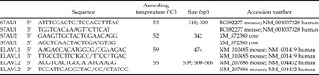
Primer sets were designed to recognize and amplify conserved nucleotide sequences encoding human and murine Staufen or ELAVL cDNAs. cDNA sequences and/or homologue(s) were identified using BLAST (Basic Local Alignment Search Tool) computer program (NCBI). Primers were designed using the ‘Primer3’ computer program (Whitehead Institute) and the corresponding oligonucleotides (Table 1), were synthesized (Invitrogen).
Primers used to detect RBP mRNAs in cow oocytes and embryos

RNA extraction, reverse transcription and PCR
Total RNA was extracted from bovine embryos (pools of 20 embryos/stage at 1-, 2-, 4-, 8-cell, morula and blastocyst stages) as described (Madan et al., Reference Madan, Calder and Watson2005). The total RNA extracts were digested with deoxyribonuclease (DNase)-1 to eliminate possible contamination from genomic DNA. The RT reactions were conducted using oligo-dT primers (Gibco BRL) as previously described (Barcroft et al., Reference Barcroft, Hay-Schmidt, Caveney, Gilfoyle, Overstrom, Hyttel and Watson1998; Offenberg et al., Reference Offenberg, Barcroft, Caveney, Viuff, Thomsen and Watson2000; Calder et al., Reference Calder, Caveney, Westhusin and Watson2001, Reference Calder, Caveney, Smith and Watson2003, Reference Calder, Caveney, Sirard and Watson2005; Natale et al., Reference Natale, Paliga, Beier, D'Souza and Watson2004). Samples were incubated for 90 min at 42 °C in a 20 μl volume of 50 mM Tris–HCl (pH 8.3), 75 mM KCl, 3 mM MgCl2, 10 mM DTT, 0.5 mM dNTPs and 200 units of Superscript II (Gibco BRL) followed by heating the samples to 95 °C for 5 °C min reaction termination.
PCR was conducted as described (Calder et al., Reference Calder, Caveney, Westhusin and Watson2001, Reference Calder, Caveney, Smith and Watson2003, Reference Calder, Caveney, Sirard and Watson2005). Briefly, two embryo equivalents for each stage of development under investigation were used per PCR reaction, which was repeated a minimum of three times from pools of three different developmental series of embryos. PCR products were resolved on 2.0% agarose gels containing 0.5 μg/ml ethidium bromide (Invitrogen, Burlington, ON). To confirm the specificity of each PCR product, representative amplicons were extracted from the gels and purified using a QIAquick Gel Extraction Kit (Qiagen) and submitted for nucleotide sequencing (DNA Sequencing Facility, Robarts Research Institute). The nucleotide sequences were compared to sequences available in GenBank to confirm the specificity of each PCR product.
Whole-mount indirect immunofluorescence
To localize Staufen and ELAVL proteins in bovine oocytes and preimplantation embryos we employed whole-mount immunofluorescence methods as outlined in (Calder et al., Reference Calder, Caveney, Westhusin and Watson2001, Reference Calder, Caveney, Smith and Watson2003, Reference Calder, Caveney, Sirard and Watson2005; Madan et al., Reference Madan, Calder and Watson2005). Embryos (oocytes, 2-, 4- 8-cell, morula and blastocyst stages) were washed in 1× phosphate-buffered saline (PBS) and then fixed in 2% paraformaldehyde (PFA) in PBS for 20 min at room temperature. These fixed embryos were washed in 1× PBS and either processed immediately for immuno-labelling or stored at 4 °C in PBS + 0.09% sodium azide for a maximum of 3 weeks. Fixed embryos were permeabilized and blocked in 1× PBS + 5% goat serum + 0.01% Triton X-100 for 1 h at room temperature. Embryos were washed in 1× PBS and incubated with primary antibody diluted 1:100 in 1× PBS + 1% goat serum + 0.005% Triton X-100 for 1 h at room temperature followed by additional washes totaling 1 h at 37 °C. Primary antibodies were detected by exposure for 1 h to FITC-conjugated secondary antibodies (Jackson Immuno Laboratories) diluted 1:200. Embryos were then treated with rhodamine-conjugated phalloidin (5 μg/ml; 1:20) and DAPI (1 mg/ml; 1:2000) for 30 min at 37 °C followed by two washes for 2 h each at 37 °C. Embryos were mounted in Fluoro-Guard Antifade Mounting Reagent (BioRad). Fluorescence patterns were examined using a Zeiss LSM 410 (laser scanning microscope) with an inverted Axiovert 100 microscope under 20–40× magnification. The images were then captured and stored as TIFF files by the Zeiss LSM software package. Rabbit polyclonal anti-human STAU1, ELAVL1 and ELAVL2 antibodies were obtained from (Chemicon).
Results and Discussion
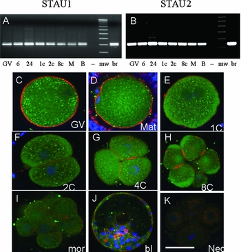
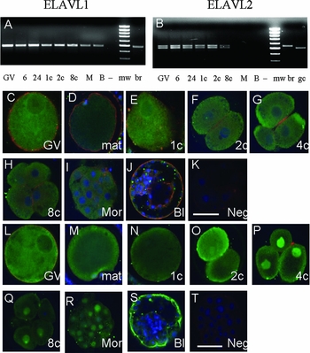
STAU1 mRNA was detected from the germinal vesicle (GV) oocyte to the blastocyst stage (Fig. 1a). The primers used to amplify STAU1 revealed two mRNA isoforms expressed in oocytes and embryos, which differ due to a splicing event resulting in an insertion of 18 bp (6 aa) in RNA-binding domain 3, similar to isoforms reported in mouse (Duchaine et al., Reference Duchaine, Wang, Luo, Steinberg, Nabi and DesGroseillers2000). STAU1 protein was detected in the cytoplasm of bovine oocytes and embryos at all stages examined (Fig. 1c–k). Staufen was observed in the GV, pronuclei or nuclei up to the morula stage but this localization was rarely stronger than cytoplasmic fluorescence (Fig. 1c–k). STAU2 mRNA was also detected in all cow preimplantation stages (Fig. 1b). ELAVL1 mRNA was detected continuously from the GV oocyte to the blastocyst stage (Fig. 2a). ELAVL1 protein was cytoplasmic at all stages examined, but was also present in the GV and most nuclei at all stages, but was less evident at the blastocyst stage (Fig. 2c–k). ELAVL2 mRNAs were detected from the GV oocyte to the 8-cell stage in all replicates (Fig. 2b). ELAVL2 mRNA was detected only in some replicates at the morula stage and not at all at the blastocyst stage (Fig. 2b). Two ELAVL2 mRNAs were detected. The larger band corresponds to a 539-bp product common to human and mouse sequences in GenBank (mouse, NM_207685). In the mouse, there is an ELAVL2 mRNA splice variant lacking 36 nt, this isoform would amplify as 503 bp (NM_207686). Another isoform lacks the bases AGG (R) before this splice junction, the 36 bp and has the addition of AGT (S) at the end of the splice junction, making it also 503 bp (AY035379). Yet another splice variant reported contains the 5′ R, misses the 36 nt and contains the 3′ S, making it 506 bp (AY035378). However, a human variant contains only 500 bp and the 5′ R, the 36 nt and the 3′ S are missing (BC030692). ELAVL2 immunofluorescence was cytoplasmic at all stages examined, but was also detectable in the GV, pronuclei and most nuclei and was brighter in the nucleus than cytoplasm at particularly the 4- and 8-cell stages (Fig. 2l–t). At the blastocyst stage, ELAVL2 also appeared at the borders of trophectoderm cells (Fig. 2s).
Staufen mRNA and protein during cow preimplantation development. (a) Staufen 1; (b) Staufen 2; (c–k) Staufen protein in cow oocytes and embryos. Green is FITC-labelled secondary antibody against rabbit anti-staufen. Red is rhodamine–phalloidin stain for F-actin. Blue is DAPI-stained nuclei. GV, germinal vesicle stage; 24 h, in vitro matured 24 h; Mat, matured 24 h in vitro; 1c, 1-cell stage; 2c, 2-cell stage; 4c, 4-cell stage; 8c, 8-cell stage; Mor, morula stage; Bl, blastocyst stage; Neg, negative control; MW, 100 bp molecular weight standard; +, positive control tissue, br, brain. Bar is approximately 50 μm. n = 3 replicates, 25–39 embryos examined at each stage.

ELAVL mRNA and protein during cow preimplantation development. (a) ELAVL1; (b) ELAVL2; (c–k) ELAVL1 protein, n = 3 replicates, 29–48 embryos examined at each stage; (l–t) ELAVL2 protein, n = 3 replicates, 25–43 embryos examined at each stage. Green is FITC-labelled secondary antibody against rabbit anti-ELAVL1 or 2. Red is rhodamine–phalloidin stain for F-actin. Blue is DAPI-stained nuclei. GV, germinal vesicle stage; 24 h, in vitro matured 24 h; Mat, matured 24 h in vitro; 1c, 1-cell stage; 2c, 2-cell stage; 4c, 4-cell stage; 8c, 8-cell stage; Mor, morula stage; Bl, blastocyst stage; Neg, negative control; MW, 100 bp molecular weight standard; +, positive control tissue, br, brain; gc, granulosa cells. Bar is approximately 50 μm.

As the vast majority of mammalian in vitro matured oocytes are meiotically competent, deficiencies in cytoplasmic maturation are proposed as a primary reason for their low developmental rates. Transcription and storage of maternal mRNAs occurs during follicular growth and slows as the oocyte reaches mature size (Fair et al., Reference Fair, Hyttel and Greve1995). The embryo is dependent on stored maternal mRNAs until at least the maternal–zygotic transition (MZT), when transcription of embryonic genes begins in earnest with a major burst of embryonic transcription initiating at the 8-cell stage in the cow (Telford et al., Reference Telford, Watson and Schultz1990). Many maternal mRNAs become deadenylated during oocyte maturation and early cleavage (Brevini-Gandolfi et al., Reference Brevini-Gandolfi, Favetta, Mauri, Luciano, Cillo and Gandolfi1999). Over 200 RBP candidate genes have been identified in Drosophila (Lasko, Reference Lasko2000). Regulated mRNA stability occurs in mammalian cells in response to nutrient levels, hormones and environmental stresses such as hypoxia and heat stress (reviewed by (Guhaniyogi & Brewer, Reference Guhaniyogi and Brewer2001). It has been suggested that variations in transcriptional activity largely govern changes in embryonic mRNA abundance associated with exposure to sub-optimal environments (Niemann & Wrenzycki, Reference Niemann and Wrenzycki2000). However, oocyte and embryo competence could also be affected by environmental effects acting through RBPs to alter transcript stability or translation.
During oogenesis, oocytes acquire a depository of maternally encoded transcripts that must be stored and remain dormant until their translation is required following oocyte activation and insemination. This delay between their production and translation is important as the oocyte becomes transcriptionally silent during the final stages of folliculogenesis and transcription is not renewed until post-insemination and early embryogenesis (Schultz, Reference Schultz2005). Yet few studies have investigated mRNA stability during preimplantation development. Instead, the majority of studies have focused upon understanding transcriptional regulation (Wrenzycki et al., Reference Wrenzycki, Herrmann, Carnwath and Niemann1999, Reference Wrenzycki, Herrmann, Lucas-Hahn, Korsawe, Lemme and Niemann2005; Niemann & Wrenzycki, Reference Niemann and Wrenzycki2000; Knijn et al., Reference Knijn, Wrenzycki, Hendriksen, Vos, Herrmann, Van Der Weijden, Niemann and Dieleman2002, Reference Knijn, Wrenzycki, Hendriksen, Vos, Zeinstra, Van Der Weijden, Niemann and Dieleman2005; Rinaudo & Schultz, Reference Rinaudo and Schultz2004). Our study provides the first indications that gene products encoding Staufen and ELAVL RBPs are expressed during bovine preimplantation development.
Preimplantation mammalian embryos attempt to respond positively to the pressures that sub-optimal culture environments place upon them (Ho et al., Reference Ho, Doherty and Schultz1994, Reference Ho, Wigglesworth, Eppig and Schultz1995; Niemann & Wrenzycki, Reference Niemann and Wrenzycki2000; Watson et al., Reference Watson, De Sousa, Caveney, Barcroft, Natale, Urquhart and Westhusin2000). The embryo, (at least partially), compensates for missing components or offsets the presence of deleterious components, by regulating its developmental programme (Niemann & Wrenzycki, Reference Niemann and Wrenzycki2000). This capacity however must operate within a defined range of tolerances, (Bolton, Reference Bolton1992; Bavister, Reference Bavister2000; Gardner et al., Reference Gardner, Lane and Schoolcraft2002; Leese, Reference Leese2002; Schultz & Williams, Reference Schultz and Williams2002) and current media, are still sub-optimal for all species. We are aware that current media are still sub-optimal since cultured embryos from all species display a reduced pregnancy rate following embryo transfer and are also prone to metabolic and growth disorders that may find their origin in a culture induced ‘metabolic re-programming’ during the preimplantation development period as compared to their in vivo-derived counterparts (Sinclair et al., Reference Sinclair, McEvoy, Maxfield, Maltin, Young, Wilmut, Broadbent and Robinson1999, Reference Sinclair, Rooke and McEvoy2003; Boerjan et al., Reference Boerjan, Daas and Dieleman2000; McEvoy et al., Reference McEvoy, Sinclair, Young, Wilmut and Robinson2000, Reference McEvoy, Robinson and Sinclair2001; van Wagtendonk-de Leeuw et al., Reference van Wagtendonk-de Leeuw, Mullaart, de Roos, Merton, den Daas, Kemp and de Ruigh2000; Barker, Reference Barker2003; Adamiak et al., Reference Adamiak, Mackie, Watt, Webb and Sinclair2005).
Due to these concerns great effort has been focused on understanding how early embryos adjust their developmental programme to compensate for exposure to sub-optimal culture environments. Approaches in this research area have included studies directly comparing the levels of ‘marker’ gene expression, also variations in embryo metabolism between in vitro- and in vivo-derived preimplantation embryos and most recently epigenetic re-programming as signified by variation in DNA methylation patterns (De Sousa et al., Reference De Sousa, Westhusin and Watson1998; Brevini-Gandolfi et al., Reference Brevini-Gandolfi, Favetta, Mauri, Luciano, Cillo and Gandolfi1999; Wrenzycki et al., Reference Wrenzycki, Herrmann, Carnwath and Niemann1999, Reference Wrenzycki, Herrmann, Lucas-Hahn, Korsawe, Lemme and Niemann2005; Watson et al., Reference Watson, De Sousa, Caveney, Barcroft, Natale, Urquhart and Westhusin2000; Leese, Reference Leese2002; Ecker et al., Reference Ecker, Stein, Xu, Williams, Kopf, Bilker, Abel and Schultz2004; Mann et al., Reference Mann, Lee, Doherty, Verona, Nolen, Schultz and Bartolomei2004; Knijn et al., Reference Knijn, Wrenzycki, Hendriksen, Vos, Zeinstra, Van Der Weijden, Niemann and Dieleman2005). The prevailing interpretation of the majority of these studies is that gene transcription is influenced by exposure to varying culture environments. Only a very few studies have begun to examine the influences of varying culture environments on mRNA deadenylation and mRNA stability (Temeles & Schultz, Reference Temeles and Schultz1997; Brevini-Gandolfi et al., Reference Brevini-Gandolfi, Favetta, Mauri, Luciano, Cillo and Gandolfi1999; Gandolfi & Gandolfi, Reference Gandolfi and Gandolfi2001). In addition, there is a need to understand the role of RBPs in regulating preimplantation mRNA stability as these proteins represent reasonable targets for culture induced influences on embryonic mRNAs (Yu et al., Reference Yu, Hecht and Schultz2003; Yang et al., Reference Yang, Medvedev, Reddi, Schultz and Hecht2005a, Reference Yang, Medvedev, Yu, Tang, Agno, Matzuk, Schultz and Hechtb, Reference Yang, Medvedev, Yu, Schultz and Hecht2006). This study therefore provides a foundation for investigating the roles of RBPs during early development and will allow for investigations regarding their putative roles in regulating preimplantation development and more importantly, eventually, their collective roles in regulating mRNA transcripts during early embryogenesis.
Acknowledgements
The authors would like to thank Barry Fong and Jenny Hickson for help with transporting ovaries and ovarian aspiration. We would like to thank Dr Allan King's and Dr Dean Betts' laboratories at the University of Guelph for assisting with ovary collections. These studies were supported by operating funds from the Natural Sciences and Engineering Research Council of Canada (NSERC) Discovery Grant # 138317-06.