Primary cardiac neoplasms are very rare and the majority is benign. The prevalence is reported to be 0.0017−0.28 with an incidence of 0.14% in foetal life.Reference Uzun, Wilson, Vujanic, Parsons and Giovanni 1 Tumours involving cardiac valves are even rarer, accounting for <10% of the overall incidence. Papillary fibroelastoma followed by myxoma and fibroma are among the most common cardiac valve tumours.Reference Benvenuti, Mansur, Lopes and Campos 2 Lipomatous hamartoma of cardiac valves is an extremely rare lesion, mostly diagnosed as incidental findings on cardiac ultrasonography. Surgical treatment of such tumours should be considered as soon as they are discovered, as they have high potential risk for haemodynamic compromise and sudden death.
A review of the literature revealed that primary valvar lipomatous hamartomas have been described in only nine patients: five with mitral hamartomas, three with tricuspid localisation, and one in the aortic valve. Usually, valve leaflets contain scarce amounts of fat cells. Lipoma, fibrolipoma, myolipoma, and angiolipoma are among fatty valvar tumours, which are well-circumscribed, encapsulated lesions, differing in their tissue composition. Together with specific cellular patterns, encapsulation is a feature of distinction from lipomatous hamartoma.Reference Karasu, Yeter, Yilmazer, Kiziltepe, Ulular and Akdemir 3
Case report
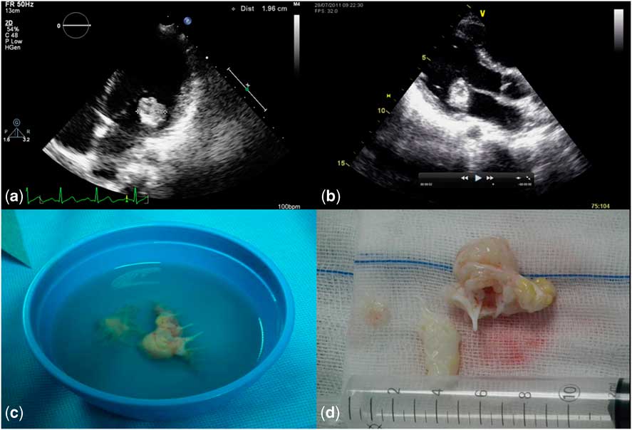
A 9-year-old girl presented at our department for heart murmur investigation. She was asymptomatic and had no relevant past medical history. Cardiac examination revealed a holosystolic murmur, grade 3/6 at the left sternal border and apex. The electrocardiogram showed sinus rhythm with normal QRS axis and no significant changes. Transthoracic echocardiography revealed a single rounded mass measuring 2 × 2 cm in diameter, mostly composed of mixed ecodensity tissue. The mass was attached to the mural leaflet's distal portion of the mitral valve by an apparent large pedicle, prolapsing to the left ventricle and causing eccentric moderate mitral regurgitation. There was no obstruction of the left ventricular inflow (Fig 1a and b) and the mass had an apparent low potential embolic risk.
( a and b ) Transthoracic echocardiography showing a 2 × 2 cm round mass apparently attached to mural leaflet of the mitral valve by a pedicle, and protruding to the left ventricle, without significant left inlet or outlet tract obstruction; ( c , d ) macroscopic appearance of the tumour and mitral valve: round-shaped mass with elastic consistency and white colour.

At surgery, the entire mural leaflet and a portion of the aortic leaflet were involved by the tumour and it was not possible to separate the tumour from the mitral valve (Fig 2b and c), and thus the valve was removed and a St Jude TM 25 mm mitral prosthesis was implanted. The surgery was uneventful and the patient was extubated 3 hours later.
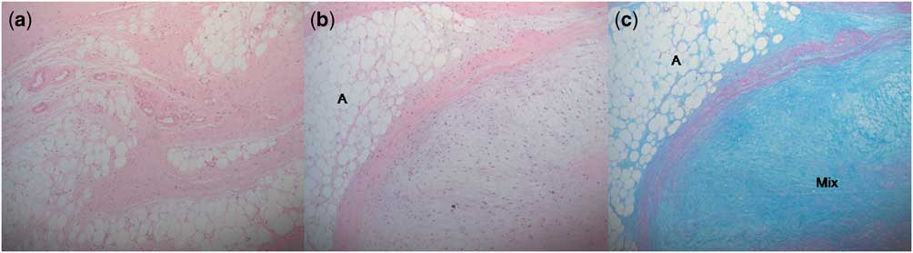
Microscopic features ( a ) lower magnification (H/E × 40). ( b ) Higher magnification highlighting mature adipose tissue aggregates [A] (H/E × 100). ( c ) Higher magnification showing both adipose tissue [A] and myxoid areas [Mix] (PAS−AB × 100).

The child recovered well and was discharged 9 days after surgery. The 1-year follow-up echocardiogram showed a well-functioning prosthesis, with no leak or stenosis and a normal cardiac function, with no recurrence of the mass.
Microscopically, the lesion consisted of abundant, disorganised aggregates of mature adipocytes separated by fibroelastic and myxoid tissue, containing scattered capillary vessels. The lesion was located inside the leaflets and there was no surrounding capsule (Fig 2). These pathological features are typical of a valvar lipomatous hamartoma.
Discussion
Barbeger et alReference Barberger-Gateau, Paquet, Desaulniers and Chenard 4 described the first lipomatous hamartoma in 1978 as a lesion composed of irregularly distributed mature fat and muscle cells that looks inside the left atrial cavity but had its origin in the mitral valve. Crotty et alReference Crotty, Edwards, Oh and Rodeheffer 5 reported the first review of literature about valvar hamartomas in 1991. In 2011, Karasu et alReference Karasu, Yeter, Yilmazer, Kiziltepe, Ulular and Akdemir 3 published a review with nine cases of lipomatous valvar hamartomas. The majority of these masses were located in the atrioventricular valves – five cases in the mitral, four in the tricuspid valve, and only one case in the aortic valve. In all, three cases had been described in children: one in the tricuspid valveReference Crotty, Edwards, Oh and Rodeheffer 5 and two cases in the mitral valveReference Barberger-Gateau, Paquet, Desaulniers and Chenard 4 , Reference Behnam, Williams, Gerlis, Walker and Scott 7 both located in the mural leaflet, as observed in our patient. The majority of the patients were asymptomatic and the tumour was detected incidentally at autopsy or during echocardiographic examination following an investigation of a heart murmur or a non-specific symptom. The symptoms reported were premature ventricular contractions, presyncope, and dyspnoea.Reference Barberger-Gateau, Paquet, Desaulniers and Chenard 4
In this case, transthoracic echocardiogram provided good definition of the lesion's shape, size, mobility, point of anchorage, and echodensity. The differential diagnosis, according to echocardiographic features, included a small number of neoplastic entities – metastatic lesions to valves and primary valvar neoplasms as papillary fibroelastoma, myxoma, fibroma, and fibrosarcoma – and non-neoplastic lesions – non-infective thrombotic vegetations, infective endocarditis, and congenital valvar cysts.Reference Dishop and O'Connor 8 All may produce echocardiographic features similar to those seen in the case reported. However, the well-defined, round-shaped mass with an apparent pedicle attachment to the mitral valve, prolapsing to the left ventricle, made the hypothesis of a benign tumour most likely.
Consistent with the previous reports of primary cardiac valve tumours, the majority of lesions were benign and confined to the valve.Reference Verliac and Morel 6 However, Anderson et alReference Anderson and Gray 9 and Jyrala et alReference Jyrala and Kay 10 described two cases of a benign cardiac tumour with extensive infiltration throughout the myocardium and subendocardium. No evidence of recurrence of benign tumours was observed in cases where follow-up was available after excision.Reference Behnam, Williams, Gerlis, Walker and Scott 7 , Reference Dishop and O'Connor 8 , Reference Jyrala and Kay 10
Less likely, primary malignant neoplasms of the cardiac valves are infiltrated by a large primary malignancy of the heart or pericardium.Reference Behnam, Williams, Gerlis, Walker and Scott 7 Metastatic neoplasms of the heart usually spread throughout the myocardium and pericardium as multiple small nodules, without valvar involvement.Reference Crotty, Edwards, Oh and Rodeheffer 5
The lesion described was composed of mature tissue that was native to the mitral valve, but present in abnormal quantities and arrangement. In addition, the lesion lacked some specific neoplastic features such as encapsulation and compression or invasion of adjacent structures. Neither did it have the appearance of an inflammatory or degenerative process. For these reasons, histological diagnosis is that of a hamartoma.
Despite the fact that the patient was asymptomatic, and the mass characteristics seemed to point to a benign origin, surgical excision was indicated given the risk of embolisation and the need for malignancy exclusion. Owing to the fact that uniform, well-defined operative protocols have not been established, even for papillary fibroelastoma,Reference Verliac and Morel 6 it is mandatory that immediate excision of valvar tumours be undertaken in order to prevent embolisation. Yet, the main goal of tumour excision is valve preservation. In our patient, resection of the valve was required because the tumour had grown and infiltrate both mural and aortic leaflets being incorporated inside the valve.
Lipomatous hamartoma of a cardiac valve is a very rare lesion, possibly a congenital malformation, mostly asymptomatic. Being an incidental diagnosis, it should be kept in mind when considering the differential diagnosis of valvar masses. The low cost and innocuous nature of echocardiography make it the technique of choice for early detection; however, the true nature of the mass and the final diagnosis can only be reached histologically. Despite the non-invasive course and benign morphological characteristics, these masses have high potential risk for haemodynamic compromise and sudden death. Surgical excision is the best approach for definitive diagnosis and effective treatment. However, owing to the rarity of cases, this tumoral entity requires strict follow-up.
Acknowledgements
The authors thank to the doctors Graça Nogueira, Miguel Abecasis and Manuela Nunes.
Financial Support
This research received no specific grant from any funding agency, commercial or not-for-profit sectors.
Conflicts of Interest
None.