Introduction
Chagas disease is an endemic parasitosis of the American continent caused by the protozoan Trypanosoma cruzi, which affects 6 million people worldwide (WHO, 2015). As a consequence of migration processes, nowadays, T. cruzi infection represents a health problem not only in the Americas but also in non-endemic countries (Rassi et al., Reference Rassi, Rassi and Marin-Neto2010).
The acute phase of T. cruzi infection is characterized by polyclonal B-cell activation, hypergammaglobulinaemia (Minoprio et al., Reference Minoprio, Burlen, Pereira, Guilbert, Andrade, Hontebeyrie-Joskowicz and Coutinho1998; Bryan et al., Reference Bryan, Guyach and Norris2010; Bermejo et al., Reference Bermejo, Amezcua Vesely, Khan, Acosta Rodríguez, Montes, Merino, Toellner, Mohr, Taylor, Cunningham and Gruppi2011) and a strong humoral response against antigenic motifs that masks key catalytic domains relevant to the infection process (Alvarez et al., Reference Alvarez, Leguizamón, Buscaglia, Pitcovsky and Campetella2001). Cumulative evidence has shown that T. cruzi expresses and sheds into the bloodstream immunomodulatory molecules which are involved in these events (Da Silva et al., Reference Da Silva, Espinoza, Taibi, Ouaissi and Minoprio1998; Leguizamón et al., Reference Leguizamón, Mocetti, Garcia Rivello, Argibay and Campetella1999; Reina-San-Martin et al., Reference Reina-San-Martin, Degrave, Rougeot, Cosson, Chamond, Cordeiro-Da-Silva, Arala-Chaves, Coutinho and Minoprio2000; Ouaissi et al., Reference Ouaissi, Guilvard, Delneste, Caron, Magistrelli, Herbault, Thieblemont and Jeannin2002). Among them, antigens belonging to the trans-sialidase (TS) superfamily have been demonstrated to participate in: (i) complement evasion (Beucher and Norris, Reference Beucher and Norris2008), (ii) B-lymphocyte proliferation (Gao et al., Reference Gao, Wortis and Pereira2002; Bermejo et al., Reference Bermejo, Jackson, Gorosito-Serran, Acosta Rodríguez, Amezcua Vesely, Sather, Singh, Khim, Mucci, Liggitt, Campetella, Oukka, Gruppi and Rawlings2013), (iii) apoptosis of immune cells (Leguizamón et al., Reference Leguizamón, Mocetti, Garcia Rivello, Argibay and Campetella1999; Mucci et al., Reference Mucci, Risso, Leguizamón, Frasch and Campetella2006), (iv) induction of non-neutralizing antibodies (Affranchino et al., Reference Affranchino, Ibañez, Luquetti, Rassi, Reyes, Macina, Aslund, Pettersson and Frasch1989; Burns et al., Reference Burns, Shreffler, Rosman, Sleath, March and Reed1992; Buscaglia et al., Reference Buscaglia, Campetella, Leguizamón and Frasch1998) and (v) evasion of protective immune responses through epitope variation (Wrightsman et al., Reference Wrightsman, Dawson, Fouts and Manning1994; Millar et al., Reference Millar, Wleklinski-Lee and Kahn1999; Martin et al., Reference Martin, Weatherly, Laucella, Cabinian, Crim, Sullivan, Heiges, Craven, Rosenberg, Collins, Sette, Postan and Tarleton2006). These actions favour the delay in the generation of specific immunity, thus allowing the parasite to spread and persist in the mammalian host (Cardoso et al., Reference Cardoso, Reis-Cunha and Bartholomeu2016).
The TS superfamily is the largest T. cruzi gene family, encoding more than 1400 genes classified into eight groups (Freitas et al., Reference Freitas, dos Santos, Rodrigues-Luiz, Mendes, Rodrigues, Gazzinelli, Teixeira, Fujiwara and Bartholomeu2011). It is worth noting that only group I contains active trans-sialidases (TcTSs), which transfer sialic residues from host sialoconjugates to the parasite cell surface (Mucci et al., Reference Mucci, Lantos, Buscaglia, Leguizamón and Campetella2017). While TcTSs contain a Tyr342 which is crucial in the enzyme activity, all the other members of the TS superfamily show a Tyr342His replacement (Cremona et al., Reference Cremona, Sánchez, Frasch and Campetella1995). These enzymatically inactive proteins (iTcTSs) retain a lectin-like activity (Cremona et al., Reference Cremona, Campetella, Sanchez and Frasch1999; Todeschini et al., Reference Todeschini, Girard, Wieruszeski, Nunes, DosReis, Mendonça-Previato and Previato2002a) and it has been hypothesized that, in natural infection, they act mainly as virulence factors (Burgos et al., Reference Burgos, Risso, Brenière, Barnabé, Campetella and Leguizamón2013). Tc13 antigens, which are members of group IV of the TS superfamily, have not been largely studied so far. They are characterized by bearing five amino-acid (EPKSA) repeats at their C-terminal region (Campetella et al., Reference Campetella, Sánchez, Cazzulo and Frasch1992), which elicit a strong humoral response in T. cruzi-infected patients and mice, especially in the acute phase of the infection (Burns et al., Reference Burns, Shreffler, Rosman, Sleath, March and Reed1992; Peralta et al., Reference Peralta, Teixeira, Shreffler, Pereira, Burns, Sleath and Reed1994; García et al., Reference García, Arnaiz, Esteva, Laucella, Garavaglia, Ibarra and Ruiz2008; Santamaría et al., Reference Santamaría, De Rissio, Riarte, Garavaglia, Bruballa, Rodríguez, Irazu, Ruiz and García2013). Genetic and recombinant immunizations of BALB/c mice with the Tc13 antigen of the Tulahuén strain of T. cruzi (Tc13Tul) have shown that Tc13-specific immunity does not confer protection against T. cruzi infection (García et al., Reference García, Arnaiz, Laucella, Esteva, Ainciart, Riarte, Garavaglia, Fichera and Ruiz2006; García et al., Reference García, Arnaiz, Esteva, Laucella, Garavaglia, Ibarra and Ruiz2008). Moreover, the high production of non-protective anti-EPKSA IgG in the acute phase of the infection, the presence of a weak anti-Tc13 memory T-cell response and the lack of Tc13-specific CD4+ T cells with an ability to release interferon-γ (IFN-γ) in the chronic phase of the infection suggest that Tc13 antigens are involved in mechanisms of evasion from host immunity (García et al., Reference García, Arnaiz, Esteva, Laucella, Garavaglia, Ibarra and Ruiz2008). To test this hypothesis and obtain further information about primary immune responses triggered by Tc13 antigens on B and T lymphocytes, the aim of the present study was to evaluate the effect of Tc13Tul on the viability and lymphoproliferation of splenocytes from naïve mice in vitro. We also aimed to study Tc13Tul ability to induce polyclonal Ig production and cytokine secretion by in vitro and in vivo stimulation of these cells.
Materials and methods
Ethical statement and mice
Animal studies were carried out with the approval of the Institutional Ethical Committee and conformed to the ethical treatment of animals established by the Argentinean Animal Protection Society. Six-week-old male naïve mice were used in all the experiments. BALB/c mice were maintained at the animal facility of INP ‘Dr. Fatala Chaben’ in ventilated plastic cages with free access to tap water and enough rodent food. C3H/HeJ mice were kindly provided by Dr Oscar Campetella from Instituto de Investigaciones Biotecnológicas-Universidad de San Martín (IIB-UNSAM), Buenos Aires, Argentina. For euthanasia, mice were subjected to cervical dislocation under anaesthesia with 100 μL of a mixture of ketamine (50 mg kg−1; Ketafine, Brouwer, CABA, Argentina) and xylazine (5 mg kg−1; Kensol, König, Avellaneda, BA, Argentina) delivered by intraperitoneal (i.p.) injection.
Recombinant proteins
Tc13Tul protein (GenBank Accession no. AF091620) was expressed as a maltose-binding protein (MBP) fusion, purified by amylose resin and filtrated with a 100 kDa MW cut-off centrifugal filter device (Centricon®, Millipore, Darmstadt, Germany) (García et al., Reference García, Joensen, Bua, Ainciart, Perry and Ruiz2003). The clone coding for EPKSA (GenBank Accession no. M92046) repeats fused to glutathione-S-transferase (GST) was kindly provided by Dr O. Campetella (IIB-UNSAM). The GST-EPKSA recombinant protein was purified by glutathione-agarose resin. The homogeneity of the proteins was evaluated by sodium dodecyl sulfate-7.5% polyacrylamide gel electrophoresis (SDS-7.5% PAGE).
In vitro culture of splenocytes
Mouse splenocytes depleted of erythrocytes were seeded in 96-well culture plates at 4 × 10⁵ cells per well in 200 μL of RPMI-10% fetal bovine serum (FBS) in the presence of either Tc13Tul (6 μg per106 cells), MBP (2.2 μg per106 cells), EPKSA (6 μg per 106 cells) or GST (1.5 μg per 106 cells) and incubated at 37°C with 5% CO2 for 24–72 h depending on each experiment conditions. For Toll-like receptor 4 (TLR4) activation assays, lipopolysaccharide (LPS) (5 μg mL-1) (Sigma-Aldrich, Saint Louis, MO, USA) and colistin (10 μg mL−1) (Alficetin, Argentia, CABA, Argentina) were used.
Cell viability evaluation
Cell viability was assessed by staining with trypan blue or propidium iodide (PI). Trypan blue staining was evaluated by optical microscopy counting non-stained cells (live) in a Neubauer camera and the percentage of live cells was calculated considering the initial cell inoculum of 2 × 10⁶ cells mL-1 as 100%. PI staining was analysed by flow cytometry and the percentage of live cells (PI negative) was calculated considering the total of 20 000 events acquired as 100%.
Flow cytometry acquisition and analysis
Splenocytes were acquired with a BD FACS Calibur flow cytometer (BD Biosciences, Franklin Lakes, NJ, USA) and analysed with FlowJo software (Tree Star, Ashland, OR, USA). A total of 20 000 events were acquired within a primary gating based on cell scatter properties. Lymphocytes were gated based on their forward and side scattering parameters (FSC-H vs SSC-H). The singlets were analysed by the use of forward scatter area vs forward scatter height dot-plot (FSC-A vs FSC-H) and the frequencies (%) of CD19+ and CD3+ were analysed with specific antibodies (CD19-phycoerythrin (PE), CD19-allophycocyanine (APC) or CD3-PE vs FSC-H) (Supplementary Fig. S1). Unstained samples and single-stain samples were used as controls for subsequent software compensation using the FlowJo analysis software compensation module. Carboxyfluorescein diacetate succinimidyl ester (CFSE) proliferation assays were analysed with the FlowJo Proliferation Tool which finds the original population (100% of max CFSE), models the division by looking for peaks with diminishing fluorescence and calculates the percentage of divided cells.
Annexin V and propidium iodide labelling
After 48 h of in vitro stimulation, splenocytes were washed, resuspended in RPMI and were double-stained for annexin V (AV)-fluorescein isothiocyanate (FITC) and PI using the ‘Annexin-V Apoptosis Detection Kit I’ (BD Pharmingen™, San Diego, CA, USA) according to the manufacturer's instructions. Incubation of cells in the presence of phytohaemagglutinin (PHA) (1.25 μg per 106 cells) was used as controls. Stained cells were analysed by flow cytometry.
CFSE proliferation and lymphocyte phenotyping assays
To track cell division, prior to in vitro culture, splenocytes were stained with 2.5 μ m of CFSE at 37°C for 10 min, using the CellTrace™ CFSE Cell Proliferation Kit (Life Technologies, Eugene, OR, USA). CD3 cross-linking was used as a positive control for T-cell proliferation. For this control, anti-CD3 monoclonal antibody (Purified Hamster anti-mouse CD3e clone145-2C11, BD Pharmingen™) was adsorbed to the culture plate (10 μg mL−1) for 1 h at room temperature (RT) before the addition of splenocytes. After 48–72 h of in vitro stimulation, cells were collected by centrifugation and washed with PBS 5% FBS. Then, cells were stained for 30 min on ice with anti-mouse CD19-APC and anti-mouse CD3-PE (BD Pharmingen™) or anti-mouse CD19-PE (Thermo Fisher, Rockford, IL, USA), washed with PBS 5% FBS and fixed with 3.6% formaldehyde for 15 min at RT. Fixed cells were analysed by flow cytometry.
Measurement of Ig secretion
Total Ig and IgM were measured in supernatants of in vitro stimulated splenocytes and sera of inoculated mice by sandwich Enzyme-Linked ImmunoSorbent Assay (ELISA). Ninety-six-well flat bottomed plates were coated with goat anti-mouse IgG (which also reacts with the light chains of IgM and IgA) (Zymed Laboratories Inc., South San Francisco, CA, USA) 5 μg mL−1 at 4°C overnight. After blocking with 5% skimmed milk in PBS, plates were incubated for 1 h at RT with samples (50 μL): supernatants in 2-fold serial dilutions from 1:16 to 1:128 and sera diluted 1:105. The mouse monoclonal antibody 2C5D6 (Bontempi et al., Reference Bontempi, García, Buschiazzo, Henriksson, Pravia, Ruiz, Pettersson and Pszenny2000) was used as quantification standard. Biotinylated goat anti-mouse IgG and biotinylated goat anti-mouse IgM followed by incubation with streptavidin peroxidase (Vector Labs, Burlingame, CA, USA) were used to detect total Ig and IgM, respectively. Colour was developed with o-phenylenediaminedihydrochloride and optical density was read at 490 nm using an ELISA microplate reader BioTek EL 808 (BioTek Instruments, Winooski, VT, USA).
For detecting anti-Tc13Tul and MBP-specific antibodies, plates were coated with MBP or Tc13Tul 5 μg mL−1 (50 μL) at 4°C overnight and the above protocol was followed from the blocking step onwards. Supernatants and sera were tested in a dilution of 1:2 and 1:100, respectively.
Measurement of cytokine secretion
Supernatants of stimulated splenocytes were assayed for cytokine secretion by sandwich ELISA using the OptEIA™ Set for mouse interleukin 4 (IL-4) and IFN-γ (BD Biosciences, San Jose, CA, USA) and the mouse IL-17A (homodimer) ELISA Ready-SET-Go!® (e-Biosciences, San Diego, CA, USA) according to the manufacturer's instructions.
Tc13Tul binding assays to splenocytes
Splenocytes were incubated for 30 min at 4°C with FITC-conjugated Tc13Tul or MBP (50 μg mL−1). After binding assay, lymphocyte phenotyping was performed by staining with anti-mouse CD19-PE (Thermo Fisher) and anti-mouse CD3-PE (BD Pharmingen™). Finally, cells were washed with PBS 5% FBS and fixed with 3.6% formaldehyde for 15 min at RT. Fixed cells were analysed by flow cytometry.
Studies of Tc13Tul effects in vivo
Six-week-old male naïve BALB/c mice were inoculated intraperitoneally (i.p.) with a daily dose of 1 μg per mouse per dose in PBS of Tc13Tul or MBP for 3 days. A control group was injected with the vehicle PBS. Sera were collected prior to inoculation and on days 4, 8 and 11 after the first inoculation from the facial vein. Eleven days after the first inoculation, mice were sacrificed and splenocytes were collected to be cultured in vitro.
Statistical analysis
Data are expressed as means + standard error (s.e.) and were derived from at least duplicate observations per experiment. All experiments were repeated at least twice. Differences among groups were evaluated by one- or two-way analysis of variance (ANOVA), as appropriate, followed by Bonferroni as post-test. Data were analysed using GraphPad Prism 5.0 software (Graph Prism, San Diego, CA, USA) and differences were considered significant when the P value was <0.05.
Results
Tc13Tul increases the viability of in vitro cultured splenocytes from BALB/c naïve mice and affects their lymphocyte population
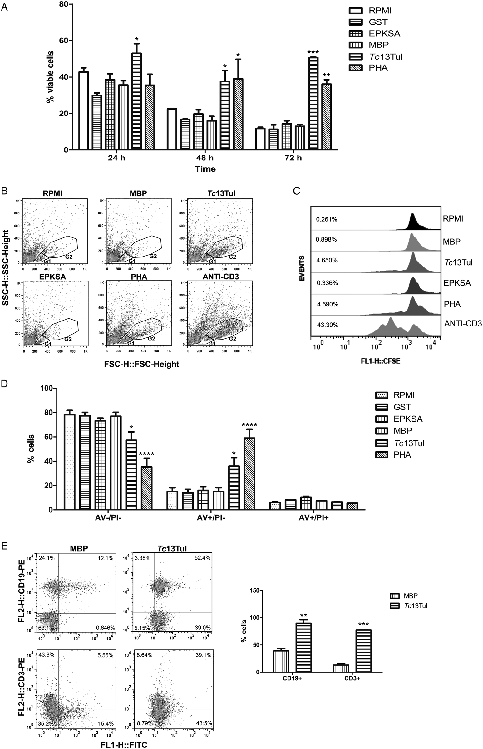
To evaluate primary immune responses triggered by Tc13Tul, primary splenocytes from naïve BALB/c mice were incubated for 24, 48 and 72 h with recombinant Tc13Tul or EPKSA repeats and cell viability was assessed by the exclusion of trypan blue staining. As Tc13Tul and EPKSA are fused to MBP and GST, respectively, incubation with these carrier proteins, in equivalent amounts to those present in the respective fusion proteins, was used to rule out their basal effects. Splenocytes cultured in the absence of stimulus (RPMI) and in the presence of the mitogen compound PHA were used as negative and positive controls, respectively. Splenocyte viability decreases over time because lymphocytes are incapable of dividing unless they are stimulated by a mitogen (Sharon and Lis, Reference Sharon and Lis2004). Similarly to PHA, incubation with Tc13Tul showed a higher number of viable cells than controls. The effect of Tc13Tul was observed as early as 24 h post-stimulation. Incubation with EPKSA repeats yielded a percentage of viable cells similar to that obtained with controls. The lack of effect with the EPKSA C-terminal portion of Tc13Tul suggests that the region responsible for increasing splenocyte viability may be located at the N-terminal segment of the protein (Fig. 1A). These findings were confirmed by PI staining analysed by flow cytometry (Supplementary Fig. S2).
Effect of Tc13Tul on the viability of in vitro cultured splenocytes from naïve BALB/c mice and their lymphocyte population. Splenocytes were cultured for the indicated times with Tc13Tul (6 μg per106 cells), EPKSA (6 μg per106 cells) or the equivalent amounts of their respective carrier proteins, MBP (2.2 μg per 106 cells) and GST (1.5 μg per 106 cells). Cells cultured in the absence of stimulus (RPMI) and in the presence of PHA (1.25 μg per 106 cells) or anti-CD3 monoclonal antibody (10 μg ml−1) were used as controls. (A) Surviving cells were evaluated by the trypan blue exclusion assay and analysed by optical microscopy. The initial cell inoculum of 2 × 10⁶ cells mL−1 was considered as 100% of viable cells. (B) Splenocytes stimulated with the antigens for 72 h were analysed by flow cytometry. Lymphocytes were gated based on forward scattering (FSC-H) and side scattering (SSC-H). Populations of basal (G1) and enlarged (G2) lymphocytes are indicated in the dot plots. (C) Splenocytes were stained with CFSE prior to stimulation, stimulated with antigens for 72 h and analysed by flow cytometry. Histograms of CFSE-stained cells in the gated population G1 + G2 showing the percentages of divided cells calculated with the FlowJo Proliferation Tool. (D) After 48 h of stimulation, splenocytes were double-stained with annexin V-FITC (AV) and propidium iodide (PI) and analysed by flow cytometry in the gated population G1 + G2. (E) Splenocytes from naïve BALB/c mice were incubated with FITC-Tc13Tul or FITC-MBP, subsequently stained with anti-CD19-PE or anti-CD3-PE and analysed by flow cytometry gated on lymphocyte population. Dot plots (CD19+ and CD3+) of FITC-stained lymphocytes and their quantification are shown. Representative data of at least two independent experiments are shown. Data are the means + s.e. Significance was determined by two-way ANOVA and Bonferroni as post-test. *P < 0.05, **P < 0.01, ***P < 0.001 and ****P < 0.0001 respect to negative controls (RPMI, MBP and/or GST).

To determine whether the stimulation of splenocytes with Tc13Tul induced changes in lymphocyte populations, cells stimulated for 72 h were subjected to flow cytometry. The analysis of side (SSC-H) vs forward (FSC-H) scatter dot plots showed that stimulation with Tc13Tul induced a lymphocyte population of increased size and granularity (named G2), which was similar to that produced by the mitogen PHA and a CD3-specific cross-linking antibody. On the other hand, unstimulated cells or cells treated with MBP or EPKSA repeats only showed the basal lymphocyte population (named G1) (Fig. 1B). To investigate whether the G2 population was due to a lymphoproliferative effect of Tc13Tul, cellular proliferation of stimulated splenocytes was monitored by CFSE staining. The flow cytometry analysis of CFSE-labelled cells indicated cellular proliferation after stimulation with Tc13Tul but not with EPKSA repeats or the carrier proteins. The percentage of divided cells induced by Tc13Tul was significantly higher than that induced by MBP and the EPKSA repeats (Fig. 1C). Next, to evaluate the ability of Tc13Tul to induce apoptosis, stimulated splenocytes were double-stained with PI and AV and analysed by flow cytometry. The analysis of quadrants AV–/PI–, AV+/PI– and AV+/PI+, performed on the lymphocyte population (G1 + G2), showed that stimulation with Tc13Tul, like PHA, induced an increase in the number of cells in early apoptosis (AV+/PI−) at the expense of a decrease in viable cells (AV–/PI–). According to previous results, EPKSA repeats failed to show this effect (Fig. 1D). These findings suggest that the increase in cell viability induced by Tc13Tul observed by trypan blue and PI staining may correspond to viable and early apoptotic cells.
Given the evidence that Tc13Tul acts on the lymphocyte population, we then studied whether this antigen is able to interact with the lymphocyte surface. With this aim, binding of FITC-labelled Tc13Tul or MBP to these cells was evaluated by flow cytometry. Results showed a 2.6-fold higher labelling in the lymphocyte population with Tc13Tul-FITC than in that with MBP-FITC. To identify which lymphocyte population Tc13Tul binds to, bound cells were dyed for markers specific to B (CD19) and T (CD3) cells. Tc13Tul-FITC bound to 96.06% of CD19+ cells and to 78.73% of CD3+ cells, while the control protein MBP-FITC bound to 34.6% of CD19+ and to 11.1% of CD3+ cells (Fig. 1E). These results suggest that Tc13Tul interacts with surface antigens on both B and T cells.
Tc13Tul induces B cell proliferation, non-specific IgM and IFN-γ production in cultured splenocytes from naïve mice
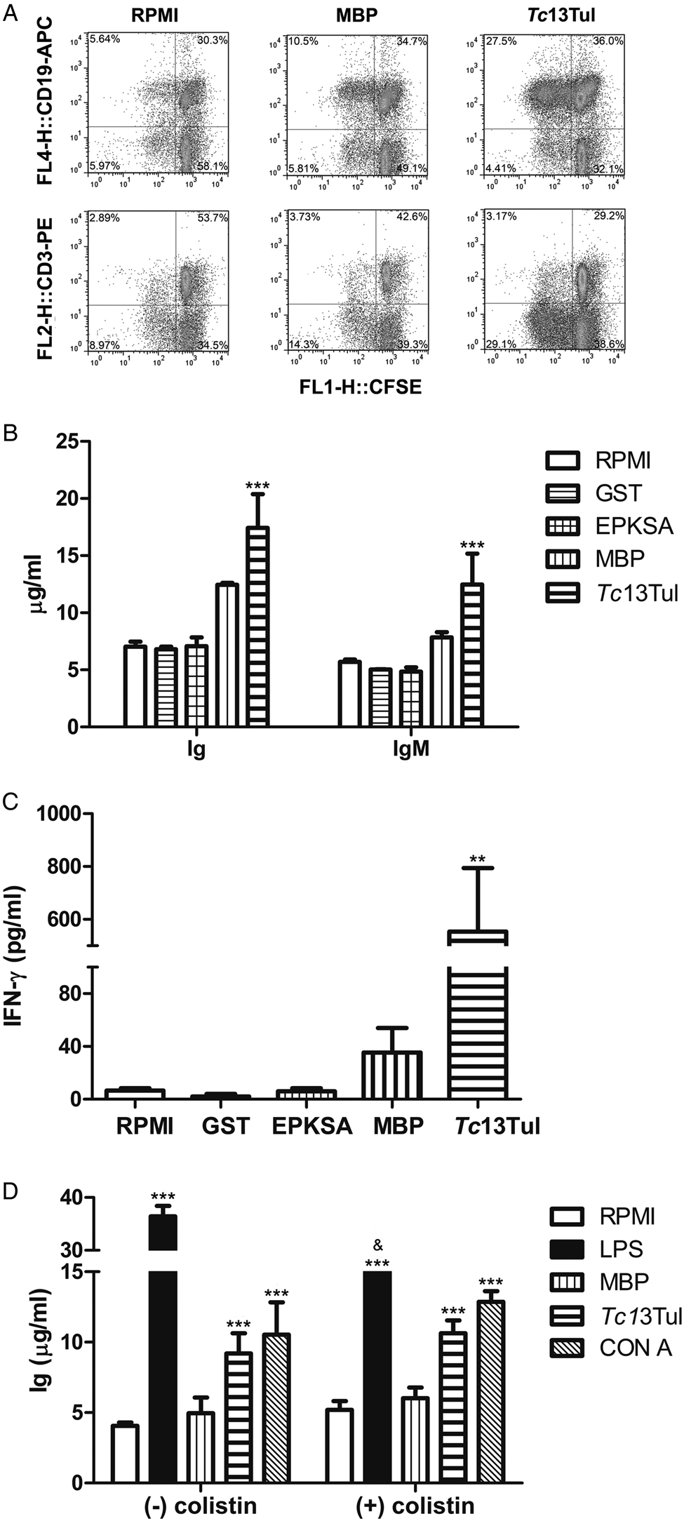
To characterize the lymphoproliferative effect of Tc13Tul, CFSE-labelled and stimulated splenocytes were phenotyped. The CFSE profile showed that Tc13Tul induced proliferation of B cells (CD19+) but not that of T cells (CD3+). Cross-linking with an anti-CD3 monoclonal antibody was used as a positive control for T-cell proliferation, which confirms the ability of these cells to divide when an appropriate stimulus is used. In contrast, MBP stimulation showed no significant differences respect to unstimulated cells (Fig. 2A and Supplementary Fig. S3).
Effect of Tc13Tul on lymphocyte proliferation, polyclonal Ig and IFN-γ secretion in cultured splenocytes from naïve BALB/c mice. Splenocytes were stained with CFSE and cultured for 72 h with Tc13Tul (6 μg per 106 cells), MBP (2.2 μg per 106 cells) or medium (RPMI). After stimulation, cells were stained with anti-CD19-APC and anti-CD3-PE and analysed by flow cytometry. (A) Dot plots showing double-stained (CFSE/CD19-APC and CFSE/CD3-PE) cells on lymphocyte gate. (B) Total Ig and IgM secretion evaluated by ELISA in splenocyte supernatants. (C) IFN-γ secretion evaluated by ELISA in splenocyte supernatants. **P < 0.01 and ***P < 0.001 respect to negative controls (RPMI, MBP and/or GST). (D) Total Ig secretion evaluated by ELISA in supernatants of splenocytes induced for 72 h with the antigens (MBP or Tc13Tul), lipopolysaccharide (LPS) (5 μg mL−1) or concanavalin A (CON A) (10 μg mL−1) in the absence or presence of colistin (10 μg mL−1), an agent which interferes with LPS activity. Representative data of at least two independent experiments are shown. Data are the means + s.e. Significance was determined by ANOVA and Bonferroni as post-test. ***P < 0.001 respect to negative controls (RPMI, MBP and/or GST) and &, P < 0.01 compared with the absence of colistin.

Since Tc13Tul stimulation induced B-lymphocyte proliferation, we next evaluated whether this effect occurred concomitantly with either non-specific or Tc13Tul-specific Ig secretion. Supernatants of Tc13Tul-stimulated splenocytes showed higher total non-specific Ig levels than those of MBP-, EPKSA-, GST- or non-stimulated cells. Non-specific IgM levels were slightly lower than total Ig levels, indicating that these recombinant antigens mainly induced this Ig isotype (Fig. 2B). Tc13Tul-induced total Ig did not react with Tc13Tul and MBP by ELISA (Supplementary Fig. S4), demonstrating that these antibodies are not specific for these antigens.
To study the cytokine profile induced by Tc13Tul, the levels of IFN-γ (Th1), IL-4 (Th2) and IL-17 (Th17) were measured in the supernatants of Tc13Tul-, MBP- and non-induced splenocytes. Neither IL-17 nor IL-4 were detected in the supernatants from MBP- and Tc13Tul-stimulated cells (Supplementary Fig. S5). In contrast, significant IFN-γ production was detected in the supernatants of Tc13Tul-induced splenocytes. No increase in IFN-γ was observed when cells were stimulated with recombinant EPKSA repeats (Fig. 2C).
To rule out the possibility that the increase in non-specific Ig is due to potential endotoxin (LPS) contamination in the Tc13Tul preparation, splenocytes were treated with antigens in the absence or presence of colistin, an agent which disrupts bacterial LPS. Colistin affected neither MBP- nor Tc13Tul-induced Ig, indicating that the level of endotoxin in these recombinant proteins has no impact on this effect. As expected, colistin neither had an effect on concanavalin A-induced Ig secretion, but significantly diminished the total Ig induced by LPS (Fig. 2D).
Tc13Tul stimulation is also observed in cultured naïve splenocytes from the LPS-resistant C3h/HeJ mouse strain
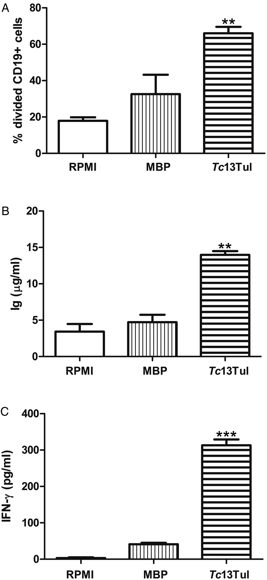
The effect of Tc13Tul on the proliferation of B cells and secretion of non-specific Ig and IFN-γ was also observed in splenocytes from naïve C3H/HeJ mice (Fig. 3). The particularity of the C3H/HeJ mouse strain is a mutation in the TLR-4, which makes it very resistant to LPS stimulation (Poltorak et al., Reference Poltorak, He, Smirnova, Liu, Van Huffel, Du, Birdwell, Alejos, Silva, Galanos, Freudenberg, Ricciardi-Castagnoli, Layton and Beutler1998). Therefore, these results indicate that these effects are mediated by neither TLR-4 nor potential LPS contamination of recombinant proteins.
Effect of Tc13Tul in cultured naïve splenocytes from the LPS-resistant C3H/HeJ mouse strain. Splenocytes from naïve C3H/HeJ mice were stained with CFSE and cultured for 72 h with Tc13Tul (6 μg per 106 cells), MBP (2.2 μg per 106 cells) or medium (RPMI). (A) After stimulation, cells were stained with anti-CD19-PE and proliferation of CD19+ cells was analysed by flow cytometry on lymphocyte gate. The percentage of divided cells was calculated with the FlowJo Proliferation Tool. (B) Total Ig and (C) IFN-γ evaluated in splenocyte supernatants by ELISA. Representative data of at least two independent experiments are shown. Data are the means + s.e. Significance was determined by one-way ANOVA and Bonferroni as post-test. **P < 0.01 and ***P < 0.001 respect to RPMI and MBP.

In vivo administration of Tc13Tul to naïve BALB/c mice induces polyclonal Ig and IFN-γ secretion
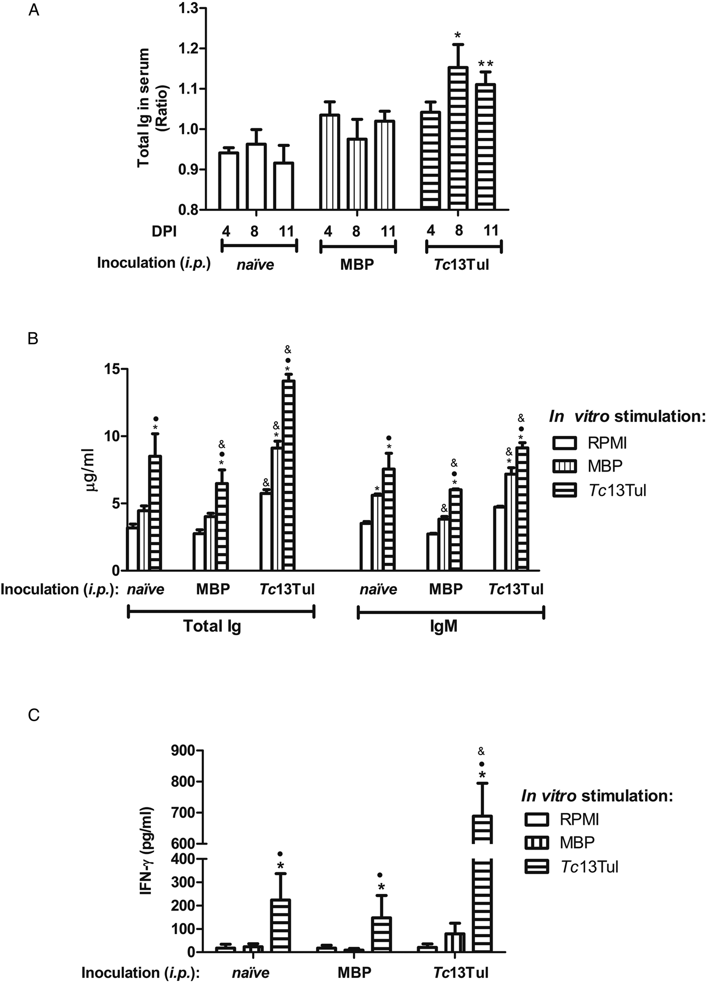
To evaluate whether the effects of Tc13Tul observed in vitro are also relevant in vivo, BALB/c mice were inoculated with three doses of Tc13Tul (or MBP as control) and total Ig and IgM levels were evaluated in sera on days 4, 8 and 11 after the first inoculation. Sera of the Tc13Tul-inoculated group showed increased levels of total Ig from day 8 post-inoculation (Fig. 4A). IgM levels were low and remained constant throughout the measurements (Supplementary Fig. S6), suggesting that in vivo Tc13Tul stimulation may induce mainly Igs of the IgG isotype.
Effect of Tc13Tul after in vivo administration to naïve BALB/c mice. (A) Total Ig levels evaluated by ELISA in mouse sera injected with buffer (naïve) and inoculated with MBP or Tc13Tul (a daily i.p. dose of 1 μg/mouse for 3 days). Sera were collected for evaluation prior to the injection and 4, 8 and 11 days post-injection (DPI). Individually for each mouse (n = 3), the ratio between Ig amounts in serum at the indicated days and their amounts prior to antigen injection was calculated. *P < 0.05 and **P < 0.01 respect to the naïve group. (B) Total Ig and IgM and (C) IFN-γ levels in supernatants of pooled splenocytes from naïve, MBP- and Tc13Tul-inoculated mice cultured in vitro for 72 h without stimulation (RPMI) or stimulated with MBP or Tc13Tul. * and •, P < 0.05 respect to in vitro cultured splenocytes without stimulation and stimulated with MBP, respectively. &, P < 0.05 respect to splenocytes from naïve mice. Data represent the means + s.e. Significance was determined by two-way ANOVA and Bonferroni as post-test.

Eleven days after the inoculation, mice were sacrificed and their splenocytes were cultured in vitro for 72 h without stimulus (RPMI) or in the presence of MBP or Tc13Tul. Basal Ig secretion of splenocytes from Tc13Tul-inoculated mice was higher than that of splenocytes from control or MBP-inoculated animals (Fig. 4B white bars). Splenocytes from MBP-inoculated mice increased Ig secretion only when they were in vitro stimulated with Tc13Tul; however, Tc13Tul stimulation was not as high as that exerted on naïve splenocytes. This observation allowed us to speculate that the in vivo administration of MBP might have an inhibitory effect on B cells. Conversely, splenocytes from Tc13Tul-inoculated mice and in vitro stimulated with Tc13Tul secreted higher Ig levels than splenocytes from naïve mice (Fig. 4B horizontal hatched bars), indicating an additive effect of in vivo and in vitro Tc13Tul administration. Splenocytes from Tc13Tul-inoculated mice also increased Ig secretion when they were in vitro stimulated with MBP (Fig. 4B vertical hatched bars). This suggests that Tc13Tul administered in vivo may have induced B-cell activation, and therefore, these cells were more reactive when in vitro stimulated also with the carrier protein MBP. The lack of Ig secretion by splenocytes from MBP-inoculated mice and in vitro stimulated with MBP validates that the one responsible for the stimulation of Ig production is the Tc13Tul portion of the recombinant protein, and not the carrier protein. Although the evaluation of polyclonal IgM secretion showed a profile similar to that of total Ig, basal IgM secretion in Tc13Tul-inoculated mice was not higher than that in control groups, suggesting that Tc13Tul administered in vivo might have induced IgG rather than IgM secretion by splenocytes (Fig. 4B). It is worth mentioning that neither Tc13Tul- nor MBP-specific antibodies were detected in sera or supernatants (Supplementary Figs S6 and S7). IFN-γ production was only observed when splenocytes were in vitro stimulated with Tc13Tul. However, splenocytes from Tc13Tul-inoculated mice secreted higher IFN-γ levels than splenocytes from naïve and MBP-inoculated mice, indicating that the in vivo Tc13Tul administration enhanced the effect of the in vitro Tc13Tul stimulation (Fig. 4C horizontal hatched bars).
Discussion
The spleen is an important lymphoid organ that produces most of the non-specific immune responses characteristic of the acute phase of T. cruzi infection (Bermejo et al., Reference Bermejo, Amezcua Vesely, Khan, Acosta Rodríguez, Montes, Merino, Toellner, Mohr, Taylor, Cunningham and Gruppi2011). Several authors have documented the involvement of the TS group I member TcTS in triggering innate and innate-like responses in this organ (Gao et al., Reference Gao, Wortis and Pereira2002; Todeschini et al., Reference Todeschini, Nunes, Pires, Lopes, Previato, Mendonça-Previato and DosReis2002b; Bermejo et al., Reference Bermejo, Jackson, Gorosito-Serran, Acosta Rodríguez, Amezcua Vesely, Sather, Singh, Khim, Mucci, Liggitt, Campetella, Oukka, Gruppi and Rawlings2013). Our previous researches on Tc13 antigens in a BALB/c experimental model were focused on memory Tc13-specific immune responses. Studies of humoral and cellular responses induced by these antigens, either in the course of T. cruzi infection or as a result of genetic and recombinant immunization with Tc13Tul antigen, suggest that the Tc13 family protein triggers mainly non-protective memory immunity (García et al., Reference García, Arnaiz, Laucella, Esteva, Ainciart, Riarte, Garavaglia, Fichera and Ruiz2006, Reference García, Arnaiz, Esteva, Laucella, Garavaglia, Ibarra and Ruiz2008). Therefore, the aim of the present work was to study primary immune responses induced by Tc13 antigens by evaluating their effect on splenocytes from naïve mice.
All the experiments were performed with recombinant Tc13Tul antigen, which is fused to MBP from E. coli. As MBP and other bacterial components have effects on innate immunity (Akira et al., Reference Akira, Uematsu and Takeuchi2006; Liu et al., Reference Liu, Zhang, Zhang, Ni, Jie, Jiang and Tai2017), special controls were used to avoid wrong conclusions. Purified Tc13Tul was subjected to a further filtration step to eliminate possible low-molecular-weight contaminants. MBP induced from the wild-type pMalp2 vector and purified in the same conditions as that used for recombinant Tc13Tul purification was thoroughly used to rule out the effect of the carrier protein. To rule out the effect of contaminant LPS: (i) control assays were performed in the presence of colistin, a cationic polypeptide which binds and neutralizes the LPS molecule (Gupta et al., Reference Gupta, Govil, Kakar, Prakash, Arora, Das, Govil and Malhotra2009) and (ii) in vitro Tc13Tul stimulation was also evaluated in splenocytes from naïve C3H/HeJ mice, a mouse strain resistant to LPS activation (Poltorak et al., Reference Poltorak, He, Smirnova, Liu, Van Huffel, Du, Birdwell, Alejos, Silva, Galanos, Freudenberg, Ricciardi-Castagnoli, Layton and Beutler1998). Neither of these strategies abrogated the effects observed, supporting that they are Tc13Tul-induced.
Tc13Tul stimulation of naïve splenocytes showed mitogenic-like effects: it increased cell viability and, in the lymphocyte population, induced proliferation as well as apoptosis. These effects were even stronger than those induced by the mitogen PHA, a lectin that binds to lymphocyte membranes (Sharon and Lis, Reference Sharon and Lis2004). Therefore, Tc13Tul, like PHA, induced a net increase in cell number as a result of the lymphoproliferative effect. However, after 48 h of stimulation, the AV/PI analysis demonstrated that a great percentage of these cells had initiated the apoptotic process. Although cell viability and apoptosis are seemingly contradictory events, it should be taken into account that early apoptotic cells (AV+/PI−) still maintain the integrity of the plasma membrane and, therefore, are detected as viable by techniques using dyes that are excluded from viable cells, such as trypan blue and PI. It is important to note that although the definition of the term mitogen comes from its ability to stimulate lymphocyte mitosis (Nowell et al., Reference Nowell1960), it is known that mitogenic stimulation also leads to Fas/FasL-mediated apoptosis, thus limiting cell expansion (Miyawaki et al., Reference Miyawaki, Uehara, Nibu, Tsuji, Yachie, Yonehara and Taniguchi1992; Tu et al., Reference Tu, Cheung and Lau2000).
In vitro stimulation with Tc13Tul of splenocytes from naïve BALB/c and C3H/HeJ mice induced B-cell proliferation and polyclonal IgM secretion. IgM production by Tc13Tul in vitro stimulation is consistent with the low frequency of switching from IgM to IgG secretion detected in single B-cell clones after treatment with mitogenic compounds (Andersson et al., Reference Andersson, Coutinho and Melchers1978). These effects were not observed when cells were stimulated with recombinant EPKSA repeats, suggesting that C-terminal repeats are not involved in these events. However, as the recombinant EPKSA used in this study belongs to the CAI strain of T. cruzi and has some variations respect to the EPKSA portion of Tc13Tul (García et al., Reference García, Joensen, Bua, Ainciart, Perry and Ruiz2003), it cannot be ruled out that the variations in the EPKSA units present in Tc13Tul are responsible for these effects and/or that the EPKSA portion as part of the whole Tc13 molecule has a different conformation that favours its activity. Similar studies using TcTS have indicated that the polyclonal B-cell mitogenic activity is present in its C-terminal SAPA repeats rather than in its N-terminal domain (Gao et al., Reference Gao, Wortis and Pereira2002). These results indicate that SAPA and EPKSA repeats might diverge in their role in early immunity, although they share other features such as immunodominance and the ability to increase the half-life of proteins in blood (Burns et al., Reference Burns, Shreffler, Rosman, Sleath, March and Reed1992; Buscaglia et al., Reference Buscaglia, Alfonso, Campetella and Frasch1999).
Tc13Tul also induced polyclonal non-specific Ig stimulation by in vivo administration. It is worth mentioning that, based on previous studies on TcTS (Da Silva et al., Reference Da Silva, Espinoza, Taibi, Ouaissi and Minoprio1998; Gao et al., Reference Gao, Wortis and Pereira2002; Aridgides et al., Reference Aridgides, Salvador and Pereira Perrin2013; Salvador et al., Reference Salvador, Aridgides and Pereira Perrin2014), in vivo administration of Tc13Tul was carried out in the absence of any adjuvant that could mask or potentiate its effect. Tc13Tul-administered mice showed increased non-specific IgG in sera from day 8 post-inoculation and increased total Ig secretion when their splenocytes were in vitro cultured for 72 h. Neither of these antibodies showed specificity for Tc13Tul or MBP. Other T. cruzi antigens also failed to induce specific antibodies when administered to mice with similar protocols (Da Silva et al., Reference Da Silva, Espinoza, Taibi, Ouaissi and Minoprio1998; Gao et al., Reference Gao, Wortis and Pereira2002). In these studies, the lack of specific responses was ascribed to the absence of adjuvant and insufficient time to trigger antigen-specific antibodies through T–B-cell interaction. These explanations can also be applied to the non-specific effect observed with Tc13Tul and MBP in vivo administration. In the spleen, B-1 and marginal zone B cells have shown to be the main source of T-independent production of IgM antibodies in the early stage of the immune response, although they are also able to undergo IgG class switching (Martin et al., Reference Martin, Oliver and Kearney2001). These B lymphocyte subsets, classified as ‘innate-like’ B cells, act as sentinels to rapidly respond to blood-borne antigens by expressing polyreactive less specific B-cell receptors and having rapid proliferation rates (Romero-Ramírez et al., Reference Romero-Ramírez, Navarro-Hernandez, Cervantes-Díaz, Sosa-Hernández, Acevedo-Ochoa, Kleinberg-Bild, Valle-Rios, Meza-Sánchez, Hernández-Hernández and Maravillas-Montero2019). The basal level of non-specific IgM secretion by splenocytes from Tc13Tul-inoculated mice and the additive effect observed when they are in vitro stimulated with Tc13Tul suggest that the primary immune mechanism triggered by Tc13Tul may include stimulation of innate-like B cells. Although splenocytes of Tc13Tul-administered mice showed increased antibody production, the B/T ratios in spleens were not increased at the time of sacrifice (1.45 ± 0.0981 and 1.21 ± 0.0641 in MBP- and Tc13Tul-administered mice, respectively, P = 0.066). This could be due to the fact that the i.p. inoculation route could generate a greater immune response in lymph nodes than in the spleen (Schmidt et al., Reference Schmidt, Khadke, Korsholm, Perrie, Rades, Andersen, Foged and Christensen2016).
In vitro stimulation with Tc13Tul of splenocytes from naïve BALB/c induced IFN-γ but not IL-4 or IL-17 secretion. Moreover, the in vivo Tc13Tul administration potentiated the secretion of IFN-γ induced by the in vitro Tc13Tul stimulation. Previous studies have shown that in vitro stimulation of splenocytes with TcTS induces the secretion of IFN-γ, IL-6 (Gao et al., Reference Gao, Wortis and Pereira2002) and IL-17 (Bermejo et al., Reference Bermejo, Jackson, Gorosito-Serran, Acosta Rodríguez, Amezcua Vesely, Sather, Singh, Khim, Mucci, Liggitt, Campetella, Oukka, Gruppi and Rawlings2013). TcTS has been identified as the sole T. cruzi molecule required for an innate-like IL-17 production by B cells, a process that involves TS activity (Bermejo et al., Reference Bermejo, Jackson, Gorosito-Serran, Acosta Rodríguez, Amezcua Vesely, Sather, Singh, Khim, Mucci, Liggitt, Campetella, Oukka, Gruppi and Rawlings2013). Therefore, the lack of IL-17 induction by Tc13Tul is an expected result, also due to the absence of Tyr342, which is essential for enzymatic activity. Regarding IFN-γ, it is known that this cytokine plays an important role in experimental T. cruzi infection by inducing macrophage activation, which controls intracellular parasite replication (Gazzinelli et al., Reference Gazzinelli, Oswald, Hieny, James and Sher1992). In T. cruzi-infected mice, NK and NK T cells are the main source of initial IFN-γ production (Cardillo et al., Reference Cardillo, Voltarelli, Reed and Silva1996; Duthie et al., Reference Duthie, Kahn, White, Kapur and Kahn2005). Although splenic IFN-γ-producing B cells have not yet been studied in T. cruzi infection, they have been identified in mice challenged with several pathogens as Listeria monocytogenes and Escherichia coli (Bao et al., Reference Bao, Liu, Han, Xu, Xie, Zhang, Gu, Hou, Qian, Qian, Han and Cao2014). Thus, it would be interesting to perform further studies to define the population involved in IFN-γ production by Tc13Tul induction. In relation to the mechanism of IFN-γ production by Tc13Tul, the fact that this antigen also induced IFN-γ secretion in naïve splenocytes from C3H/HeJ mice suggests that this effect is not mediated by TLR4. This is an interesting result because TLR4 signalling is required for optimal IFN-γ production as part of the innate immune response against T. cruzi (Oliveira et al., Reference Oliveira, de Alencar, Tzelepis, Klezewsky, da Silva, Neves, Cavalcanti, Boscardin, Nunes, Santiago, Nóbrega, Rodrigues and Bellio2010).
In conclusion, our studies support that the possible participation of Tc13Tul in the early phase of the immune response against T. cruzi is mainly exerted in phenomena related to the evasion of the immune system, such as polyclonal B-cell expansion and non-specific Ig production. In contrast, as IFN-γ is an important factor involved in the resistance to T. cruzi, this effect may be considered in favour of the host. However, it has been suggested that the excess of nitric oxide induced by IFN-γ may be involved in downregulating the immune responses against the parasite (Martins et al., Reference Martins, Vieira, Cunha and Silva1999). The mechanism by which Tc13Tul produces these effects was not studied in this work. Affinity assays showed that Tc13Tul binds the B-cell surface but also, to a lesser extent, the T-cell surface. As this antigen has no TS activity, a possible hypothesis to be further evaluated is that all these effects could be mediated by the lectin motif present in the amino terminal region of this antigen (Buschiazzo et al., Reference Buschiazzo, Amaya, Cremona, Frasch and Alzari2002; Todeschini et al., Reference Todeschini, Girard, Wieruszeski, Nunes, DosReis, Mendonça-Previato and Previato2002a, Reference Todeschini, Dias, Girard, Wieruszeski, Mendonça-Previato and Previato2004). In this regards, studies on TcTS have demonstrated its ability to induce stimulatory responses on T and B cells through the engagement of host sialylated glycoproteins, such as CD43 and CD45, respectively (Todeschini et al., Reference Todeschini, Girard, Wieruszeski, Nunes, DosReis, Mendonça-Previato and Previato2002a, Reference Todeschini, Nunes, Pires, Lopes, Previato, Mendonça-Previato and DosReis2002b; Bermejo et al., Reference Bermejo, Jackson, Gorosito-Serran, Acosta Rodríguez, Amezcua Vesely, Sather, Singh, Khim, Mucci, Liggitt, Campetella, Oukka, Gruppi and Rawlings2013). Therefore, these molecules are good candidates to take into account for future studies on the mechanism of action of Tc13Tul.
Supplementary material
The supplementary material for this article can be found at https://doi.org/10.1017/S0031182020000864.
Acknowledgements
MCA and GAG are members of the Research Career of the Consejo Nacional de Investigaciones Científicas y Técnicas of Argentina (CONICET). We specially thank Dr Susana Laucella for critically reviewing the manuscript.
Financial support
This work was supported by the Consejo Nacional de Investigaciones Científicas y Técnicas of Argentina (CONICET) (grant PIP 2012-14 no 114-201101 00197 to GAG) and the Instituto Nacional de Parasitología ‘Dr. Mario Fatala Chaben’ – ANLIS.
Ethical standards
Animal studies were carried out with the approval of the Institutional Ethical Committee and conformed to the ethical treatment of animals established by the Argentinean Animal Protection Society.
Conflict of interest
None.