Introduction
Nervus intermedius neuralgia is a rare form of cranial nerve neuralgia. The diagnostic criteria described by the International Headache Society are: brief paroxysms of stabbing pain felt deep in the auditory canal, lasting anywhere from seconds to minutes; an established trigger area in the posterior wall of the auditory canal; and the absence of other pathology.
Previously, patients with nervus intermedius neuralgia were likely to be diagnosed with geniculate neuralgia. Various treatments have been used for this latter condition, including section of the nervus intermedius, nervus petrosus major and/or geniculate ganglion. Such radical surgical management has resulted in varying outcomes and obvious morbidity.Reference Pulec1–Reference Rupa, Saunders and Dudley3 Nowadays, patients are treated medically, with varying degrees of success. Carbamazepine is frequently prescribed to treat neuralgias, often with a good initial response.Reference McQuay, Carroll, Jadad, Wiffen and Moore4 However, the mild to severe adverse effects, medication tolerance and decreasing effectiveness after prolonged use often eventually lead to treatment failure. It is therefore essential to find alternative treatment options.
In many cases of trigeminal neuralgia, a vascular conflict is revealed to be the cause. Microvascular decompression has become a well established surgical treatment not only for trigeminal neuralgia but also for hemifacial spasm and glossopharyngeal neuralgia.Reference Kabatas, Albayrak, Cansever and Hepgul5–Reference Patel, Kassam, Horowitz and Chang9
Here, we present a patient with nervus intermedius neuralgia who was successfully treated by microvascular decompression. We hope that this case report will contribute to the literature regarding both this syndrome and this specific form of treatment.
Case report
A 24-year-old woman with a nine-year history of otalgia was referred to our tertiary medical centre. She complained of stabbing otalgia felt deep within her left ear. The pain came in attacks varying in severity and length, lasting from minutes to hours, several times a day. She did not complain of decreased hearing, tinnitus or vertigo. During an attack, the pain scored 9 on the Numerical Rating Scale.
The patient had already tried a wide range of therapies, both surgical and medical, including carbamazepine, gabapentine and amitriptyline, all without any effect. Cervical blockade had also failed to offer any relief.
Remarkably, during our examination it became apparent that palpating the posterior wall of the meatus acousticus externus actually provoked the pain.
A standard ENT examination and cranial nerve screening revealed no other abnormalities.
Electronystagmography and audiometric testing also showed no abnormalities.
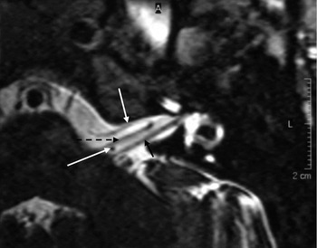
Diagnostic imaging by means of magnetic resonance imaging showed the anterior inferior cerebellar artery running vertically between the nervus facialis and the nervus vestibulocochlearis, at the location of the nervus intermedius (Figure 1).
Axial, T2-weighted magnetic resonance imaging scan showing the left cerebellopontine angle at the level of the acousticofacial bundle. The anterior inferior cerebellar artery (white arrows) approaches the acousticofacial bundle from the anterior, then loops into the internal auditory canal and runs horizontally along the nervus facialis (broken black arrow). Here, it bends upwards and runs vertically between the nervus facialis and the nervus vestibulocochlearis (black arrow), at the site of the nervus intermedius.

Given the suspected diagnosis of nervus intermedius neuralgia, and the fact that all treatments thus far had proven ineffective, our patient subsequently underwent microvascular decompression of the nervus intermedius.
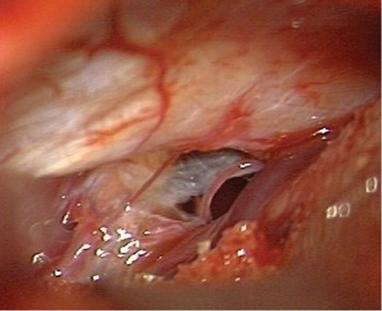
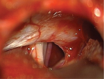
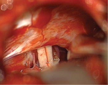
During surgery, we found several blood vessels together with thin strings of arachnoidal connective tissue layered over the nervus facialis and the nervus vestibulocochlearis complex. The connective tissue was removed and the vessels were separated from the nerves using Gelita-Spon® (Baxter, Vienna, Austria) and Tissucol Duo 500® (Gelita Medical BV, Amsterdam, The Netherlands) (Figures 2 to 4).
Intra-operative view of the cerebellopontine angle; several arachnoidal strings of connective tissue lie adhered to the acousticofacial bundle.

View of the cerebellopontine angle after removal of the tissue.

View of the cerebellopontine angle after placement of Spongostan® between the acousticofacial bundle and the anterior inferior cerebellar artery.

Post-operatively, the patient reported immediate and total relief of her otalgia. Moreover, it immediately became clear that there was no otological morbidity and that she had retained normal facial nerve function. One year later, she was still free from otalgia.
Discussion
The Vth, VIIth, IXth and Xth cranial nerves and the second and third dorsal cervical roots supply sensory innervation to the ear. Pathology of any these nerves may lead to otalgia. Thus, a thorough knowledge and understanding of the anatomy and pathophysiology of this area are essential when managing the patient with otalgia.
The nervus intermedius is frequently described as a part of the nervus facialis. However, part of the proximal segment of the nervus intermedius runs close to the brainstem together with the nervus vestibulocochlearis. From this point, the nervus intermedius then runs free in the cerebellopontine angle over a variable length between the nervus facialis and the nervus vestibulocochlearis. In approximately 20 per cent of all cases, the nervus intermedius together with the nervus vestibulocochlearis runs all the way into the meatus acousticus internus. Pressure on the nerve from an outside source, for example an artery, is probably an essential factor in the development of nervus intermedius neuralgia. Although the anterior inferior cerebellar artery normally lies closest to the acousticofacial bundle, the posterior inferior cerebellar artery and the branches of the arteria vertebralis or the vena petrosum may also exert pressure upon this nerve complex.
Contact between cranial nerves and cranial blood vessels is common; however, symptoms of neurovascular compression are rare.Reference Sunderland10 Therefore, factors other than neurovascular compression alone must influence the development of neuralgia. The exact location or site of contact between a blood vessel and a nerve appears to be of importance. Research shows that pressure on the central nervous system segment of the nerve or on the transitional segment (the so-called root entry zone) may more readily lead to nerve demyelinisation.Reference De Ridder, Moller, Verlooy, Cornelissen and De Ridder11 Pressure on a demyelinised nerve, possibly in combination with a local inflammatory reaction, may lead to higher levels of excitability and pain perception.
Analysis of patients presenting with otalgia should start with a thorough conventional ENT examination in order to exclude more common causes. There are a large variety of known causes for referred otalgia, e.g. otitis, temporomandibular dysfunction, Eagle syndrome, herpes zoster oticus, and carcinoma of the nasopharynx and larynx. Whenever nervus intermedius neuralgia is suspected, a neuro-otological examination is important in order to establish the possible involvement of the nervus vestibulocochlearis and/or the nervus facialis.
In our opinion, it is of vital importance to verify possible neurovascular compression by means of diagnostic imaging. Occasionally, diagnostic imaging may show neurovascular contact in asymptomatic patients; however, such neurovascular contact will be present in virtually all symptomatic patients.Reference Yap, Pothula and Lesser12 Magnetic resonance angiography appears to be the diagnostic imaging tool of choice in establishing the presence of neurovascular contact.Reference Adler, Bernardi, Bosley, Savino, Sergott and Zimmerman13, Reference Bernardi, Adler, Savino and Zimmerman14 When magnetic resonance angiography is used in conjunction with computerised reconstruction, enabling simultaneous imaging of both nerves and blood vessels, it becomes possible to obtain a diagnostic sensitivity of 83–100 per cent and a specificity of 83–96 per cent.Reference Furuya, Ryu, Uemura, Sugiyama, Isoda and Hasegawa15, Reference Meaney, Dunn, Eldridge, Miles, Nixon and Whitehouse16
• Diagnostic criteria for nerve intermedius neuralgia comprise: brief paroxysms of stabbing pain felt deep in the auditory canal, lasting seconds to minutes; an established trigger area in the posterior auditory canal wall; and absence of other pathology
• Neurovascular compression at the root entry zone of the nervus may lead to demyelinisation; pressure on a demyelinised nerve may lead to a higher level of excitability and pain perception
• In patients with nerve intermedius neuralgia, it is important to verify neurovascular compression using diagnostic imaging; magnetic resonance angiography appears to be the best diagnostic tool in establishing neurovascular contact
• Patients who do not respond to medical treatment may benefit from microvascular decompression
Our patient complained of a stabbing pain felt deep in the auditory canal and lasting minutes to hours. The diagnostic criteria for nervus intermedius neuralgia described by the International Headache Society (in the second edition of International Headache Classification) includes paroxysmal pain that lasts seconds to minutes.17 According to this classification, our patient did not meet the criteria for diagnosis of nervus intermedius neuralgia. However, we believe that our patient did indeed suffer from this condition, and that this diagnosis was verified by the results of surgical decompression. We therefore recommend that the diagnostic criterion describing the duration of the attacks of pain in nervus intermedius neuralgia be adjusted accordingly.
Conclusion
We conclude that patients with nervus intermedius neuralgia who do not respond to medical treatment may benefit from microvascular decompression.