Introduction
Proteolytic enzymes have been shown to participate in multiple phases of mammalian ovulation, oocyte maturation (Mondéjar et al., Reference Mondéjar, Acuña, Izquierdo-Rico, Coy and Avilés2012a; Liu et al., Reference Liu, Liu, Nin, Shi and Chen2013), fertilization, including the acrosome reaction, sperm binding to the zona pellucida (ZP), ZP penetration, zona hardening during activation (Zhang et al., Reference Zhang, Rutledge, Khamsi and Armstrong1992) and ZP-specific proteolysis (Funahashi et al., Reference Funahashi, Ekwall, Kikuchi and Rodriguez-Martinez2001).
Plasminogen activators (PAs)/plasmin, belong to one of the most broadly expressed systems, and catalyze extracellular proteolysis (Ny et al., Reference Ny, Wahlberg and Brandstrom2002; Martínez-Hernández et al., Reference Martínez-Hernández, Baiza-Gutman, Castillo-Trápala and Armant2011). PAs can be produced by many cell types in order to convert the widely distributed zymogen plasminogen to plasmin. Plasmin degrades most extracellular proteins either directly or by activating other proteases, thus affecting cell–cell and cell–matrix interactions. Urokinase-type plasminogen activator (uPA) appears to be involved in the pericellular proteolysis required for cell migration and tissue remodelling. The localization of uPA seems to be controlled through specific cell-surface receptors (Blasi & Sidenius, Reference Blasi and Sidenius2010). The urokinase-type plasminogen activator receptor (uPAR) regulates the activity of the plasminogen activation system by binding to uPA. As it is associated with the external surface of the plasma membrane by a glycosylphosphatidylinositol anchor, uPAR localizes uPA activity to the cell surface (Smith & Marshall, Reference Smith and Marshall2010). The activity of uPA is controlled by the specific inhibitor type 1 (PAI-1), a member of the serpin proteinase inhibitor superfamily. PAI-1 binds uPA, forming a complex that is recognized by uPAR and permits the internalization of the uPAR/uPA/PAI-1 complex. This complex is degraded in lysosomes, while uPAR itself is recycled back from endocytic compartments to the cell surface (Dass et al., Reference Dass, Ahmad, Azmi, Sarkar and Sarkar2008).
Plasminogen, the uPA substrate, has also been found in the oviductal lumen of pigs and cows (Mondéjar et al., Reference Mondéjar, Grullón, García-Vázquez, Romar and Coy2012b) as well as in the plasma membrane and ZP of hamster oocytes (Jiménez-Díaz et al., Reference Jiménez-Díaz, Roldán and Miceli2002), immature porcine oocytes (Roldán-Olarte et al., Reference Roldán-Olarte, Jiménez-Díaz and Miceli2005) and in in vitro matured oocytes of pigs and cows (Mondéjar et al., Reference Mondéjar, Grullón, García-Vázquez, Romar and Coy2012b). PAs activity has also been detected in bovine cumulus–oocyte complexes (COCs), with variable uPA mRNA levels during in vitro maturation (Park et al., Reference Park, Choi, Song, Funahashi and Niwa1999; D’Alessandris et al., Reference D’Alessandris, Canipari, Giacomo, Epifano, Camaioni, Siracusa and Salustri2001). All this evidence suggests that PAs could allow the generation of plasmin on the surface of the oocyte as proposed previously (Roldán-Olarte et al., Reference Roldán-Olarte, García, Jiménez-Díaz, Valdecantos and Miceli2012).
The objective of this study was to examine uPA, uPAR and PAI-1 gene expression in immature and in vitro matured oocytes and cumulus cells. As uPAR could act as a key molecule for the localization of uPA activity, the presence of this receptor in cumulus–oocyte complexes was also studied.
Materials and methods
Recovery of cumulus–oocyte complexes
Ovaries were collected from young beef cows (Bos taurus) at a local abattoir within 20 min of death and transported to the laboratory in sodium phosphate buffer, pH 7.4 (PBS) at 25°C for processing within 3h after collection. COCs were collected by aspiration from the follicles (3–6 mm in diameter) with an 18-gauge needle connected to a 10-ml disposable syringe. Oocytes with uniform ooplasm and a compact cumulus cell mass were selected by stereoscopic microscope observation. After two washes in PBS, COCs were randomly distributed into two groups: COCs in the first group were used at the immature stage and those in the second group were processed to obtain in vitro matured (IVM) COCs.
In vitro maturation
Groups of 30–40 selected COCs were matured in vitro in 400 μl drops of TCM-199 medium (Gibco 12340, Grand Island, NY, USA) supplemented with 10% fetal bovine serum (Internegocios, Buenos Aires, Argentina), 0.05 IU/ml FSH (Puregon, Organon, Dublin, Ireland), 0.23 mmol/l of sodium pyruvate (Sigma P4562, St. Louis, MO, USA) and 50 μg/ml of gentamicin for 22–24 h at 38.5°C under a 5% CO2 in air atmosphere. Only COCs with a compact cumulus mass of at least three layers of cumulus cells and a homogeneous cytoplasm were used (Papanikolaou et al., Reference Papanikolaou, Amiridis, Dimitriadis, Vainas and Rekkas2008). At the end of incubation, the COCs were assessed for cumulus expansion and extrusion of the first polar body; 90–95% of the COCs reached these maturation criteria.
RNA isolation and reverse transcription from oocytes and cumulus cells
Fifty immature and IVM COCs were treated with hyaluronidase (1 mg/ml) at 37°C (n = 4). Denuded oocytes and cumulus cells were selected separately. Total RNA from each sample was obtained by RNeasy Micro Kit (Qiagen, Valencia, CA, USA) according to the manufacturer's instructions. Reverse transcription assays were carried out using 11 μl of total RNA from each sample. The 25 μl reaction mixture contained 50 mM Tris–HCl (pH 8.3), 75 mM KCl, 3 mM MgCl2, 0.5 mM of each dNTP, 25 pmol random primers, 10 mM dithiothreitol and 200 units of reverse transcriptase enzyme M-MLV (Promega, Madison, WI, USA). Reactions were incubated at 42°C for 1 h followed by a reverse transcriptase inactivation at 94°C for 5 min. The resulting cDNAs were stored at –20°C until polymerase chain reaction (PCR) amplification.
PCR amplification
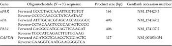
PCR analyses of cDNA were performed with 1 μl of each reverse transcription (RT) reaction, 2.0 U of Taq DNA polymerase (Invitrogen, Burlington, ONT, Canada) in a 25 μl reaction volume containing 10mM Tris–HCl (pH 9.0), 50 mM KCl, 0.1% Triton X-100, 2.5 mM MgCl2, 0.2 mM of each dNTP and 1 μM of each oligonucleotide specific primer for the gene of interest (see Table 1). PCR conditions were assayed as described above for amplification of uPAR, uPA, PAI-1 and glyceraldehyde phosphate dehydrogenase (GAPDH). PCR cycles consisted of: (1) an initial denaturing step at 94°C for 1 min followed by (2) 35 cycles of denaturation for 30 s at 94°C, annealing for 30 s at the appropriate annealing temperature for each pair of primers (uPA: 60°C; uPAR, GAPDH: 58°C and PAI-1: 55°C), and extension for 50 s at 72°C; (3) a final extension step at 72°C for 7 min. Each PCR assay was performed at least twice for each sample. Primer pairs were designed with Primer3 software from published Bos taurus cDNA sequences. The GenBank accession numbers, primer sequences and amplification fragment size are shown in Table 1. GAPDH mRNA was used as a control for RNA quantity and PCR reaction efficiency. PCR products were resolved on 1.5% agarose gel containing 0.06 μg/ml Sybr® Safe DNA Gel Stain (Invitrogen, Burlington, ON, Canada). The nature of the PCR products obtained was confirmed by sequencing. Gels images were captured with a digital camera (Olympus C-5060). Non-template controls were included in each experiment.
Primers used to amplify specific bovine transcripts from oocyte and cumulus cells

Immunochemical assays by indirect immunofluorescence
One hundred and fifty immature COCs were fixed in 4% formaldehyde in PBS, pH 7.4, for 1–2 h at room temperature and stored in ethanol 70% at 4°C for no more than 7 days. Then samples were washed five times with PBS. COCs were incubated in PBS containing 10 mg/ml BSA for 30 min to reduce the extent of nonspecific binding of the primary antibody. Then fifty COCs were incubated overnight at 4°C with 8 μg/ml goat anti-human uPAR polyclonal antibody (R&D Systems, AF807) in the blocking solution. COCs were rinsed three times in PBS and subsequently incubated with rabbit anti-goat IgG (whole molecule) biotin conjugated antibody (Sigma, B7024) at a 1:50 dilution for 1 h at room temperature. After rinsing three times with PBS, samples were incubated with extravidin–fluorescein isothiocyanate (FITC) (Sigma E2761, St. Louis, MO, USA) at a dilution of 1:200 for 30 min at room temperature in a dark chamber. Fifty COCs were used as negative controls, carried out by omitting the primary antibody treatment and adding 3% (vol/vol) preimmune goat serum instead of the primary antibody to assess nonspecific background fluorescence. Also, controls were assessed by incubating 50 COCs with extravidin–FITC alone, without primary or secondary antibodies. Photomicrographs were taken using a confocal microscope (Olympus FV300).
Statistical studies
The statistical analysis for mRNA expression was performed on values from densitometry analyses normalized to corresponding GAPDH mRNA. Relative mRNA expression values were compared with Student's t-test. A P-value of less than 0.05 was considered significant.
Results
Expression of uPAR, uPA and PAI-1 in bovine oocytes and cumulus cells
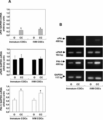
uPAR and PAI-1 mRNAs were detected in both immature and IVM oocytes and in cumulus cells of both types of oocytes (Fig. 1). No uPA mRNA expression was observed in immature or IVM oocytes although a low uPA mRNA expression was observed in cumulus cells of both immature and IVM COCs (Fig. 1).
(A) Comparison of relative density of uPA/GAPDH, uPAR/GAPDH and PAI-1/GAPDH RT-PCR products in immature and IVM oocytes and cumulus cells. Arbitrary units were determined by relating band density to the corresponding GAPDH for each sample. Values are mean ± SE (n = 4). *Indicates significant difference (P < 0.05) between oocytes and cumulus cells in uPA and PAI-1 expression. (B) Specific RT-PCR products for uPA (498 bp), uPAR (164 bp), PAI-1 (406 bp) and GAPDH (117 bp) in immature and IVM oocytes and cumulus cells separated by agarose gel electrophoresis. CC: cumulus cells; O: Oocytes.

uPAR immunolocalization in cumulus–oocyte complexes
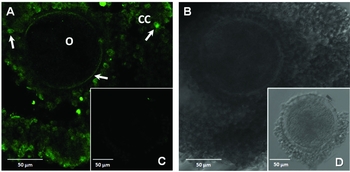
The localization of uPAR in bovine immature COCs using the specific anti-uPAR antibody showed this receptor in the oocyte plasma membrane and in cumulus cells (Fig. 2 A). Controls carried out to detect molecules with affinity for the secondary labeled antibodies yielded negative results (Fig. 2 C).
Immunofluorescence staining showing uPAR receptor. (A) Representative cumulus–oocyte complex, darkfield. (B) Cumulus–oocyte complex, brightfield. (C) Preimmune goat serum instead of the primary antibody, darkfield. (D) Brightfield, observed by confocal microscopy.

Discussion
The plasminogen activation system has been the focus of many investigations carried out in the female reproductive tract of different mammalian species (Huarte et al., Reference Huarte, Vassalli, Belin and Sakkas1993; Kouba et al., Reference Kouba, Alvarez and Buhi2000; Rekkas et al., Reference Rekkas, Besenfelder, Havlicek, Vainas and Brem2002; Mondéjar et al., Reference Mondéjar, Grullón, García-Vázquez, Romar and Coy2012b; Roldán-Olarte et al., Reference Roldán-Olarte, García, Jiménez-Díaz, Valdecantos and Miceli2012), showing that its components could be involved in several steps of the reproductive process.
Several authors have studied the expression and activity of uPA in oocytes of different species. Zhang et al. (Reference Zhang, Kidder, Zhang, Khamsi and Armstrong1994) found no uPA expression in rat oocytes. Bieser et al. (Reference Bieser, Stojkovic, Wolf, Meyer and Einspanier1998) analyzed uPA expression in matured COCs stimulated with growth factors and Park et al. (Reference Park, Choi, Song, Funahashi and Niwa1999) concluded that bovine COCs do not show PA activity just after collection from antral follicles in either cumulus-enclosed oocytes or in denuded oocytes during in vitro maturation. In the present work, uPA was detected in cumulus cells but not in immature or IVM oocytes by RT-PCR. Mondéjar et al. (Reference Mondéjar, Grullón, García-Vázquez, Romar and Coy2012b) showed uPA protein located in bovine oocytes and suggested the association of uPA to its receptor. In the present work, we demonstrate the presence of uPAR in the oolemma and plasma membrane of cumulus cells. As cumulus cells express uPA, we propose that oocytes may receive uPA from cumulus cells and/or oviductal fluid. Interaction of secreted uPA into the oviduct with uPAR present in the oocyte could allow proteolysis and activation of intracellular signaling pathways. Taking into account the present results and the previously reported evidence of uPA activity in bovine COCs, it seems clear that uPA detected by Mondéjar et al. (Reference Mondéjar, Grullón, García-Vázquez, Romar and Coy2012b) in the bovine oocyte surface could originate from cumulus cells, which probably synthesize uPA that binds to its specific receptor localized in the oolemma.
It is known that the proteolytic activity of the plasminogen activation system is regulated by several inhibitors involved during different steps of plasmin generation (Coy et al., Reference Coy, Jiménez-Movilla, García-Vázquez, Mondéjar, Grullón and Romar2012; Lu et al., Reference Lu, Lee, Hwu, Lin, Yeh, Chen, Lin and Li2013). One of them, PAI-1, participates in the specific inhibition of both uPA and tPA plasminogen activators. The detection of PAI-1 transcripts in oocytes and cumulus cells suggests that both could secrete PAI-1 regulating plasmin generation. A finely tuned extracellular proteolysis possibly facilitates cumulus expansion thus preventing oocyte damage.
In conclusion, the presence of uPA/uPAR in COCs has been demonstrated, although the role of these molecules remains to be studied. While they could be involved directly in promoting fertilization and early embryonic development, as discussed by Papanikolaou et al. (Reference Papanikolaou, Amiridis, Dimitriadis, Vainas and Rekkas2008), it is possible that they may also be involved in the proteolytic activation of growth factors and cytokines and/or in triggering intracellular autocrine and paracrine signaling pathways. These molecular processes could be responsible for the behaviour of oviductal cells as well as of gametes and embryos, allowing the normal development of these events.
Acknowledgements
The authors wish to thank Frigorífico Calchaquí for allowing reproductive tract collection from their slaughtered cows.
Conflict of interest statement
The authors declare that they have no conflict of interests.
Financial support
This work was supported by Agencia Nacional de Promoción Científica y Tecnológica, ANPCYT, Argentina (D.C.M., grant BID 1201 OC/AR PICT 2006 no. 00502 and M.R-O., grant BID PICT 2013 no. 0770), Consejo de Investigaciones de la Universidad Nacional de Tucumán, Argentina (D.C.M., CIUNT, grant 26/D438) and CONICET (M.R-O., PIP 2011 no. 025).