




No CrossRef data available.
Article contents
Pulmonary regurgitation after percutaneous balloon valvoplasty for isolated pulmonary valvar stenosis in childhood
Published online by Cambridge University Press: 24 May 2005
Abstract
Objectives: A retrospective study was undertaken to determine the degree of pulmonary regurgitation following percutaneous balloon valvoplasty for isolated pulmonary valvar stenosis. Background: Percutaneous balloon valvoplasty is the recognised treatment of choice in pulmonary valvar stenosis with effective relief of gradient. Few studies have reviewed the degree of pulmonary regurgitation after the balloon valvoplasty. Methods: We reviewed all patients with isolated pulmonary valvar stenosis undergoing percutaneous balloon valvoplasty at a tertiary centre between December 1984 and August 1996. Those with an associated haemodynamically insignificant atrial septal defect or patent oval foramen were also included. Their medical records, echocardiograms and angiograms were studied. Colour flow Doppler was used as a semi-quantitative way of assessing the pulmonary incompetence. Results: Over the period of review, 57 procedures had been performed in 49 patients. The median age at the time of the procedure was 1.08 years, with a range from 0.01 to 13.3 years. The median period of follow-up was 5.64 years, with a range from 3.00 to 14.26 years. Immediately following the dilation of the pulmonary valve, the peak-to-peak instantaneous systolic pressure gradient was significantly reduced. Seven patients required a second dilation of the valve. Their median age at the first valvoplasty, at 0.49 year, was significantly lower than those who required only one procedure, at a median of 1.50 years. Following the valvoplasty, mild pulmonary incompetence was noted in 26 out of 42 patients the day after the procedure, but only 7% had moderate incompetence. On follow-up, there was an increase in the number of patients with moderate to severe pulmonary incompetence, from 7% to 29%. Those patients in whom the procedure was performed at a younger age had more significant pulmonary incompetence. Neither the initial gradient across the pulmonary valve, nor the size of the balloon used, were related with statistical significance to the subsequent development of pulmonary incompetence. Conclusions: The majority of the patients with congenital isolated pulmonary valvar stenosis had only a mild increase in the degree of pulmonary incompetence following a single pulmonary valvoplasty. Patients who required the procedure early in life were more likely to develop significant pulmonary incompetence. The ratio of the size of the balloon to the diameter of the valve did not significantly affect the outcome. We suggest that those patients who had more severe stenosis because of a more abnormal pulmonary valve, and hence required early intervention, were more likely to develop greater pulmonary incompetence after the valvoplasty.
Information
- Type
- Original Article
- Information
- Copyright
- © 2003 Cambridge University Press
Pulmonary stenosis accounts for between one-twentieth and one-tenth of congenital abnormalities of the heart.1 Over the past decade, balloon valvoplasty has become the recognized therapeutic alternative to surgery for treatment of valvar pulmonary stenosis, for children as well as adults. It was Semb et al.,2 in 1979, who reported the first transcatheter intervention in a child with valvar pulmonary stenosis. Subsequently, Kan et al.3 in 1982 reported the first series of procedures. Since then, valvoplasty has been shown to be effective in reducing the obstruction across the abnormal valve.4 Compared with surgical valvotomy, the catheter technique has been reported to provide nearly equivalent long-term relief of obstruction, with less valvar incompetence, and less late ventricular ectopic activity.5 This technique has now been widely used for the relief of stenosis of the pulmonary valve in neonates,6 infants, children,7, 8 and even adults.1, 9 Immediate, short and long term results have been well documented,1, 6, 10, 11 and the procedure has now largely replaced surgical valvotomy as the treatment of choice. Though many studies have shown the long-term effectiveness, and persistence of relief of obstruction, after the intervention,6, 12 other long-term complications have been less well addressed. The purpose of our study was to review the degree of pulmonary incompetence induced by valvoplasty carried out for isolated valvar pulmonary stenosis in childhood. We also studied the relationship between the age of the patients, the size of the balloon used relative to the diameter of the abnormal pulmonary valve, and the degree of valvar incompetence at late follow-up.
Methods and subjects
We recruited all patients with isolated pulmonary valve stenosis who underwent balloon valvoplasties at a single tertiary centre, albeit by multiple operators, between December 1984 and August 1996. They included patients with an associated haemodynamically insignificant atrial septal defect or patent oval foramen. Only those patients who have been followed up for at least three years were enrolled. Their medical records and investigations were reviewed.
The technique used has been described in detail, and was similar to that performed by others.13, 14 The size of the balloon varied according to the measurement of the diameter of the abnormal pulmonary valve taken at their basal attachment of the hinge points of the leaflets, on echocardiography, and angiography when available. There was no uniform criterion for sizing the balloon amongst the different operators, although most used a ratio of 1.2 to 1, with the balloon being bigger than the valvar diameter. The gradient across the pulmonary valve, or the right ventricular pressures in those with critical pulmonary stenosis, were measured before and after the procedure. We used two balloons8 in 10 patients, in whom the diameter of the pulmonary valve ranged from 7 to 20 mm, with a median measurement of 12 mm. There was no consistent record of the number of inflations made in each procedure, but usually 2 or 3 inflations were made, nor was it noted if any of the balloons ruptured during the procedure. Different catheters were used over the period of time, including Omega, Berman, Accent, Meditech, Cook and Mansfield balloons. All procedures were carried out under general anaesthesia. The procedures were performed by five cardiologists over the study period, but all essentially used similar techniques and made comparable measurements.
Follow-up included clinical evaluation and echocardiographic studies, with cardiac catheterization being repeated only in exceptional circumstances. The maximum peak instantaneous gradient across the pulmonary valve, and the degree of pulmonary incompetence, were reviewed in all patients by cross-sectional echocardiography aided by pulsed and colour Doppler. Angiograms were available in 25 patients.
Since general anaesthesia can lower both the measured gradient and pressures in the right ventricle, unless they were unavailable, we used echocardiographic studies to quantitate the degree of valvar incompetence. The severity of the incompetence was then graded semi-quantitatively using the colour Doppler images obtained from cross-sectional echocardiography (Fig. 1). The incompetence was judged to be trivial when the diastolic flow reversed just below the valve, mild when reversal occurred midway between the valve and the bifurcation of the pulmonary trunk, moderate when reversal was seen at the level of the bifurcation, and severe when flow reversal occurred within the right and left pulmonary arteries.

Figure 1. We are assessing semi-quantitatively the degree of pulmonary incompetence using colour Doppler, utilizing the parasternal short axis view on cross sectional echocardiography. Figure 1a shows trivial incompetence, with diastolic reversal of flow seen for a short distance below the pulmonary valve. RA: right atrium; RVOT: right ventricular outflow tract; PT: pulmonary trunk; AO: aorta; LA: left atrium. Mild incompetence is shown in Figure 1b, with reversal occurring midway between the pulmonary valve and the bifurcation of pulmonary trunk. Moderate incompetence is shown in Figure 1c, with the reversal of flow seen at the bifurcation of the pulmonary trunk. Severe incompetence, with reversal of flow occurring within the right and left pulmonary arteries, is shown in Figure 1d. RPA: right pulmonary artery; LPA: left pulmonary artery.
Other criterions, such as right ventricular size, the extent of the regurgitant jet in relation to the diameter of the pulmonary valve, and its extent in the right ventricular outflow tract, were noted if available, though the analysis only incorporated the colour Doppler findings, these being available in all subjects.
Statistical methods
Comparisons between the two groups were performed using Student's two-tailed paired t-test. The degree of pulmonary valvar insufficiency, and the age at the procedure, were compared between the groups of patients using an unpaired t-test. The analysis of variance was used to study the relationship between the ratio of the size of the balloon to the diameter of the valve, and the severity of pulmonary valvar insufficiency. A p value of less than 0.05 was considered statistically significant.
Results
The median age at initial valvoplasty was 1.08 years, including 7 neonates (Table 1). Of the 47 patients, seven required more than one valvoplasty. They were analysed separately. The median duration of follow-up was 5.64 years, with a range from 3.00 to 14.26 years. We followed up 11 patients for more than 10 years.
Table 1. The age of patients at the initial pulmonary valvoplasty.

In all, 57 valvoplasties had been performed in 49 patients between December 1984 and August 1996. Nearly all patients had a significant gradient across the pulmonary valve, with a median gradient of 60 mmHg, and a range from 11 to 140 mmHg. Critical stenosis had been diagnosed in 4 neonates requiring an infusion of prostaglandin, which would have maintained the pulmonary arterial pressure, thereby reducing the gradient (Table 2).
Table 2. Comparison of variables between those patients who underwent a single valvoplasty as compared with those that required a second procedure.

The pulmonary valves were described as “thickened and doming” in 36 patients, and as relatively normal in 13 patients, the information being derived mainly from the echocardiograms. In 11 patients, pulmonary incompetence was noted prior to dilation, trivial in 10 patients, but moderate in one.
Pulmonary stenosis
Immediately following dilation of the valve, the peak-to-peak instantaneous systolic pressure gradient was significantly reduced (p < 0.001) as assessed by pulling the catheter back across the valve (Table 2). On follow-up, 7 patients required a second dilation at a median interval of 0.81 year, with a range from 0.19 to 2.00 years, after the initial dilation. Their median age at the initial dilation had been 0.49 year, with a range from the first day of life to 3.36 years. The median ratio of the size of the balloon to the diameter of the pulmonary valve was 1.33, with a range from 1.06 to 1.64 (Table 2).
The patients who had only one valvoplasty had ongoing improvement in the valvar gradient as judged from follow-up echocardiograms. None of the patients in this group required surgical intervention at any stage. The median age when the solitary procedure had been performed was 1.50 years, with a range from the first day of life to 13.27 years. The median gradient across the valve prior to dilation was the same as in those who required valvoplasties at 60 mmHg, with a range from 11 to 140 mmHg.
The median ratio of the size of the balloon to the diameter of the pulmonary valve in this group of patients was 1.35, with a range from 0.65 to 2.14.
Pulmonary incompetence
Following valvoplasty, 26 of 42 patients (60%), who had one procedure demonstrated trivial or mild incompetence as shown by cross-sectional echocardiography and colour Doppler performed one day after the procedure (Table 3 and Fig. 2). Moderate incompetence was seen in three patients (7%) after the procedure, these patients having had no incompetence noted before. In five patients (12%), there was no incompetence noted immediately after the procedure, while the presence of immediate incompetence after the dilation was not recorded in 8 patients (20%). The one subject with moderate incompetence noted before the procedure suffered no change in the severity of valvar incompetence after. At follow-up, there was an increase from 3 to 15 in the number of patients with moderate or severe pulmonary incompetence (Fig. 3). None of them developed significant right ventricular dilation or paradoxical septal motion, at least within the limitations of short-to-medium term follow-up, and may have reflected little change in ventricular compliance, which may precede true volume loading. The higher the initial grade of pulmonary incompetence after the valvoplasty, the more likely it was to progress with time. The patients with moderate or severe pulmonary incompetence found on follow-up were those who had the procedure done at a younger age (Fig. 4).
Table 3. The outcome for patients following a single pulmonary valvoplasty. The age at time of the procedure, and the duration of follow-up, are cited as median values, with the range in brackets. The change in the severity of pulmonary regurgitation relates to findings prior to valvoplasty and the latest follow-up. The severity of regurgitation continued to increase progressively after valvoplasty in 12 of the 42 patients (see Fig. 3).


Figure 2. The change in degree of pulmonary incompetence after a single pulmonary valvoplasty.

Figure 3. The change in degree of pulmonary regurgitation on follow-up seen in those patients where such change occurred, and excluding the remaining 30 patients where there was no change.

Figure 4. The overall change in the grading of pulmonary regurgitation. Patients with severe pulmonary regurgitation were mainly babies who had required the procedure early in life. Patients with trivial regurgitation showed a wide range of distribution in age.
The majority, however (62%), had only a mild increase in the degree of pulmonary incompetence after a single valvoplasty over the period of intermediate to long-term follow-up (Table 3 and Fig. 2).
The 7 patients who required repeated valvoplasties had no or only trivial incompetence immediately following the second procedure. At the latest follow-up, however, three of these 7 patients were found to have moderate pulmonary incompetence.
In our patients, there was no significant difference in the initial pressure gradient across the pulmonary valve, nor in the ratio of the size of the balloon to the diameter of the pulmonary valve among the groups of patients, and the ratio did not significantly alter the outcome (Fig. 5).

Figure 5. The severity of pulmonary regurgitation after valvoplasty is compared with the ratio of the size of the balloon to the diameter of the pulmonary valve.
Discussion
We have charted the long-term results of relief of pulmonary valvar stenosis in 49 patients who underwent balloon valvoplasty. Our findings are generally comparable to previous reports, with several studies documenting the ongoing relief of obstruction at follow-up.6, 11, 12 These studies, for the most part, showed that the incompetence seen after the procedure was mild or haemodynamically insignificant. In part that may be related to reduced right ventricular compliance immediately after valvoplasty. In a series studying the results of percutaneous balloon valvoplasty in adults, it was reported that the mild incompetence noted immediately after the procedure may even resolve on follow-up.9
McCrindle and Kan11 reported the results of a follow-up of about five years in 46 patients. They found a residual peak instantaneous Doppler gradient of 20 mmHg, with mild incompetence seen in four-fifths. Rao et al.6 reported the follow-up from 3 to 10 years in 80 children, with 51 having other associated cardiac lesions. They showed a residual peak instantaneous Doppler gradient of 17 mmHg. Pulmonary incompetence immediately after the procedure had increased in half, and was worse in four-fifths on long term follow-up, albeit that none of their patients was considered to have significant pulmonary incompetence.
In our study, 10 patients had trivial to mild incompetence prior to the procedure. It is noteworthy in this respect that trivial incompetence has been reported as a normal finding in healthy persons.15 The pulmonary valve coapts less well than the aortic valve, and the retrograde pressure gradient through the pulmonary valve in diastole may not be enough to close the valve as tightly as the aortic valve. Although four-fifths of our patients had an increase in the severity of pulmonary incompetence, only 7% showed moderate incompetence immediately after the procedure. In 13 patients (30%), we found a gradual increase in the severity of pulmonary incompetence at late follow-up, these findings being comparable to those reported by Rao et al.6 This funding may be due to changes in the pulmonary vascular resistance and right ventricular compliance with time.
Indeed, pulmonary incompetence has been found to be increased in most studies after balloon valvoplasty. It was increased in three-quarters of the patients studied by Ray et al.16 and in over seven-eighths of the cohort reported by McCrindle and Kan.11 We should emphasize, nonetheless, that our system for grading pulmonary incompetence is only semi-quantitative. As stated by Feigenbaum,17 it is difficult to validate the degree of pulmonary regurgitation with Doppler echocardiography.15, 18, 19 William et al.20 recently proposed more sophisticated methods for assessing pulmonary incompetence. Thus, angiograms can be considered as an adequate way for grading incompetence if the catheter can be placed in the distal pulmonary trunk, or one of its branches, with the catheter ports well away from the pulmonary valve. Serial noninvasive transthoracic echocardiographic studies, nonetheless, remain the mainstay for evaluation of these patients. Besides measuring the diastolic reversal in flow in the branches of the pulmonary trunk as we have described, others have used the ratio of the width of the regurgitant colour jet to the diameter of the pulmonary valve in early diastole as an objective method of assessing pulmonary incompetence severity using colour Doppler images.20 Unfortunately clear documentation of this feature was not routinely available in our patients. Besides the regurgitant jet in the right ventricle, spectral Doppler deceleration, magnetic resonance imaging or even three-dimensional echocardiography, might also be used to assess the regurgitation and the right ventricular volume. But determination of right ventricular volumes is technically difficult because of right ventricular geometry. New technologies employing magnetic resonance imaging and assessing regurgitant fractions may provide the answer, though there is always the problem of studying an infant in a scanner. Prospective use of these techniques may, in future, provide a more accurate picture of outcome.
Be that as it may, we found that patients with more significant pulmonary incompetence on follow-up were those who had required the initial procedure at a younger age. That is probably because they had a more “abnormal” pulmonary valve, leading to significant stenosis and hence the need for earlier intervention. The period of follow-up was too short to determine the long-term impact of more severe pulmonary incompetence on right ventricular function, as is now known to occur in those adolescents and adults with moderate-to-severe pulmonary incompetence following surgical repair of tetralogy of Fallot.
In our study, we found no significant relationship between the grade of pulmonary incompetence and the ratio of the size of the balloon to the diameter of the pulmonary valve. The ratio varied from 0.65 to 2.14. Even in those in whom big balloons were used, there was no overt evidence of complications. There are reports, however, showing that larger balloons may cause damage to the right ventricular outflow tract,21 without conferring any additional benefits in relieving the valvar stenosis.15 Oversized balloons, with the ratio larger than 1.5, therefore, are not recommended. Those patients in whom the largest balloons were used relative to the diameter of the valve were those treated early in the series, when there was little information concerning the optimal size of balloon needed to relieve the stenosis.
In our study, therefore, we failed to demonstrate a significant relationship between the severity of the pulmonary incompetence as assessed by colour flow Doppler following the valvoplasty and the degree of incompetence noted before the procedure, nor the size of the balloon used relative to the diameter of the pulmonary valve. The severity of incompetence tended to increase with increasing years of follow-up. Age was the only significant factor in determining the severity of pulmonary incompetence noted after the valvoplasty in our patients. We postulated that those patients who required early intervention for pulmonary valvar stenosis tended to have more abnormal valves, and hence developed more severe pulmonary incompetence, after the percutanous balloon valvoplasty.
Footnotes
Presented in part at the Cardiac Society of Australia and New Zealand, August 2000.
References
Teupe CHJ, Burger W, Schrader R, Zeiher AM. Late (five to nine years) follow up after balloon dilation of valvar pulmonary stenosis in adults. Am J Cardiol 1997; 80: 240–242.Google Scholar
Semb BHK, Tjonneland S, Stake G. “Balloon valvotomy” of congenital pulmonary valve stenosis with tricuspid valve insufficiency. Cardiovasc Radiol 1979; 2: 239.Google Scholar
Kan JS, White RI Jr, Mitchell SE, Gardner TJ. Percutaneous balloon valvoplasty: a new method for treating congenital pulmonary-valve stenosis. N Engl J Med 1982; 307: 540–542.Google Scholar
Rocchini AP, Kveselis DA, Crowley D, Dick M, Rosenthal A. Percutaneous balloon valvoplasty for treatment of congenital pulmonary valvar stenosis in children. J Am Coll Cardiol 1984; 3: 1005–1012.Google Scholar
Brian KO, Robert HB, Amy L, Alert R. Intermediate-term outcome after pulmonary balloon valvoplasty: comparison with a matched surgical control group. J Am Coll Cardiol 1992; 20: 169–173.Google Scholar
Rao PS, Galal O, Patnana M, Buck SH, Wilson AD. Results of three to 10 year follow up of balloon dilatation of the pulmonary valve. Heart 1998; 80: 591–595.Google Scholar
Sullivan ID, Robinson PJ, Macartney FJ, et al. Percutaneous balloon valvoplasty for pulmonary valve stenosis in infants and children. Br Heart J 1985; 54: 435–441.Google Scholar
DiSessa TG, Alpert BS, Chase NA, Birnbaum SE, Watson DC. Balloon valvoplasty in children with dysplastic pulmonary valves. Am J Cardiol 1987; 60: 405–407.Google Scholar
Chen CR, Cheng TO, Huang T, et al. Percutaneous balloon valvoplasty for pulmonic stenosis on adolescents and adults. N Engl J Med 1996; 335: 21–25.Google Scholar
Rao PS. Further observations on the effect of balloon size on the short term and intermediate term results of balloon dilatation of the pulmonary valve. Br Heart J 1988; 60: 507–511.Google Scholar
McCrindle BW, Kan JS. Long-term results after balloon pulmonary valvoplasty. Circulation 1991; 83: 1915–1922.Google Scholar
Rao PS, Fawzy ME, Solymar L, Mardini MK. Long-term results of balloon pulmonary valvoplasty of valvar pulmonic stenosis. Am Heart J 1988; 115: 1291–1296.Google Scholar
Lababidi Z, Wu JR. Percutaneous balloon pulmonary valvoplasty. Am J Cardiol 1983; 52: 560–562.Google Scholar
Kan JS, White RF, Mitchell SE, Anderson JH, Gardner TJ. Percutaneous transluminal balloon valvoplasty for pulmonary valve stenosis. Circulation 1984; 69: 554–560.Google Scholar
Takao S, Miyatake K, Izumi S, et al. Clinical implications of pulmonary regurgitation in healthy individuals: detection by cross sectional pulsed Doppler echocardiography. Br Heart J 1988; 59: 542–550.Google Scholar
Ray DG, Subramanyan R, Titus T, et al. Balloon pulmonary valvoplasty: factors determining short- and long-term results. Int J Cardiol 1993; 40: 17–25.Google Scholar
Feigenbaum H. Accurate valvar heart disease. Echocardiography. Lea & Febiger, Philadelphia, 1994, pp 297–349.
Patel AK, Rowe GG, Dhanani SP, Kosolcharoen P, Lyle LE, Thomsen JH. Pulsed Doppler echocardiography in diagnosis of pulmonary regurgitation: its value and limitations. Am J Cardiol 1982; 49: 1801–1805.Google Scholar
Vermilion RP, Snider AR, Meliones JN, Peters J, Merida-Asmus L. Pulsed Doppler evaluation of right ventricular diastolic filling in children with pulmonary valve stenosis before and after balloon valvoplasty. Am J Cardiol 1990; 66: 79–84.Google Scholar
Williams RC, Minich L, Shaddy RE, Pagotto LT, Tani LY. Comparison of Doppler echocardiography with angiography for determining the severity of pulmonary regurgitation. Am J Cardiol 2002; 89: 1438–1441.Google Scholar
John CR, Thomas JK, Barbara AB, James EL. Morphologic changes induced by dilatation of the pulmonary valve annulus with overlarge balloons in normal newborn lambs. Am J Cardiol 1984; 55: 210–214.Google Scholar

We are assessing semi-quantitatively the degree of pulmonary incompetence using colour Doppler, utilizing the parasternal short axis view on cross sectional echocardiography. Figure 1a shows trivial incompetence, with diastolic reversal of flow seen for a short distance below the pulmonary valve. RA: right atrium; RVOT: right ventricular outflow tract; PT: pulmonary trunk; AO: aorta; LA: left atrium. Mild incompetence is shown in Figure 1b, with reversal occurring midway between the pulmonary valve and the bifurcation of pulmonary trunk. Moderate incompetence is shown in Figure 1c, with the reversal of flow seen at the bifurcation of the pulmonary trunk. Severe incompetence, with reversal of flow occurring within the right and left pulmonary arteries, is shown in Figure 1d. RPA: right pulmonary artery; LPA: left pulmonary artery.
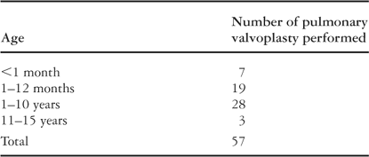
Table 1.
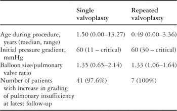
Table 2.

Table 3.

The change in degree of pulmonary incompetence after a single pulmonary valvoplasty.

The change in degree of pulmonary regurgitation on follow-up seen in those patients where such change occurred, and excluding the remaining 30 patients where there was no change.

The overall change in the grading of pulmonary regurgitation. Patients with severe pulmonary regurgitation were mainly babies who had required the procedure early in life. Patients with trivial regurgitation showed a wide range of distribution in age.

The severity of pulmonary regurgitation after valvoplasty is compared with the ratio of the size of the balloon to the diameter of the pulmonary valve.
 You have
Access
- 21
- Cited by