Introduction
Congenital cholesteatomas are epithelial-lined cysts filled with desquamated keratin debris. Congenital cholesteatomas of the temporal bone are thought to be acquired embryologically from squamous epithelial ‘rests’ in the temporal bone. They are a relatively unusual clinical entity, accounting for approximately 2–5 per cent of all cholesteatomas.Reference Paparella and Rybak1 Congenital cholesteatoma of the temporal bone can be found intradurally, most commonly at the cerebellopontine angle, or extradurally in the middle ear. However, congenital cholesteatoma of the infratemporal fossa has not previously been documented (as researched by an Ovid Medline search for the medical subject heading ‘congenital cholesteatoma’).
We report here the first case, to our knowledge, of a patient with an infratemporal fossa cholesteatoma and intact tympanic membrane. Clinical, radiological and intra-operative findings are presented.
Case report
A five-year-old girl presented several times to the otological clinic of our hospital with a history of left-sided hearing impairment. At each presentation, the left tympanic membrane was carefully examined otoscopically and was found to be intact and retracted.
Consecutive pure tone audiograms showed a mild to moderate conductive hearing loss in the affected ear, and tympanometry revealed a type B configuration consistent with middle-ear effusion.
The patient was initially managed with oral antibiotics and nasal corticosteroids, with limited benefit. There was no previous history of ear pain, otorrhoea or contralateral otitis media.
During the last of the patient's three monthly, regular visits, a possible retrotympanic mass in the anteroinferior quadrant was suspected. This whitish mass showed continuing growth over time.
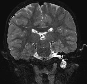
A high resolution computed tomography scan of the temporal bone demonstrated a soft tissue mass located in the hypotympanum and infratemporal fossa, with localised bone erosion of the carotid canal and jugular bulb (Figure 1). The mastoid was well pneumatised. Magnetic resonance imaging showed a large mass occupying the region of the left infratemporal fossa and protruding into the hypotympanum. The mass was hypo-intense in T1-weighted images and hyper-intense in T2-weighted images (Figure 2), suggesting a cholesteatoma.
Axial computed tomography scan of the temporal bone showing a rounded, soft tissue mass occupying the left infratemporal fossa, with bony erosion of the carotid canal and jugular bulb.

Coronal, T2-weighted magnetic resonance imaging scan showing a hyper-intense mass in the left infratemporal fossa with limited extension into the middle ear.

The patient underwent surgical treatment via a tympanomastoid approach. A complete mastoidectomy was performed. The bony canal wall was drilled down as low as possible and the external auditory canal was enlarged for better visualisation. A pearly white mass was encountered in the hypotympanum, which extended deeply into the infratemporal fossa inferiorly and into the eustachian tube anteriorly. The ossicular chain was found to be intact and free of disease. Resection of disease was commenced at the centre of the mass to diminish the lesion. The surrounding matrix was then removed carefully with the aid of a 30° oto-endoscope, from the surface of the carotid artery and the jugular bulb in its entirety. After complete removal of the mass, the remnant cavity was packed with a muscle plug, and temporalis fascia was used as an overlay graft for tympanic membrane reconstruction.
Histopathological examination of the mass confirmed the diagnosis of cholesteatoma.
After surgery, the patient initially showed some improvement of her hearing. However, two months later her hearing returned to its pre-operative level. Otoscopic examination revealed a retracted tympanic membrane. The mastoid cavity was well epithelialised. During the patient's eight-month post-operative follow up, her tympanic membrane remained intact and her hearing was unchanged.
Discussion
According to Derlacki and Clemis, the diagnosis of congenital cholesteatoma is based mainly on the previous medical history and the clinical presentation.Reference Derlacki and Clemis2 These include a pearly white mass medial to an intact tympanic membrane, a normal pars flaccida and tensa, and no prior history of otorrhoea, tympanic membrane perforation or previous otological procedures. Levenson et al. proposed that prior bouts of otitis media were not grounds for the exclusion of congenital cholesteatoma as a possible diagnosis.Reference Levenson, Parisier, Chute, Wenig and Juarbe3 Our patient had an intact tympanic membrane and no previous otological procedures, which suggested a congenital origin of her cholesteatoma.
Several theories have been proposed to explain the existence of congenital cholesteatoma, of which the epithelial rest theory is the most commonly accepted. This theory is based on Michaels's observation of squamous epithelial cells in the lateral wall of the embryonic tympanic cavity.Reference Michaels4 Usually, these squamous cells have disappeared by 33 weeks' gestation. Incomplete resorption is thought to lead to the formation of congenital cholesteatoma.Reference Bennett, Warren, Jackson and Kaylie5 Recent studies have found that the location of epithelial rests, and of the consequent congenital cholesteatomas, varies. Reported sites of origin within the temporal bone include the middle-ear space and mastoid, the external auditory canal, the petrous apex, and the tympanic membrane.
Up to two-thirds of congenital cholesteatomas in the middle ear have been reported to occur within the anterosuperior quadrant, followed by the posterosuperior quadrant.Reference Kazahaya and Potsic6 Unlike most other reports, our patient's cholesteatoma was detected otoscopically in the anteroinferior quadrant.
As shown in Figures 1 and 2, radiological studies revealed that the mass was located mainly in the infratemporal fossa, with only limited extension into the hypotympanum, and this was confirmed by subsequent surgical exploration. These findings strongly indicate that the infratemporal fossa rather than the tympanic cavity was the likely site of origin in our patient. If the tympanic cavity is the site of origin, then growth of the mass would be expected to follow a posterosuperior direction, as described by Koltai et al., with other parts of the middle ear being involved (e.g. mesotympanum and attic) before extension into the infratemporal fossa.Reference Koltai, Nelson, Castellon, Garabedian, Triglia and Roman7 In our patient, however, most parts of the middle ear were free of disease.
• This paper reports a rare case of congenital cholesteatoma of the infratemporal fossa
• If there is suspicion of cholesteatoma behind an intact eardrum, and unexplained middle-ear effusion, then diagnostic imaging of the temporal bone should be undertaken
• In this patient, a tympanomastoid approach with canal wall down technique was selected, instead of an infratemporal fossa approach, for better cosmetic effect
The most common presentation of congenital cholesteatoma is a white, retrotympanic mass with or without hearing impairment.Reference Darrouzet, Duclos, Portmann and Bebear8 In our patient, conductive hearing loss was the only presenting symptom at her initial few visits. This, combined with a type B tympanogram, led us to diagnose otitis media with effusion. The cholesteatoma was not suspected until we observed an enlarging mass behind an intact tympanic membrane. Unlike the many other reports of congenital cholesteatoma of the middle ear in which conductive hearing loss was usually caused by ossicular destruction, our patient presented with hearing loss due to local expansion of the mass into the eustachian tube, as indicated by the intra-operative findings.Reference Kojima, Tanaka, Shiwa, Sakurai and Moriyama9
In such a case, the location of the mass makes the decision regarding surgical options somewhat challenging. Instead of an infratemporal fossa approach, a tympanomastoid approach with canal wall down technique was selected for better cosmetic effect. Complete eradication of disease was facilitated by intra-operative oto-endoscopy, without complications. A disadvantage of this procedure is reportedly poorer hearing outcomes.Reference Kazahaya and Potsic6
Conclusion
Congenital cholesteatoma of the infratemporal fossa is a very rare condition; to the best of our knowledge, our patient represents the first reported case. The otologist should be alert to this problem because of its insidious nature. If there is a suspicion of cholesteatoma behind an intact tympanic membrane, and an unexplained middle-ear effusion, then diagnostic imaging of the temporal bone should be undertaken.
Acknowledgement
This work was supported by a grant from Jiangsu (China) Health Administration to G Xing (grant number RC2007064).