

Introduction
From the perspective of both individuals and populations, it is widely accepted that birth size is a main indicator of fetal and neonatal health. In particular, birth weight is strongly related to fetal, neonatal and post-neonatal mortality and with infant and child morbidity. Low birth weight (LBW) is related to health outcomes later in life and could be a major independent risk factor for the development of hypertension and obesity in school children (Zarrati et al., Reference Zarrati, Shidfar, Razmpoosh, Nezhad, Keivani, Hemami and Asemi2013). A growing body of evidence has linked LBW to the risk of developing coronary heart disease, chronic kidney disease and metabolic syndrome in later life (Boney et al., Reference Boney, Verma, Tucker and Vohr2005; Huxley et al., Reference Huxley, Owen, Whincup, Cook, Rich-Edwards, Smith and Collins2007; White et al., Reference White, Perkovic, Cass, Chang, Poulter and Spector2009; Haas et al., Reference Haas, Liepold and Schwandt2015).
Birth weight also reflects inequities in the conditions affecting women. It can provide information on which population groups may benefit from improved antenatal and neonatal care (Khatun & Rahman, Reference Khatun and Rahman2008). Birth length and head circumference are other indicators of neonatal size that may provide important diagnostic and prognostic information beyond that provided by birth weight alone. The measurement of head circumference is an important screening procedure for detecting abnormalities in head growth (Sniderman, Reference Sniderman2010). Also, it has been reported that impaired fetal linear growth is a major predictor of early infant linear growth failure (Berngard et al., Reference Berngard, Berngard, Krebs, Garcés, Miller and Westcott2013). The findings of a nationwide longitudinal survey have indicated a relationship between birth length and risk of hospitalization. Short babies, as well as long babies with low weight, were found to be at increased risk of hospitalization (Kato et al., Reference Kato, Yorifuji, Inoue, Doi and Kawachi2013). Thus, it appears that health practitioners, health care providers and health policymakers can derive benefits from undertaking neonatal anthropometric measurements to assess a population’s health needs and subsequent policy development and programming.
Numerous factors, such as maternal genetic factors, nutritional status, smoking, age, disease, complications of pregnancy and physical, social and economic environments, have been suggested as causes of suboptimal fetal growth (Utpala & Gregory, Reference Utpala and Gregory2004; Martinez & Simmons, Reference Martinez and Simmons2005). Moreover, maternal height, pre-pregnancy weight and gestational weight gain are considered as important determinants of LBW that may play an important aetiological role (Weller & Romney, Reference Weller and Romney1988; Nahar et al., Reference Nahar, Mascie-Taylor and Housne2007). In another study, birth order and maternal height were found to be the most important maternal parameters impacting birth weight (Elshibly & Schmalisch, Reference Elshibly and Schmalisch2008). In addition, the presence of long-lasting poor nutrition and health, infections, pregnancy complications, demanding work during pregnancy and deprived socioeconomic conditions of mothers are the other most influential factors affecting birth weight (UNICEF/WHO, 2004).
According to the last nationwide survey conducted from 2010 to 2011, the nationwide prevalence of LBW in Iran was 7.7% (7.7% in urban and 7.8% in rural areas; Rashidian, Reference Rashidian2010; UNICEF Child Info, 2013), indicating a remarkable increase compared with the former national integrated monitoring and evaluation survey (IMES study) conducted in 2004 (7.2% in the whole country, 7.2% and 7.1% in urban and rural areas, respectively). Studies from different geographical areas of the country, all with Persian ethnicity and urban residence, indicate that the LBW rate varies from 5.2% to 11.8% (Roudbari et al., Reference Roudbari, Yaghmaei and Soheili2007; Vahdaninia et al., Reference Vahdaninia, Tavafian and Montazeri2008; Golestan et al., Reference Golestan, Akhavan Karbasi and Fallah2011). Collectively, the findings indicate that LBW remains a growing public health concern in the country.
The present study was conducted in two districts in north-west Iran. Tabriz, the capital city of the East Azerbaijan province, with a population of more than 1,600,000, acts as a regional hub for the trade and industrial plants of north-west Iran. Its population are ethnically (Azeri or Turk) and linguistically (Turkish) different from the other major cities of Iran. Heriss is a small town near Tabriz city with a population of 68,000, and sharing the same ethnic and language characteristics.
Although LBW is affected by diverse genetic, ethnic, environmental and socioeconomic status characteristics of families, little has been done to elucidate the contribution of these factors to the prevalence of LBW in Iran. Therefore, this study aimed to evaluate the possible maternal factors affecting newborns’ anthropometric measurements, in particular LBW, in a community with an Iranian–Azeri population.
Methods
Field of study and participants
This cross-sectional, population-based study was conducted in Tabriz, the capital city of East Azerbaijan province, and Heriss town, located in the north-west of Iran, from June to August 2014. Of 27 public primary health centres located in Tabriz, six were selected randomly and these, together with two centres in Heriss district, were used for data collection. The study was approved by the ethical committee of the Tabriz University of Medical Sciences.
Data for 807 urban and Azeri mothers delivering a singleton live infant, and their offspring during the last two years up to August 2014, were collected from the databases of eight health care facilities located in the two districts (n=707, 87.61%) or from a university hospital (n=100, 12.39%), which is a major referral centre for obstetric and gynaecological services. In the hospital, newly delivered mothers were recruited using the convenience sampling method and interviewed face-to-face by four well-trained individuals. In addition, the anthropometric measures of mothers and their neonates were obtained from hospital medical records. Multiple deliveries were excluded. The data included all potential maternal-related factors, including anthropometry, age, smoking, drinking, education, economic status, nutrient supplements, gestational weight gain, gestational age and birth order, which were hypothesized to affect the anthropometric characteristics of newborns. Data on smoking and alcohol consumption were collected as ‘yes’ and ‘no’. Informed consent was obtained from the mothers for themselves and on behalf of their neonates.
Infants fell into four birth weight categories: very LBW (<1500 g), moderate LBW (1500–2499 g), normal weight (2500–4200 g) and high birth weight (>4200 g). In another definition, birth weights less than 2500 g were defined as LBW and those more than 2500 g as normal weight. The binary version was used for logistic regression analysis. Maternal age at the time of delivery was categorized as follows: <18, 18–34.9 and ≥35 years. Receiver Operating Characteristic (ROC) curves were drawn to determine optimal cut-off values for maternal height. Then, the mothers were defined as short stature (<160 cm), middle height (160–169.9 cm) and taller mothers (≥170 cm). Mothers were nutritionally classified into four groups using the cut-off points recommended by the World Health Organization (WHO) as follows: underweight (BMI<18.5 kg/m2), normal weight (18.5≤BMI<24.99 kg/m2), overweight (25≤BMI<29.99 kg/m2) and obese (BMI≥30 kg/m2). Gestational age was classified into four groups: babies born alive at ≤36 weeks of gestation were defined as preterm, 37–38 weeks as early term, 39–40 weeks as full term and ≥41 weeks as late term. Mothers who had attended a national maternity care programme at least 5 times and received iron or folic acid supplements were defined as ‘regular recipients=always’; those with 2–5 times and less than 2 times were considered as ‘occasionally recipients’ and ‘never recipients’, respectively.
Illiterate women, along with those who were educated to primary level, were combined into one category defined as ‘low education’. Those with junior or senior high school level education were classified as ‘moderate’ and mothers with at least college education or higher were grouped as ‘high education’ level. In addition, items such as educational level and occupational status were used to estimate the economic status of the study subjects and categorized in four groups as follows: very low (unemployed or jobless with low education), low (construction workers, handicraft makers, housekeepers with low or moderate education), middle (single parent/government staff, farmer with medium education) and high (dual worker parents/government staff, large trader and farmer with medium or high education).
Statistics
All analyses were performed using SPSS Version 16.0. Means and standard deviations (SD) were computed for continuous data, medians (percentile 25, 75) for non-continuous data and frequency (%) for proportional data. Differences between the groups were assessed using χ 2 for categorical variables, and Student’s t-test for continuous variables. For variables with three or more categories, differences between groups were assessed by one-way analysis of variance (ANOVA), followed by post-hoc comparisons using the Tukey test. Linear regression was performed for associations between variables. Univariate and multivariate logistic regression models were used to estimate the odds ratios (OR) and 95% confidence intervals (95% CI). Low birth weight was the dependent variable, maternal factors (age, height, BMI, iron intake) and birth order were used as independent variables and maternal age 18–34.9, height 160–169.9 cm, BMI 25–29.99 kg/m2, always consumed iron and second birth order were used as reference categories. The data were adjusted for appropriate confounding factors (gestational age, maternal age, height, BMI and birth order). Statistically significance was considered to be at p<0.05.
Results
Characteristics of newborns and their mothers
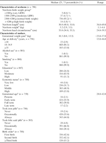
Table 1 shows the characteristics of the newborn infants and their mothers. Of the 790 newborns, 5.1% were LBW. The median (interquartile range, IQR) of neonates’ weight, length and head circumference were 3.2 (2.9, 3.5) kg, 50.0 (48.0, 51.0) cm and 35.0 (34.0, 35.5) cm, respectively. Of the 794 mothers, 2.9% were in the younger age group (<18 years) against 13.0% of mothers ≥35 years. Further, 99.9% of the mothers did not smoke or drink alcohol, 63.9% had a moderate level of education and 37.1% were from families with low or very low incomes. The percentages of mothers regularly receiving iron and folic acid from health centres were 44 and 28.4%, respectively. Of neonates, 55.9% were born early term and 2.1% preterm. Of all babies, 42.6% were first born.
Characteristics of newborn infants and their mothers, urban north-west Iran, 2014

a Frequency (%)
b Median (25, 75 percentiles).
Correlation of maternal factors and birth order with newborns’ anthropometric measurements
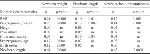
Table 2 shows the correlation between maternal factors and birth order with newborn anthropometric measurements using linear regression analysis. There was a significant positive correlation between mothers’ pre-pregnancy weight and BMI and newborn weight (both p=0.0001), length (p=0.002 and p=0.01, respectively) and head circumference (p=0.001 and p=0.003, respectively). A significant positive correlation was also observed between gestational folic acid consumption and newborn length (p=0.04). There were no significant correlations between mother’s gestational multivitamin intake, educational level and economic status and their newborns’ anthropometric measurements (data not shown). A significant negative correlation was observed between maternal age and newborn weight (p=0.004) and a positive association was determined between birth order with newborn birth weight (p=0.009). Receiver Operating Characteristic (ROC) curves were drawn and the discriminative power of the maternal characteristics and birth order to estimate the risk of LBW was assessed by the area under the curve (Table 3). Only maternal pre-pregnancy BMI, weight and iron intake had statistically significant discriminative ability to distinguish between normal and LBW infants. A strong correlation was observed between neonates’ length and their weight (r=0.61, p=0.0001) and head circumference (r=0.48, p=0.0001).
Correlation of maternal factors with their newborns’ anthropometric measurements using linear regression analysisFootnote a
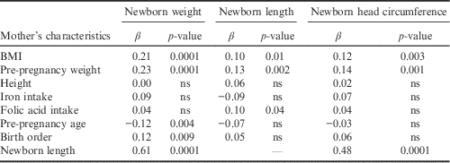
a Confounding factors were gestational age, maternal age, BMI, pregnancy weight gain and birth order. Dependent variables were newborn weight, length and head circumference.
Receiver operating characteristic (ROC) analysis of maternal parameters and birth order in the estimation of the risk for LBW

a AUC is the area under the ROC curve.
Effect of birth order and maternal factors on their newborns’ anthropometric measurements
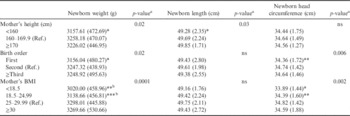
Table 4 shows the effect of maternal factors and birth order on newborn anthropometric measurements using ANOVA. Among mothers of short height (<160 cm) mean newborn weight (p=0.02) and length (p=0.03) were significantly less than those of newborns of middle-height mothers. The birth weight and head circumference of first-born babies were significantly lower than those of babies with second- or higher-born rank (p=0.02 and p=0.006, respectively). Mother’s pre-pregnancy BMI had significant effect on newborn weight and head circumference. Newborn birth weight (p=0.0001) and head circumference (p=0.002) were significantly lower in underweight (BMI<18.5) or normal weight (18.5≤BMI<25) mothers compared with overweight mothers (25≤BMI≥30). Gestational iron, folic acid and multivitamin intakes did not affect newborn anthropometric measurements (data not shown, p>0.05).
Effect of birth order and maternal factors on their newborns’ anthropometric measurements
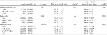
All data were expressed as mean (SD).
a Differences between groups were assessed by ANOVA for variables with three or more categories.
b p<0.05 compared with BMI≥30 category.
*p<0.05; **p<0.01; ***p<0.001, compared with reference category using the Tukey test.
Effect of birth order and maternal factors on LBW rate
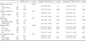
Table 5 shows the effect of birth order and maternal factors on LBW rate using ANOVA and on LBW odds ratio using univariate and multivariate logistic regression analysis. A significantly higher LBW rate was observed in the newborns of mothers aged ≥35 years (12.0%) compared with those aged 18–34.9 (3.9%) or under 18 years (8.7%) (p=0.002). After adjusting for confounding factors (maternal BMI, gestational age and birth order), the odds of being LBW in the newborns of mothers aged≥35 years were 3.82 (95% CI 1.54–9.45, p=0.004) times higher than those aged 18–34.9 years. The LBW rate was significantly higher in the newborns of tall (height≥170 cm, 11.1%) and short mothers (6.1%) compared with mothers of middle height (3.3%) (p=0.03). Additionally, logistic regression analysis showed that, after adjusting for confounding factors (maternal BMI, age, gestational age and birth order), the chances of having LBW newborns among tall and short mothers were 4.00 (95% CI 1.38–11.60, p=0.01) and 2.09 (95% CI, 0.98–4.45, p=0.06) times more than among mothers of middle height, respectively.
The effect of birth order and maternal factors on LBW rate and odds ratio using univariate and multivariate logistic regression analysis
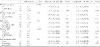
a Differences between groups were assessed by ANOVA for variables with ≥three categories.
b Exp(B) (lower bound, upper bound).
c With and
d without adjusting for confounding factors (gestational age, maternal age, height, BMI and birth order).
e p<0.05 compared with BMI reference category using the Tukey test.
*p<0.05; **p<0.01, compared with second category using the Tukey test.
A significantly higher LBW rate was observed in first-born infants (7.4%) compared with second-birth infants (2.4%) (p=0.01). The odds of being LBW in first- and ≥third-born infants were 5.97 (95% CI, 2.06–17.33, p=0.001) and 3.67 (95% CI, 1.03–13.06, p=0.04) times greater than those of second-birth infants, respectively, after adjusting for confounding factors (maternal BMI, age, and gestational age).
Underweight, normal weight and obese mothers had significantly higher rates of LBW offspring (14.3%, 6.3%, and 6.7%, respectively) compared with overweight mothers (2.0%; p=0.009). The odds of having LBW newborns among underweight, normal weight and obese mothers were 9.07 (95% CI, 2.12–38.85, p=0.003), 3.63 (95% CI, 1.29–10.22, p=0.01) and 3.50 (95% CI, 1.07–11.40, p=0.04) times more, respectively, than for overweight mothers after adjusting for confounding factors (maternal age, gestational age and birth order).
A significantly higher LBW rate was observed among the newborns of mothers who never consumed (10.5%) or irregularly took (6.9%) gestational iron supplements compared with mothers who regularly consumed the supplement (2.6%) (p=0.01). After adjusting for confounding factors (maternal BMI, age, gestational age and birth order), the chances of having LBW babies among mothers who had never or only occasionally received iron supplements were 5.00 (95% CI, 0.54–46.27, p=0.16) and 2.50 (95% CI, 1.13–5.52, p=0.02) times more than for those who received the supplements regularly. Gestational folic acid and multivitamin intake did not affect LBW rate (data not shown).
According to the univariate analysis, the odds of having LBW neonates were significantly higher among mothers who were of advanced age (≥35 years) (OR=3.36; 95% CI, 1.63–6.89; p=0.001), tall stature ≥170 cm (OR=3.62; 95% CI, 1.30–10.11; p=0.01), primiparous (OR=3.26; 95% CI, 1.45–7.33; p=0.004), underweight (OR=8.30; 95% CI, 2.09–32.99; p=0.003), normal weight (OR=3.34; 95% CI, 1.24–9.03; p=0.01), obese (OR=3.59; 95% CI, 1.15–11.22; p=0.03) and occasional iron consumers (OR=2.77; 95% CI, 1.29–5.90; p=0.009).
Discussion
The significant contribution of the low birth weight burden to neonatal and childhood survival, and its adverse impact on the health of adolescents, and even adults, has encouraged global efforts to reduce this burden. This study describes the contribution of prenatal maternal anthropometric characteristics, age, iron intake and birth order on newborns’ anthropometric status and its substantial contribution to the burden of LBW in Iran. The majority of LBW is avoidable by addressing modifiable risk factors, and an in-depth identification of the primary factors contributing to LBW remains a core component of any effective and sustained interventions aimed at improving maternal health and pregnancy outcomes.
The present study found the incidence of LBW to be 5.1% in this urban sample in Iran. Birth weight is a potent indicator of maternal health, quality of maternal health care and nutritional status, as well as newborns’ chances for survival, growth and psychosocial development (Sutton & Darmstadt, Reference Sutton and Darmstadt2013). A reduction of LBW has made a substantial contribution to the achievement of the Millennium Development Goal for the reduction of child mortality. According to UNICF estimates, 16% of all babies born globally in 2013 were of LBW (UNICEF Statistics, no date). In developing countries, the level of LBW was 16.5%, which was more than double that in developed regions (7%) in the year 2004 (UNICEF/WHO, 2004). According to a national survey conducted in 2011, 7.7% of all newborns in Iran were LBW (Rashidian, Reference Rashidian2010; WHO, 2013). The observed decline in the incidence of LBW in the country is a reflection of improved maternal and child health care policies.
This study found that maternal and neonatal anthropometric characteristics were closely inter-correlated. Mother’s nutritional status has long been known to be associated with adverse birth outcomes. Previous studies have also indicated that neonatal birth weight is affected to a great extent by the mother’s body composition at conception. Moreover, mother’s low BMI, pre-pregnancy weight and height are nutrition-related indicators of intrauterine growth restriction (Elshibly & Schmalisch, Reference Elshibly and Schmalisch2008; Voigt et al., Reference Voigt, Rochow, Jährig, Straube, Hufnagel and Jorch2010; Britto et al., Reference Britto, Florêncio, Benedito Silva, Sesso, Cavalcante and Sawaya2013). In a study conducted on 1104 pregnant women, maternal weight and height at antenatal registration were found to be the best predictors of birth weight (Nahar et al., Reference Nahar, Mascie-Taylor and Housne2007). In this study, low BMI was significantly associated with increased LBW rate and had a markedly greater effect than other maternal characteristics. A meta-analysis of 78 studies involving 1,025,794 women indicated that both in developed and developing countries underweight women are at enhanced risk of having a LBW newborn (Han et al., Reference Han, Mulla, Beyene, Liao and McDonald2011).
Overweight mothers in the current study had better neonate size (weight, length and head circumference) and a lower LBW rate. It has previously been established that women of short stature are more likely to have smaller babies (UNICEF/WHO, 2004; Elshibly & Schmalisch, Reference Elshibly and Schmalisch2008). In a study on 2382 neonates and mothers, it was shown that maternal height ≤145 cm was associated with delivery of a greater number of LBW newborns (Mumbare et al., Reference Mumbare, Maindarkar, Darade, Yenge, Tolani and Patole2012). Interestingly, in this work the relative risk of LBW was higher not only among mothers of short stature (although the figure was not significant, p=0.09) but also among taller women (≥170 cm) (Britto et al., Reference Britto, Florêncio, Benedito Silva, Sesso, Cavalcante and Sawaya2013). Differences in cut-off points for height across different studies make inter-study comparisons difficult. However, consistent with the present findings, Kramer et al. (Reference Kramer, Olivier, McLean, Dougherty, Willis and Usher1990) demonstrated that taller mothers tended to have thinner babies. Taken all together, the findings of the present study confirm that anthropometric measurements are helpful in identifying mothers at high risk of delivering LBW newborns, as found by other researchers (Mohanty et al., Reference Mohanty, Prasad, Srikanth Reddy, Ghosh, Singh and Das2006; Ogbonna et al., Reference Ogbonna, Woelk, Ning, Mudzamiri, Mahomed and Williams2007).
Maternal age is another factor influencing the rate of LBW. The results of this study revealed that the rate of LBW in infants was higher in women aged ≥40 years. The risk of LBW has been found to vary markedly by maternal age. It has long been known that the youngest and oldest mothers are more likely to deliver at-risk newborns (Martin et al., Reference Martin, Hamilton, Sutton, Ventura, Menacker and Munson2005). A study in India reported that the prevalence of LBW was 42.86% and 33.33% among mothers aged <18 years and >35 years, respectively (Agarwal et al., Reference Agarwal, Ahmad, Goel, Kumar, Goel, Garg and Punj2012). The newborns of teenage mothers have been shown to be at increased risk of LBW (Friede et al., Reference Friede, Baldwin, Rhodes, Buehler, Strauss, Smith and Hogue1987). Previous investigations have suggested that mothers under the age of 18 and those over 35 are more likely to have LBW babies than those aged 20–34 years (Reichman & Teitler, Reference Reichman and Teitler2006; Restrepo-Méndez et al., Reference Restrepo-Méndez, Lawlor, Horta, Matijasevich, Santos and Menezes2015). Furthermore, advanced maternal age has been postulated as an independent risk factor, not only for low birth weight, but also for preterm delivery and certain adverse pregnancy outcomes (Jolly et al., Reference Jolly, Sebire, Harris, Robinson and Regan2000). It is postulated that the uterus of these two age groups does not sustain pregnancy as well as during the prime childbearing years. Young age is associated with a small uterine volume and a short cervix, which may result in preterm birth and consequently LBW (Gibbs et al., Reference Gibbs, Wendt, Peters and Hogue2012). The increased likelihood of LBW with advanced maternal age has been related to poor placental perfusion or the transplacental flux of nutrients (Godfrey et al., Reference Godfrey, Breier and Cooper1999). Besides, at older ages, women are more likely to have pre-existing, possibly undiagnosed diseases or poor health, including cardiovascular insufficiency, which could lead to poor placentation and consequently LBW (Heffner, Reference Heffner2004; Luke & Brown, Reference Luke and Brown2007; Balasch & Gratacós, Reference Balasch and Gratacós2012). In addition, adverse perinatal outcomes in older mothers might be related to relative infertility, which could influence the likelihood of preterm births and LBW (Luke & Brown, Reference Luke and Brown2007).
In the present work, it was also observed that first-born infants were lighter with a smaller head circumference and increased risk of LBW than subsequent newborns. A relatively lighter weight for first-born infants has also been reported by UNICEF and WHO (UNICEF/WHO, 2004). Moreover, the present finding is in agreement with a previous report that suggested birth order was one of the most important maternal factors affecting birth weight and the incidence of LBW (Elshibly & Schmalisch, Reference Elshibly and Schmalisch2008). In another study carried out on 65,731 live-born infants, primiparity was suggested as a contributing factor in decreasing newborns’ birth weight (Terada et al., Reference Terada, Matsuda, Ogawa, Matsui and Satoh2013). The results of a study in India found the prevalence of LBW to be 42.86% among primiprous mothers (Agarwal et al., Reference Agarwal, Ahmad, Goel, Kumar, Goel, Garg and Punj2012).
The current study demonstrated that the LBW rate was much lower among mothers who had regularly received iron supplements during pregnancy in comparison with those who never or irregularly took iron supplements. The observation is consistent with previous reports that have shown maternal iron supplementation to be associated with a lower risk of LBW (Palma et al., Reference Palma, Perez-Iglesias, Prieto, Pardo, Llorca and Delgado-Rodriguez2008; Balarajan et al., Reference Balarajan, Subramanian and Fawzi2013). In the present study, the LBW rate was more than four-fold in women with no history of receiving iron pills during pregnancy compared with regular iron recipients. The result is similar to the Cleveland trial, in which iron supplementation to pregnant women led to a four-fold reduction in the risk of LBW (Cogswell et al., Reference Cogswell, Parvanta, Ickes, Yip and Brittenham2003). Biological mechanisms that might underlie the effects of iron on fetal growth and preterm birth have been extensively documented (Allen, Reference Allen2001).
The study has several limitations. Data from urban population were used due to the difficulties accessing people living in rural regions and the different surveillance system administrated in rural areas of Iran. Receiving iron and folic acid supplements, as part of the national maternal care programme, does not necessary indicate that all the mothers completely consumed the pills. Lastly, limitations in the geographic area of the study may prohibit generalization of the data to all populations living in urban areas of the country, which warrants further study with greater population size and a broader geographic coverage.
In conclusion, the results indicate that maternal anthropometric characteristics, age, iron intake and birth order affect the risk of LBW. This finding could be beneficial for the classification of areas and population groups that need to be targeted for intervention.