Type 2 diabetes mellitus (T2DM) is a chronic multifactorial metabolic disease that is prevalent in nearly all populations in the world, compromising the quality and expectancy of life in affected individuals. In addition, it is the fifth leading cause of death globally according to a World Health Organization estimate (Ref. Reference Roglic1). T2DM is an important risk factor for many serious long-term complications as a result of chronic hyperglycaemia, which increases the likelihood of microvascular and macrovascular complications such as cardiovascular disease, lower-limb amputation, diabetic retinopathy, arterial hypertension, nephropathy, neuropathy, cancer and premature mortality (Refs Reference Donnelly2, Reference Morrish3). Appropriate clinical intervention, in addition to effective self-management of lifestyle factors such as diet and physical activity, is therefore important to reduce the risk of and mortality from complications.
T2DM is characterised by defects in both insulin action (insulin resistance) and insulin secretion, and typically has an onset after the age of 40 years; however, because of the increasing prevalence of obesity, it is also being observed in younger people (Refs Reference Song and Hardisty4, Reference Hatunic5, Reference Steinberger6).
The heritability of T2DM is well established by twin and adoption studies. It has been clearly demonstrated that genetic factors, as well as interaction with environmental factors, have a key role in predisposition to T2DM (reviewed in Refs Reference Permutt, Wasson and Cox7, Reference Stumvoll, Goldstein and van Haeften8). Monogenic forms of T2DM follow a mendelian inheritance. However, most cases of T2DM are caused by polygenic interaction of small-effect variants within the genome as well as environmental factors. The heritability of T2DM is estimated to be high. Heritability of fasting glucose concentration and homeostatic model assessment for β-cell function (HOMA-B) were estimated to be 72% and 78%, respectively. Features of insulin resistance syndrome also had a high heritability estimate in families with increased genetic susceptibility to T2DM (Ref. Reference Mills9).
The past couple of decades have seen great developments in genetic engineering and sequencing technologies that have allowed researchers to manipulate the mouse genome precisely to improve our understanding of the development of T2DM and the possibility of clinical intervention. In humans, recent advances allowing large-scale genome-wide association studies (GWASs) have greatly accelerated the pace of discovery of susceptibility genes. In an article published in this journal earlier in the year, Vimaleswaran and Loos reviewed the use of GWASs in identifying loci and listed 18 loci for common T2DM (Ref. Reference Vimaleswaran and Loos10). Since then, additional GWAS loci have been identified for T2DM and related glycaemic traits such as haemoglobin A1C levels, which has raised this number to over 40 loci (Refs Reference Dupuis11, Reference Saxena12, Reference Voight13, Reference Soranzo14). Some of these loci contain genes that are known to be involved in monogenic forms of T2DM such as KCNJ11 (encoding Kir6.2), PPARG, HNF1A and SLC2A2 (encoding GLUT2). However, most of the loci contain genes that have never been studied in the context of diabetes, which increases the demand for good mouse models to validate the involvement of candidate genes.
The mouse as a model organism for T2DM
The laboratory mouse (Mus musculus) is considered one of the best species for modelling human disease, because compared with other model organisms such as Drosophila melanogaster and Caenorhabditis elegans, mice are more closely related to humans in evolutionary terms and thus share many biochemical pathways, physiopathological features and developmental properties with humans. Apart from general advantages such as their small size, short gestation, low cost and easy maintenance compared with other mammalian species, another reason for their popularity in biomedical research is the ability to manipulate the mouse genome in a highly precise manner using gene-knockout and transgenic technologies (Refs Reference Peters15, Reference Rosenthal and Brown16).
Over the past couple of decades, the use of mouse models has enabled numerous breakthrough discoveries in many aspects of human disease. With respect to diabetes research, mice can be phenotyped for various metabolic traits using standard procedures such as the glucose tolerance test and the insulin tolerance test. Standard protocols are also used for measuring metabolic rate (by indirect calorimetry), food intake, urine production and physical activity. The urine and plasma obtained can be biochemically analysed for modulators of blood glucose such as insulin, adrenaline, leptin and glucagon. Bone mineral density, and fat and lean mass composition, can also be obtained by clinical imaging technologies such as dual-energy X-ray absorptiometry and magnetic resonance imaging (Refs Reference Fuchs17, Reference Mallon, Blake and Hancock18).
Mouse models of T2DM have been identified by the discovery of spontaneous mutants, or created by genetic modification (by gene targeting and transgenics) to study functions of genes that are implicated from candidate gene approaches or in human association studies.
Spontaneous T2DM obese models
Monogenic models
The Lepob/ob (obese) and Leprdb/db (diabetic) mice were two of the first monogenic mouse models of T2DM found; they were characterised in the 1960s by Douglas Coleman, and the subsequent cloning of leptin by Jeffrey Friedman's research group was followed by numerous studies in laboratories worldwide on its mechanism of action (Refs Reference Coleman19, Reference Halaas20, Reference Chen21, Reference Lee22). The phenotypes of Lepob/ob and Leprdb/db are caused by spontaneous autosomal recessive mutations of leptin (Lep) and its receptor (Lepr), respectively. Both models are characterised by hyperphagia, reduced energy expenditure, obesity and insulin resistance with compensatory hyperinsulinaemia during postnatal development.
Lack of leptin and the leptin receptor in Lepob/ob and Leprdb/db mice results in abnormally high levels of the potent appetite-stimulant neuropeptide Y (NPY), and an increase in circulating cortisol levels that underlies the muscular insulin resistance. Exogenous administration of leptin in Lepob/ob mice, but not in Leprdb/db, was able to restore normal food intake and body weight (Ref. Reference Asensio23). These key mouse models opened a new field of discovery for the control of food intake and the coordination of metabolism.
Polygenic models
Several spontaneous, polygenic, obese models of T2DM exist, including KK (Kuo Kondo), NZO (New Zealand obese), TSOD (Tsumara Suzuki obese diabetes) and M16 (reviewed in Refs Reference Srinivasan and Ramarao24, Reference Chatzigeorgiou25). Usually, polygenic strains are produced by generations of selective breeding for an obesity-related trait that also results in the development of diabetes. The genes are yet to be identified, but these models will provide opportunities for identifying novel components involved in insulin signalling and glucose homeostasis.
Spontaneous T2DM nonobese models
T2DM can exist in the absence of an obese phenotype. Unlike patients with ‘diabesity’, nonobese diabetic patients have disproportionately reduced insulin secretion and only mild insulin resistance (Ref. Reference Chatzigeorgiou25). The best example of a spontaneous T2DM nonobese mouse model is the Akita mouse, derived from a colony of C57BL/6J in Japan. This model carries a spontaneous autosomal dominant mutation in the Ins2 gene, which is the mouse homologue of the human preproinsulin gene (Ref. Reference Yoshioka26). The mutation disrupts normal insulin processing in Ins2 Akita mice and results in a failure to produce mature insulin (Ref. Reference Mathews, Langley and Leiter27).
Knockout and transgenic T2DM models
Since the development of genetic modification in mice via embryonic stem (ES) cells, knockout mice have remained one of the most powerful means for studying the genetics and mechanisms of human diseases, including diabetes. To study the function of a gene or protein in a specific cell type or at a specific developmental stage, conditional gene-targeting systems are often used. The most current system is based on use of the site-specific recombinase Cre (‘cyclisation recombination’), which catalyses recombination between two recognition sites (loxP; ‘locus of crossing over in phage P1’) (Refs Reference Lakso28, Reference Orban, Chui and Marth29, Reference Akagi30). The strategy involves flanking the target gene sequence with two loxP sites (i.e. ‘floxing’) by homologous recombination with the targeting construct in ES cells. Disruption of genes in targeted tissue is activated by crossing the ‘floxed’ strain with a Cre-transgenic line that carries the tissue-specific promoter for Cre expression, thus generating a null allele in cells where Cre is active.
Glucose homeostasis relies on the normal, coordinated functioning of several tissues in the body. Use of the Cre–loxP system for tissue-specific disruption of gene expression has enabled study of the role of genes in specific tissues, including the effect of insulin resistance in specific tissues on disease progression – such as through the use of conditional insulin receptor (INSR) knockouts for muscle and adipose tissue (known as FIRKO and MIRKO). The lines that are typically used in T2DM research for tissue-specific Cre expression include Mck-Cre (muscle), Alb-Cre (liver), aP2-Cre (adipose tissue), UCP1-Cre (brown adipose tissue), RIP-Cre (pancreatic β-cell) and Nestin-Cre (neuron). Detailed information on all the available Cre lines is accessible through the Jackson Laboratory (http://cre.jax.org/index.html).
Expression of Cre can also be induced in a temporally controlled manner. A fusion protein comprising a mutated oestrogen (estrogen) receptor (ER) ligand-binding domain (LBD) and Cre (Cre–ER) is commonly used to control Cre recombinase activity through the administration of the synthetic ER antagonist tamoxifen, which is metabolised to 4-hydroxytamoxifen (4-OHT) in the mouse. The mutation in the LBD is engineered to render it insensitive to endogenous β-oestradiol, but it can be activated by 4-OHT, allowing for exogenous temporal activation of Cre by the administration of tamoxifen orally or by intraperitoneal injection. Currently, several different ERs are available. The human ERT2 (which contains the triple mutation G400V–M543A–L544A) developed by Feil et al. (Ref. Reference Feil31) is considered more sensitive to 4-OHT than the other single-mutation ER LBD alternatives (Refs Reference Feil31, Reference Kim, Nakashima and de Crombrugghe32). In the absence of a ligand, the Cre–ER recombinase is suppressed by heat shock protein (HSP) and is inactive. On binding of 4-OHT to the mutant ER LBD, the Cre–ER fusion protein undergoes a conformational change that causes the release of HSP and results in the translocation of Cre–ER into the nucleus, causing dose-dependent loxP excision of all floxed alleles. This system enables study of the effect of gene disruption in adults in a temporally controlled and dosage-dependent manner, bypassing embryogenesis and other developmental stages (Refs Reference Akagi30, Reference Hayashi and McMahon33, Reference Remedi34, Reference Koch35).
Apart from loss-of-function mutations, human diseases are often associated with variants or mutations that result in the upregulation of a particular gene. Overexpression of a gene or a certain allele (e.g. a dominant negative allele) in the wild-type or mutant mouse is typically generated by pronuclear injection of a transgene, which inserts randomly into the genome. The site of integration and the number of copies present in the host genome are unknown, resulting in variations in expression level and pattern; thus, more than one founder is usually required to verify the result. To obtain better control of transgene expression, transgene integration can be targeted to a predetermined site of the genome, such as the Rosa26 locus (Refs Reference Remedi34, Reference Ernst36, Reference Girard37, Reference Soriano38, Reference Belgardt39). This allows strong and predictable expression of inserted transgenes from exogenous global or tissue-specific promoters, using a Cre–loxP system to remove spacer DNA between promoter and transgene.
Chemically induced (N-ethyl-N-nitrosourea) T2DM models
Since the completion of the mouse genome sequence, an alternative way of mutagenesis has re-emerged as a powerful tool for both forward and reverse genetics approaches. N-ethyl-N-nitrosourea (ENU) is a highly efficient mutagen (with a per locus hit rate of around 1 in 1000, depending on dose and mouse strain) that is used to induce random point mutations in mouse spermatogonia (Refs Reference Justice40, Reference Hitotsumachi, Carpenter and Russell41). ENU mutants might be obtained by screening for an interesting phenotype or screening for mutations in a gene of interest in an archive of F1-mutagenised mice, representing forward (phenotype-driven) and reverse (gene-driven) genetics approaches, respectively.
Unlike the generation of conventional knockouts and transgenics, ENU mutagenesis does not involve any genomic recombination events, leaving the genomic landscape intact and unaltered. Because the location of ENU mutations is random, it requires no pre-assumptions regarding the molecular mechanisms involved in an observed phenotype. Moreover, the effect of a functional change is likely to be subtle compared with a complete disruption by gene targeting, and is therefore likely to resemble the human situation more closely (Ref. Reference Justice40).
Gene-driven approach
This is demonstrated in studies on a protein that has recently been identified in a GWAS for T2DM susceptibility (Ref. Reference Frayling42). Variants within the first intron of the fat mass and obesity-associated (FTO) gene were found to associate with increased obesity risk in humans. However, it was unclear whether the effect could be attributed to FTO or was a possible regulatory effect of the variant on another gene. Using a gene-driven ENU mutagenesis approach, Church et al. isolated an Fto mutant with a dominant I367F substitution. Both heterozygotes and homozygotes show reduced fat mass and increased energy expenditure, suggesting that FTO is the casual gene underlying that particular obesity GWAS locus in humans by regulating energy homeostasis (Ref. Reference Church43). At around the same time, another group had published a targeted Fto knockout that abolished FTO function completely (Ref. Reference Fischer44). Consistent with the Fto-I367F mouse, Fto−/− mice display a lean phenotype. However, there is also a marked developmental defect in Fto−/− with significant perinatal lethality, which is not observed in Fto-I367F, showing that a complete removal of Fto is likely to affect postnatal development as well as metabolism in adults. This demonstrates that ENU mutagenesis is efficient in generating alleles that more closely mimic conditions in humans resulting from gene variations.
Phenotype-driven approach for identifying hyperglycaemic models
ENU mutagenesis is also commonly used for novel gene identification (Refs Reference Nolan45, Reference Hrabe de Angelis46). After mutagenesis, mutant mouse lines might be assessed for a desired phenotype. The causative mutation can then be mapped by generating recombinants from interesting mutants, with subsequent genotyping to narrow down the region containing the mutation. In the near future, the process of identifying genes is likely to be greatly accelerated by the advent of affordable high-throughput whole-exome and -genome sequencing for identifying nucleotide changes within candidate regions, including noncoding conserved elements and regulatory regions.
Recently, ENU mutagenesis projects using plasma glucose as a screening parameter have identified a range of hyperglycaemia lines harbouring dominant and recessive mutations with various degrees of disease severity (Ref. Reference Aigner47), most of which are yet to be mapped. One of the mutants identified from a phenotype-driven screen for free-fed (nonfasted) plasma glucose was GENA348, which has been mapped to an I366F substitution in Gck (Ref. Reference Toye48), the enzyme implicated in human MODY2 (maturity onset diabetes of the young). Unlike the homozygous Gck-knockout mouse (Refs Reference Grupe49, Reference Bali50), the GckI366F/I366F mouse is viable, demonstrating the power of ENU mutagenesis in generating models of complex phenotypes that are directly relevant to human disease. Using this approach, novel genes involved in glucose homeostasis can be discovered.
Sensitised ENU mutagenesis
Apart from novel gene identification and allelic series generation, ENU mutagenesis can also be applied to dissecting genetic interactions. Recently, gene targeting and ENU mutagenesis have been combined to identify modifier genes of mutant phenotypes in sensitised mutagenesis screens. For example, a recent study has used an existing knockout model, Insr+/− mice, which are insulin resistant but do not develop diabetes, as a sensitising condition in an ENU mutagenesis screen. This study has identified an autosomal dominant mutation in the SRY-related high-mobility group box 4 gene (Sox4) that causes hyperglycaemia in the mutants only in the presence of the Insr+/− allele (Ref. Reference Goldsworthy51). This model represents a novel digenic model of insulin resistance coupled with an insulin secretory defect, which closely resembles the genetic heterogeneity underlying inheritance of T2DM. Using this approach, it is possible to identify interacting partners of the insulin signalling pathway, in which the presence of the Insr+/− allele might enhance or modify the diabetic phenotype.
Use of mouse models in dissecting T2DM pathways
Mouse models of insulin resistance
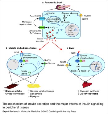
The effects of insulin on glucose homeostasis depend largely on the responsiveness of target peripheral tissues – mainly muscles and liver. Insulin resistance is a primary feature of T2DM and is characterised by impaired glucose uptake in muscle and adipose tissue, and the lack of hepatic inhibition of gluconeogenesis or glucose output (Refs Reference Nandi52, Reference Pirola, Johnston and Van Obberghen53) (Fig. 1). Insulin acts through a cell-surface receptor that belongs to a subfamily of growth factor receptor tyrosine kinases, comprising three members: INSR, type 1 insulin-like growth factor receptor (IGF1R) and the INSR-related receptor (Ref. Reference Nakae, Kido and Accili54).
The mechanism of insulin secretion and the major effects of insulin signalling in peripheral tissues. (a) Glucose uptake into the pancreatic β-cell by GLUT2 is processed by several glycolytic enzymes such as GCK, which catalyses the phosphorylation of glucose. The subsequent glycolysis and mitochondrial metabolism lead to an increase in the ATP:ADP ratio, causing the KATP channel to shut. This produces a membrane depolarisation that activates the voltage-gated Ca2+ channel. The increase of Ca2+ influx leads to a rise in intracellular [Ca2+], which triggers the release of insulin into the bloodstream. This process is regulated by several transcription factors such as HNF4α, HNF1α and PDX1, which have been reported to regulate the expression of genes involved in insulin secretion. (b,c) The effect of insulin in glucose homeostasis is executed by insulin-responsive tissues such as skeletal muscle, adipose tissue and liver. Insulin binds to the extracellular domain of INSR and causes a conformational change, leading to autophosphorylation and tyrosine phosphorylation of intracellular protein substrates such as IRS1, which then binds to and activates the heterodimeric phosphoinositide 3-kinase (PI3K) formed by regulatory (p85) and catalytic (p110) subunits. This leads to the activation of phosphatidylinositol trisphosphate [PtdIns(3,4,5)P 3]-dependent kinases such as AKT (PKB) and the modulation of various enzyme activities, as detailed separately for muscle, adipose tissue and liver below. The combined effects of these actions contribute to the maintenance of glucose homeostasis in the organism. (b) In myocytes and adipocytes, activation of AKT results in the insulin-stimulated translocation of GLUT4 from intracellular vesicles to the plasma membrane and increased glucose uptake. Apart from the immediate actions, insulin also induces the increase of glucose storage in muscle and adipose tissues in the form of glycogen and triglycerides, respectively, and inhibits lipolysis. (c) In hepatocytes, glucose transport is through GLUT2, and is not regulated by insulin. Here, insulin promotes AKT-dependent phosphorylation of the transcription factor FOXO1, and results in decreased expression of gluconeogenic enzymes, such as PCK2 and G6PC, leading to a reduction in gluconeogenesis and hepatic glucose output. In addition to these immediate actions, insulin also increases glucose storage in the liver in the form of glycogen. Abbreviations: ATP/ADP, adenosine triphosphate/ diphosphate; FOXO1, forkhead box O1; G6P, glucose-6-phosphate; G6PC, glucose-6-phosphatase, catalytic subunit; GCK, glucokinase; GLUT, glucose transporter; HNF, hepatocyte nuclear factor; INSR, insulin receptor; IRS1, insulin receptor substrate 1; NEUROD1, neurogenic differentiation 1; PDX1, pancreatic and duodenal homeobox 1; PCK2, phosphoenolpyruvate carboxykinase 2; PKB, protein kinase B.

The principal substrate for INSR and IGF1R is the cytoplasmic protein insulin receptor substrate 1 (IRS1). After activation by tyrosine phosphorylation, IRS1 binds to and activates the regulatory subunit of phosphoinositide 3-kinase (PI3K). This leads to the activation of phosphatidylinositol trisphosphate [PtdIns(3,4,5)P 3]-dependent kinases such as the serine/threonine protein kinase AKT (also known as protein kinase B, PKB), protein kinase C isoforms and the serum- and glucocorticoid-inducible kinase, resulting in the translocation of the glucose transporter GLUT4 from cytoplasmic vesicles to the plasma membrane and subsequent glucose uptake into muscle and adipose tissue (Ref. Reference Shepherd, Withers and Siddle55).
The expression of GLUT4 is therefore upregulated by insulin. It is essential in the postprandial uptake of excess glucose from the bloodstream into muscle and adipose tissue, where glucose is stored in the form of glycogen and triglyceride, respectively. In hepatocytes, however, glucose uptake is through GLUT2 (encoded by Slc2a2) and is not mediated by insulin. Because the liver functions as a ‘glucostat’ for the organism, the rate of glucose entry into hepatocytes is proportional to blood glucose levels (Ref. Reference Thorens56).
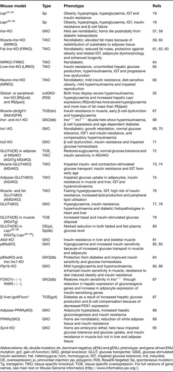
To understand how the insulin signalling pathway mediates insulin sensitivity in peripheral tissues, mouse models with targeted disruption in components of the insulin signalling pathway have been generated to allow the dissection of disease mechanisms. The mouse models of insulin resistance described in this review are listed in Table 1.
Mouse models of insulin resistance

Abbreviations: db, double mutation; dn, dominant negative; pENU and gENU, phenotype- and gene-driven ENU mutation; gof, gain-of-function; GKO, global knockout; GLUT, glucose transporter; GSIS, glucose-stimulated insulin secretion; het, heterozygous; hom, homozygous; IGT, impaired glucose tolerance; ind, inducible; OE, overexpression; pi, pronuclear injection; pp, polygenic; R26, Rosa26-targeted; Sp, spontaneous mutation; Tg, transgenic; TiKO, tissue-specific knockout; TiOE, tissue-specific overexpression. For full versions of gene names, see main text or Mouse Genome Informatics (http://www.informatics.jax.org/).
Use of mouse models in studying insulin resistance in skeletal muscle
Disruption of Insr in mice results in early neonatal lethality in homozygotes characterised by severe hyperglycaemia, diabetic ketoacidosis and a marked reduction of white adipose tissue, whereas heterozygous mice display normal glucose tolerance (Refs Reference Accili57, Reference Joshi58). To study the effect of the complete loss of INSR in specific tissues, several tissue-specific knockout mouse models of Insr have been generated.
Skeletal muscle is regarded as the most important tissue in maintaining glucose homeostasis because it accounts for the majority of insulin-dependent glucose disposal in the body. Mice deficient in INSR in muscles (MIRKO) have normal glucose uptake and plasma insulin levels but increased fat mass, serum triglyceride and free fatty acid levels, and a threefold increase in insulin-stimulated adipose glucose transport, suggesting that insulin resistance in muscle contributes to alterations in fat metabolism and redistribution of substrates to adipose tissue (Refs Reference Kim59, Reference Bruning60). To study the role of adipose tissue in MIRKO mice, a mouse model that is insulin resistant both in skeletal muscle and in white and brown adipose tissue (M/FIRKO) was generated. Although the combined effect results in a mild impairment of β-cell function, the mice are not hyperglycaemic, and hepatic glycogen synthesis and gluconeogenesis were unaffected (Ref. Reference Lauro64). This can be explained by the insulin-independent mechanism of glucose uptake in the muscle of MIRKO, because it was found that muscle contraction alone triggers GLUT4 translocation through the activation of AMP-activated kinase and is adequate for achieving normal resting glucose uptake without muscle INSR expression (Ref. Reference Wojtaszewski91).
The lack of hyperglycaemia in M/FIRKO mice might also be explained by the fact that apart from INSR, IGF1R might exert a compensatory role in mediating insulin-stimulated glucose transport by GLUT4 in muscle, as evident in the finding that mice with combined muscle-specific disruption of both IGF1 and insulin signalling display β-cell dysfunction (Ref. Reference Fernandez67), whereas knocking out Insr alone (MIRKO) results in a mouse with normal glucose tolerance.
In contrast to Insr knockout, removal of GLUT4 directly blocks glucose uptake into the cell. As a result, mice deficient in GLUT4 in muscle (MG4KO) lose both contraction- and insulin-stimulated glucose uptake, resulting in glucose intolerance and severe insulin resistance in adipose tissue and liver at an early stage, which are secondary to hyperglycaemia or glucose toxicity (Ref. Reference Zisman73). Interestingly, the markedly reduced glucose transport in muscle is coupled with an increase in glycogen synthase activity and a decrease in glycogen phosphorylase activity, resulting in increased glycogen content in muscle in the fasted state when glucose transport is reduced (Ref. Reference Kim74). In contrast to MIRKO mice, however, MG4KO mice do not have increased adipose mass to compensate for the reduction in muscle glucose uptake. This might explain the diabetic phenotype in MG4KO that is absent in MIRKO.
Moreover, overexpression of GLUT4 in muscle results in an increase in basal and insulin-stimulated glucose uptake, suggesting that skeletal muscle GLUT4 is rate limiting for whole-body glucose disposal and that GLUT4 is a potential target for pharmacological or genetic manipulation to correct metabolic abnormalities associated with noninsulin-dependent T2DM (Ref. Reference Ren79). Consistent with this finding, overexpression of GLUT4 in Leprdb/db also results in an improvement in glucose tolerance and insulin-stimulated glucose uptake (Refs Reference Ren79, Reference Gibbs80).
Insulin-stimulated vesicle trafficking of GLUT4 to the plasma membrane is regulated by various proteins such as syntaxin 4, which is a plasma membrane SNARE protein that mediates vesicle fusion. Mice homozygous null for Syn4 display early embryonic lethality, whereas heterozygotes show no developmental abnormalities but show glucose intolerance with an ~50% decrease in GLUT4 translocation in skeletal muscle (Ref. Reference Yang90). Nevertheless, insulin-stimulated glucose uptake and metabolism in adipose tissue and liver are normal in Syn4 +/− mice. This demonstrates that Syn4 is essential in the regulation of insulin-stimulated glucose uptake in muscle by GLUT4 translocation, and that genetic alteration in Syn4 can result in impairment in whole-body insulin-stimulated glucose metabolism.
Role of adipose tissue in glucose homeostasis and insulin resistance
Obesity is a major risk factor for T2DM. High body fat mass and high levels of circulating lipids such as free fatty acids are prominent clinical features of T2DM (Ref. Reference Samuel, Petersen and Shulman92). Therefore, much research on T2DM has focused on studying the role of adipose tissue in whole-body insulin sensitivity and the role of adipokines in regulating glucose and lipid metabolism.
Consistent with INSR global knockout (Ref. Reference Joshi58), adipose-tissue-specific ablation of INSR (FIRKO) leads to defects in insulin-stimulated glucose uptake and a defective antilipolytic effect in white adipose tissue. FIRKO mice also demonstrate a marked reduction in fat mass, and protection against age-related obesity and other obesity-related metabolic abnormalities such as glucose intolerance and insulin resistance. This is possibly due to lack of insulin-induced triglyceride storage or lack of the antilipolytic effect of insulin in white adipose tissue (Ref. Reference Bluher61). In addition, the white adipose tissue of FIRKO mice demonstrates a polarisation of adipocytes into two major populations of either large or small cells, differing in basal triglyceride synthesis, lipolysis, and the expression of key transcription factors that regulate fatty acid synthesis such as fatty acid synthase and sterol regulatory element-binding protein (Ref. Reference Bluher62). Therefore, insulin signalling in white adipose tissue is likely to be involved in adipocyte differentiation, triglyceride storage, inhibition of lipolysis, insulin-stimulated glucose uptake and development of obesity (Refs Reference Bluher61, Reference Bluher62), although it is not indispensable in the maintenance of glucose homeostasis.
In adipose tissue, removal of GLUT4 (AG4KO) abolishes insulin-stimulated glucose uptake in adipocytes, and causes insulin resistance in muscle and liver despite a normal level of GLUT4 expression in muscle. These combined effects eventually result in the development of hyperglycaemia and glucose intolerance in AG4KO mice (Ref. Reference Abel75), which is consistent with the findings from GLUT4-null mice (Refs Reference Katz77, Reference Stenbit78). Similarly, ablation of GLUT4 in both adipose tissue and muscle (AMG4KO) results in glucose intolerance, fasting hyperglycaemia and a higher incidence of insulin resistance than is seen from knocking out GLUT4 in either tissue alone (Ref. Reference Kotani76). AMG4KO mice also show an increase in glucose uptake in the liver and lipid synthesis, suggesting a compensatory mechanism to insulin resistance.
Interestingly, when GLUT4 is overexpressed in the adipose tissue of MG4KO mice, despite resulting in a slight increase in fat mass and free fatty acid level, it reverses whole-body insulin resistance in MG4KO mice without restoring glucose transport in muscle (Ref. Reference Carvalho72). This might be due to a larger surface area for glucose uptake provided by the hyperplastic mass of small adipocytes, thereby dissociating mild obesity from insulin resistance when glucose transport is high in adipose tissue. This demonstrates that adipose tissue has a central role in regulating insulin sensitivity and energy balance in the body.
Insulin resistance has long been associated with obesity and the abnormal accumulation of lipids in tissues other than adipose tissue (Ref. Reference Samuel, Petersen and Shulman92). The peroxisome proliferator-activated receptors (PPARs) are members of the nuclear receptor transcription factor superfamily and regulate many cellular and metabolic processes, including lipid uptake and catabolism. One of the PPAR members, PPARγ, is known to regulate fatty acid storage and glucose metabolism by stimulating lipid uptake and adipogenesis in adipocytes. Adipose-specific knockout of PPARγ results in progressive loss of fat, as well as hyperlipidaemia, fatty liver and hepatic insulin resistance because of lipid accumulation (Ref. Reference He88), and protection against high-fat diet-induced increase in fat mass (Ref. Reference Jones93). Selective disruption of the PPARγ2 splice isoform also results in a similar reduction in fat mass and insulin resistance, accompanied by a decreased expression of IRS1 and GLUT4 in muscle and a reduced capacity for adipogenesis (Ref. Reference Zhang89).
Modelling for insulin resistance in other tissues
Unlike MIRKO and FIRKO, liver-specific knockout of INSR (LIRKO) in the mouse resulted in whole-body insulin resistance and severe glucose intolerance because of the failure of insulin to suppress hepatic glucose production and regulate the hepatic expression of gluconeogenic enzymes. In addition, insulin resistance results in hyperinsulinaemia because of a combination of decreased hepatic insulin clearance and increased insulin secretion as a result of the hyperproliferation of pancreatic β-cells. Therefore, insulin signalling in the liver is crucial in the regulation of postprandial glucose homeostasis by regulating hepatic glucose production and maintaining whole-body insulin sensitivity and insulin clearance (Ref. Reference Michael65).
Knockout of INSR in the brain (NIRKO) results in pleiotropic effects on the normal functioning of various systems in the adult. Despite the neuron-specific removal of Insr, NIRKO mice have normal brain development but develop diet-induced obesity, showing increased body fat and plasma leptin, with female-specific hyperphagia. They also display mild insulin resistance, hyperinsulinaemia, hyperglyceridaemia and impaired fertility because of hypothalamic dysregulation of luteinising hormone. Therefore, INSR signalling in the central nervous system (CNS) has an important role in both reproduction and the regulation of energy homeostasis (Ref. Reference Bruning66). Interestingly, restoration of INSR function in the brain alone is able to rescue Insr−/− mice from neonatal death (Ref. Reference Okamoto94).
To study the consequences of insulin resistance in adult mice, mice with an inducible whole-body INSR deficiency [IR(Δwb)] and an inducible INSR knockout throughout the body except the brain [IR(Δper)] were generated (Ref. Reference Koch35). Both mice models display severe hyperinsulinaemia, hyperglycaemia and upregulation of hepatic Lepr expression. However, compared with IR(Δper) mice, IR(Δwb) mice have more severe hyperglycaemia, a more pronounced reduction in white adipose tissue mass, and severe hypoleptinaemia. Moreover, only IR(Δper) mice display increased leptin-activated hepatic STAT3 phosphorylation. This study demonstrates the role of CNS insulin action in regulating white adipose tissue mass and glucose metabolism by hepatic STAT3 activation.
Study of the intracellular events of insulin signalling using mouse models
IRS1 is the principal substrate for INSR and IGF1R, which binds to and activates PI3K and several other proteins containing SH2 domains. Knockout of IRS1 results in growth retardation, mild impairment of glucose tolerance and a decrease in insulin/IGF1-stimulated glucose uptake (Refs Reference Araki69, Reference Tamemoto70), demonstrating the role of IRS1 in mediating the growth-promoting actions of IGF1R and metabolic signalling.
The mild phenotype observed in Irs1−/− mice might be due to the presence of residual insulin/IGF1 action, which can be explained by the existence of IRS2, an alternative insulin substrate capable of activating the signalling cascade. Mice lacking IRS2 show both insulin resistance and impaired β-cell function, and a subsequent aberration in glucose homeostasis resulting in the development of T2DM. In contrast to Irs1−/− mice, the number of β-cells was reduced in Irs2−/− mice, suggesting that IRS2 has a crucial role in the regulation of β-cell mass (Refs Reference Kubota95, Reference Withers71).
As mentioned before, T2DM is commonly inherited as a nonmendelian trait as a result of interaction between polygenic alterations and various environmental factors. This is demonstrated by the observation that mice heterozygous null for either Insr or Irs1 have normal glucose homeostasis, whereas double heterozygotes of Insr and Irs1 display insulin resistance, hyperinsulinaemia and β-cell hyperplasia, and develop diabetes in an age-dependent manner. This shows that epistatic interaction between two subclinical defects in the insulin signalling pathway can result in T2DM that can be inherited in a nonmendelian manner (Ref. Reference Bruning68).
Apart from the key components involved in promoting insulin-stimulated glucose uptake, there are various other phosphatases and kinases that are involved in the negative regulation of insulin signalling in insulin-sensitive tissues, including regulators that are responsible for the termination and modulation of the pathway (reviewed in Ref. Reference Nandi52).
Most PI3K activation in insulin-responsive tissues is mediated by the regulatory subunit p85α (encoded by Pik3ca), and its splice variants p50α and p55α. Mice lacking p85α show an increase in insulin sensitivity and glucose transport in adipose tissue and skeletal muscle, leading to hypoglycaemia (Ref. Reference Terauchi82). In addition, gluconeogenesis is elevated as a compensatory mechanism to the increased glucose uptake in peripheral tissues (Ref. Reference Aoki83). Interestingly, when crossed with insulin-resistant mouse models (Insr and Irs1 knockouts), haploinsufficiency of p85α exerts protection from insulin resistance and diabetes by improving the efficiency of insulin signalling (Ref. Reference Mauvais-Jarvis84).
Mice deficient in the PtdIns(Reference Morrish3,Reference Song and Hardisty4,Reference Hatunic5)P 3-dependent kinase AKT2 display insulin resistance in the liver and skeletal muscles and a lack of normal suppression of hepatic glucose production. This results in mild fasting hyperglycaemia, which becomes more pronounced during fed states. The Akt2-null mice also display hyperinsulinaemia because of the decreased responsiveness to insulin in peripheral tissues. This shows that AKT2 is essential in insulin-dependent glucose uptake/disposal and glucose production (Ref. Reference Cho81). The protein tyrosine phosphatase 1B (PTP1B) is the key substrate for AKT. Phosphorylation of PTP1B by AKT at Ser50 is known to negatively modulate its phosphatase activity and to create a positive feedback mechanism for insulin signalling (Ref. Reference Ravichandran96). Disruption of PTP1B in the mouse results in an improvement in whole-body insulin sensitivity (improved glucose tolerance, decreased glucose and insulin levels) and protection from high-fat-diet-induced obesity and insulin resistance, because of a reduction in tyrosine dephosphorylation of INSR and its substrates in both muscle and liver (Refs Reference Elchebly85, Reference Klaman86). It has been shown that PTP1B is required for the termination of insulin action, making it a promising target for the treatment of T2DM.
The forkhead box O1 (FOXO1) transcription factor is an AKT substrate in pancreatic β-cells and insulin-responsive tissues. The activity of FOXO1 is negatively regulated by insulin through AKT-dependent phosphorylation and nuclear exclusion. Deficiency of FOXO1 is able to restore insulin sensitivity and rescue diabetes in Insr+/− mice by reducing the hepatic expression of gluconeogenic genes and increasing the expression of insulin-sensitising genes in adipocytes (Ref. Reference Nakae87). Haploinsufficiency of FOXO1 also results in protection against diet-induced diabetes in mice (Ref. Reference Nakae97). In contrast, gain-of-function FOXO1 mutation targeted to the liver and β-cells causes diabetes because of the combined effects of an increase in hepatic gluconeogenesis and impaired β-cell compensation as a result of decreased Pdx1 expression (Ref. Reference Nakae87).
Mouse models of β-cell dysfunction
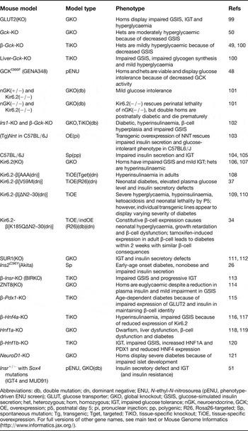
The core function of the β-cell is to secrete insulin into the bloodstream on glucose stimulation to initiate signal transduction through the activation of INSRs that induce intracellular events required for the regulation of lipid metabolism and maintenance of glucose homeostasis (Ref. Reference Chang-Chen, Mullur and Bernal-Mizrachi98). The mouse models of β-cell dysfunction described in this review are listed in Table 2.
Mouse models of β-cell dysfunction

Abbreviations: db, double mutation; dn, dominant negative; ENU, N-ethyl-N-nitrosourea (pENU, phenotype-driven ENU screen); GLUT, glucose transporter; GKO, global knockout; GSIS, glucose-simulated insulin secretion; het, heterozygous; hom, homozygous; IGT, impaired glucose tolerance; nGK, neuroendocrine, GCK; OE, overexpression; p5, postnatal day 5; pi, pronuclear injection; pp, polygenic; R26, Rosa26-targeted; Sp, spontaneous mutation; Tg, transgenic; Tget, targeted; TiKO, tissue-specific knockout; TiOE, tissue-specific overexpression. For full versions of other gene names, see main text or Mouse Genome Informatics (http://www.informatics.jax.org/).
Impairment of glucose-stimulated insulin secretion (GSIS) is an early feature of T2DM. The key events involved in normal GSIS in the β-cell are as follows: glucose is transported into the β-cell through the facilitative glucose transporter GLUT2, and is then phosphorylated to form glucose-6-phosphate (G6P), a process catalysed by glucokinase (GCK), which is also a glucose sensor. Downstream events include glycolysis and mitochondrial metabolism, which leads to the generation of ATP. The increased mitochondrial ATP:ADP ratio causes the ATP-sensitive potassium (KATP) channels to shut, producing a membrane depolarisation that activates voltage-gated Ca2+ channels. The rise in intracellular [Ca2+] leads to increased fusion of insulin granulae with the cell membrane and a subsequent increase in proinsulin secretion (Ref. Reference Chang-Chen, Mullur and Bernal-Mizrachi98) (Fig. 1).
MODY is a monogenic form of T2DM characterised by an early age of onset (around 25 years) with an autosomal dominant mode of inheritance (Refs Reference Vaxillaire and Froguel122, Reference Vaxillaire and Froguel123). Mutations in at least six different genes are known to cause MODY: one encodes the β-cell glucose-sensing glycolytic enzyme GCK (MODY2), whereas the other five encode transcription factors, including hepatocyte nuclear factor (HNF)-4α, HNF-1α, pancreatic and duodenal homeobox-1 (PDX1), HNF-1β and NEUROD1/BETA-2 (associated with MODY1, -3, -4, -5 and 6, respectively). These transcription factors participate in a regulatory network essential for adult β-cell function, including the transcription of insulin and other β-cell-specific genes. This set of genes does not explain all cases of MODY; other genes reported to cause MODY in humans include KLF11, CEL, PAX4, INS and BLK, which are related to MODY7, −8, −9, −10 and −11, respectively, as reported in the Online Mendelian Inheritance in Man (OMIM) database (http://www.ncbi.nlm.nih.gov/omim).
Glucose uptake and glucose processing
The disruption of INSR in β-cells (βIRKO) in mice has shed light on our understanding of the role of INSR in glucose sensing by the β-cell. βIRKO mice show a reduction in acute first-phase insulin secretion in response to glucose, and an age-dependent glucose intolerance, indicating that insulin has an autocrine action that stimulates its own secretion via INSR in β-cells. This suggests that defects in insulin signalling at the level of the β-cell might contribute to the impairment in insulin secretion in T2DM (Ref. Reference Kulkarni113).
The Slc2a2-knockout mouse (lacking GLUT2) was one of the first models of T2DM. Homozygotes are hyperglycaemic with impaired insulin secretion and increased plasma glucagon and free fatty acid levels. There is also a defect in the postnatal development of pancreatic islets with disproportionate α- to β-cell ratios. These data show that GLUT2 is required for GSIS in pancreatic β-cells and for the normal function and development of the pancreas (Ref. Reference Guillam99).
Mice deficient in GCK in all tissues or in β-cells alone die perinatally with severe hyperglycaemia (Refs Reference Grupe49, Reference Postic100, Reference Remedi101), while the specific expression of Gck in β-cells of Gck-knockout mice was sufficient for survival (Ref. Reference Grupe49). Mice heterozygous null for Gck, either globally or in β-cells, survive but are moderately hyperglycaemic (Refs Reference Grupe49, Reference Bali50, Reference Postic100, Reference Remedi101), which resembles MODY2 in humans. Disruption of Gck in the liver also causes milder hyperglycaemia, but, in addition, hepatic Gck-knockout mice have defects in insulin-stimulated glycogen synthesis. Surprisingly, they also have impaired GSIS. Taken together, these data show that the GCK-linked MODY2 cases likely reflect deficiencies in both β-cell and hepatic GCK activity (Ref. Reference Postic100). Interestingly, knocking out of the KATP-channel-forming subunit Kir6.2 (encoded by Kcnj11) rescues perinatal lethality in mice lacking neuroendocrine GCK [nGK(+/−)] and such mice show only mild glucose intolerance, indicating that the effect of GCK deficiency is due to altered KATP signalling (Ref. Reference Remedi101).
To study the interaction between insulin resistance and insulin secretory defects in the development of T2DM, a double knockout mouse with disruption of Irs1 and β-cell-specific Gck was generated. Both Irs1−/− and mice heterozygote null for Gck are nondiabetogenic, but when mice carry both these knockouts, they develop overt diabetes with hyperglycaemia and β-cell hyperplasia. This shows that combined minor defects can cause overt diabetes, which recapitulates the polygenic nature of T2DM in humans (Ref. Reference Terauchi102).
KATP channels and β-cell electrical activity
The KATP channels couple β-cell metabolism to electrical activity and are important in the control of insulin secretion. The β-cell KATP channel is a tetrameric complex consisting of four Kir6.2 subunits, each of which is associated with a SUR1 subunit (encoded by ABCC8) (Ref. Reference Aguilar-Bryan124). Mutations in both subunits have been found in both hypoglycaemic and hyperglycaemic patients (Refs Reference Flanagan125, Reference Shimomura126).
Over the past couple of decades, several transgenic and knockout lines have been generated for studying the function of the KATP channel in glucose homeostasis. The first animal model of diabetes caused by a mutation in the KATP channel was generated by Koster et al. in 2000 (Ref. Reference Koster109), which came before the identification of KCNJ11 mutations in human neonatal diabetes (Ref. Reference Gloyn127). This study was followed by the generation of other gain-of-function mutations in Kcnj11 (Refs Reference Remedi34, Reference Girard37, Reference Koster110), all of which cause the reduction of ATP sensitivity and result in the KATP channel remaining open and unable to respond to an endogenous stimulus. The hypoexcitability of β-cells in these mutants suppresses the electrical trigger of insulin secretion and subsequently results in hyperglycaemia.
Loss-of-function mutations of β-cell KATP subunits (both Kir6.2 and SUR1) cause the absence or closure of the KATP channel, thereby causing dysregulation of insulin release, and leading to hyperinsulinaemic hypoglycaemia of infancy (HI) in humans (Refs Reference Nestorowicz128, Reference Huopio129). In mice, complete loss of Kir6.2 causes transient hyperinsulinaemia in neonates, but both mice lacking Kir6.2 and mice lacking SUR1 show glucose intolerance and loss of insulin secretion in adults as a result of the initial excessive excitability above the hyperinsulinaemia threshold that results in β-cell dysfunction (Refs Reference Shiota111, Reference Seghers112, Reference Miki106). Partial loss of β-cell KATP channel activity, as observed in dominant-negative Kir6.2-[AAA] (Ref. Reference Koster108) and Kir6.2 (+/−) mice (Ref. Reference Remedi107), causes hyperinsulinaemia that more closely recapitulates HI in humans.
Recently, a nonsense mutation (Y12X) of Kir6.2 was generated by ENU mutagenesis in the mouse, and is the same as that found in a human infant who had inherited the Y12X allele from both parents. Although the patient displayed hyperinsulinaemic hypoglycaemia from birth (and underwent near-total pancreatectomy), Kir6.2(Y12X/Y12X) mice show hypoinsulinaemia and hyperglycaemia in the adult (Ref. Reference Hugill130). This further demonstrates a discrepancy between humans and mice in the effect of the same mutation on β-cell function and possibly on disease onset and severity, although the mouse model might represent later events in such patients.
Insulin synthesis and secretion
Recent human GWASs have identified novel gene variants highly associated with altered susceptibility to T2DM, some of which are involved in the insulin signalling and secretory pathway. One of the variants lies within SLC30A8 (Ref. Reference Zeggini131), which encodes the islet-specific zinc transporter ZNT8 that provides zinc for insulin-hexamer formation. Mice deficient in ZNT8 show hypoinsulinaemia despite normal plasma glucose levels and normal insulin sensitivity, and a reduction in GSIS (Ref. Reference Pound114). Although the loss of ZNT8 does not result in major alterations in glucose metabolism, the presence of a phenotype in Znt8−/− mice makes it conceivable that variants in SLC30A8 might be linked to T2DM risk in humans because of a secretory defect. Interestingly, the expression of SLC30A8 is reported to be regulated by TCF7L2 polymorphisms (Ref. Reference Sladek132), which are strongly associated with T2DM risk in humans (Ref. Reference Grant133).
Transcription factors involved in GSIS of β-cells
Transcription factors are vital for many important biological processes, such as development and cell cycle control. Islets in T2DM often show abnormal expression of genes that are involved in the development and normal functioning of pancreatic β-cells (Ref. Reference Shao134). Global or β-cell-specific disruption of the MODY-associated genes Hnf4a, Hnf1a (Tcf1), Pdx1, Hnf1b (Tcf2) Neurod1 and also Sox4 all result in β-cell dysfunction and have shed light on our understanding of the mechanisms of insulin secretion and glucose homeostasis (Refs Reference Ahlgren115, Reference Gupta116, Reference Miura117, Reference Lee, Sauer and Gonzalez118, Reference Pontoglio119, Reference Wang120, Reference Naya121). These transcription factors function in the nucleus of the β-cell and regulate the transcription of genes that are key to various aspects of β-cell function such as Ins2, Igf1, Gck, Kcnj11, Slc2a2, Ucp2 and several glycolytic enzymes either directly or, in the case of HNF1β, indirectly through effects on the expression of other transcription factors such as HNF1α and PDX1 (Refs Reference Vaxillaire and Froguel123, Reference Fajans, Bell and Polonsky135).
Mapping quantitative trait loci for T2DM
T2DM in humans is caused by a complicated interplay of many factors, including environmental factors and interaction between various genes with varying degrees of effect. Unlike the complexity of human T2DM, the development of diabetes in laboratory mice is genetically determined (unless challenged under controlled conditions). This allows researchers to eliminate the environmental/lifestyle factors and focus on the genetics underlying an observed phenotype. The availability of inbred mouse strains and the accessibility of abundant genomic resources have also allowed us to study the full impact of small- or moderate-effect polymorphic loci and replicate infinitely to reach adequate statistical power.
Because of their properties, inbred strains have also been used as a common tool in forward genetics approaches to identify novel susceptibility genes in complex diseases. The genetic variation between inbred strains often confers varying degrees of susceptibility or resistance to certain conditions or diseases. The availability of well-characterised inbred strains has therefore provided the possibility of mapping novel loci between two strains. Mapping for a novel disease locus involves interval mapping between an unmapped mutant with an interesting phenotype (such as a spontaneous mutant or a disease-prone inbred strain) and a strain of different background or with contrasting phenotype. This approach is powerful in identifying novel genes in mice that would not have been picked up in candidate gene approaches, or gene effects that would have been masked by environmental factors in human association studies (Ref. Reference Schmidt136).
For example, C57BL/6J is a substrain that shows mild glucose intolerance and impaired insulin secretion despite normal insulin sensitivity. By interval mapping of intercrossed progeny between C57BL/6J and C3H/HeH mice, three loci were identified, with nicotinamide nucleotide transhydrogenase (Nnt), an inner mitochondrial proton pump, being a strong candidate gene within one of the loci (Refs Reference Huang104, Reference Toye105). The phenotype in C57BL/6J is likely to be caused by the spontaneous five-exon deletion of Nnt that results in a complete absence of NNT (Ref. Reference Toye105). This is supported by the finding that transgenic expression of an intact copy of Nnt was able to rescue the impaired insulin secretion and glucose-intolerant phenotype specific to C57BL/6J (Ref. Reference Freeman103).
Although this approach of gene mapping is powerful in identifying novel loci, it involves considerable animal breeding and genotyping, which is demanding in terms of cost and effort. Moreover, the mapping resolution using this approach is low. As a result, to date, most quantitative trait loci (QTLs) have been mapped only to a region or various regions (summarised in Ref. Reference Schmidt136) containing several genes and elements. The causative variants often remain unknown for many years.
For example, Tallyho (TH) is a polygenic mouse model for T2DM with diet-induced obesity. Genetic analysis in backcross progeny from a cross between B6 and TH mice located a QTL named TH-associated body weight 2 (tabw2) on chromosome 6. Intercross of B6.TH-tabw2/+ mice further refined the region to 15 cM. Congenic homozygotes for tabw2 on standard chow diet show slightly increased body mass and fat mass and plasma leptin levels. In contrast, when put on a high-fat and high-sucrose diet, these mice show hyperleptinaemia, mild hyperinsulinaemia, impaired glucose tolerance and insulin resistance of the adipose tissue (Refs Reference Kim137, Reference Kim138). Therefore, TH represents a model of diet-induced obesity that will facilitate our understanding of gene–diet interactions.
The Mouse Genomes Project: uncovering genetic diversity between mouse strains
Recent advances in sequencing methodology have provided new promise for a more efficient way of identifying QTLs in the mouse. The Mouse Genomes Project (http://www.sanger.ac.uk/resources/mouse/genomes/), led by the Wellcome Trust Sanger Institute (UK) and collaborators, aims to sequence 17 commonly used laboratory inbred strains by ‘next-generation’ sequencing technologies using Illumina's Genome Analyzer. The aim of this project is to generate de novo assemblies of the genome of all 17 strains.
Unlike previous efforts using DNA microarray to genotype 8.27 million single-nucleotide polymorphisms (SNPs) in 11 inbred mouse strains (Ref. Reference Frazer139), next-generation sequencing captures all genetic variants including copy number variants, and repetitive sequences, revealing the whole picture of the genomic landscape. Mouse genomic DNAs for the Mouse Genomes Project are provided by MRC Harwell (UK) and the Jackson Laboratory (USA), where mouse strains have been maintained, characterised and documented for many generations and have varying degrees of susceptibility to different diseases and conditions. By sequencing these strains and fully uncovering the genetic diversity, complex trait loci can be readily identified, thus increasing the chance of discovering novel unexpected strain-specific mutations by computational genome analysis. This will accelerate the process from having a candidate region to knowing the variants in genome-wide QTL mapping, and the process of finding causal variants within small candidate regions mapped by phenotype-driven ENU screening.
Fine-mapping small-effect QTLs using outbred mouse strains
The Flint laboratory has utilised heterogeneous stock mice that are made up of mosaics of eight inbred strains to map QTLs by combining phenotype data and high-density SNP genotyping to correlate with the highly recombined DNA inherited in each stock (Ref. Reference Valdar140). This offers fine mapping of QTLs, although not necessarily at gene resolution.
The Complex Trait Consortium (http://www.complextrait.org/) has initiated a similar approach using a large-scale genetic breeding project termed the Collaborative Cross, with the aim of generating 1000 outbred strains of known ancestry from eight inbred progenitor strains (A/J, AKR/J, BALB/cJ, C3H/HeJ, C57BL/6J, CBA/J, DBA/2J and LP/J), which are among those being sequenced by the Mouse Genomes Project (Ref. Reference Churchill141). After 50 generations of strategic breeding, each mouse strain will carry chromosomes with fine-grained haplotypic mosaics of the progenitor strains, with an average distance of <2 cM per recombination, allowing high-resolution QTL mapping. More recently this is being extended to outbred stocks of mice, offering the potential of even higher genetic resolution that might allow a trait to be mapped to single genes in one step (Refs Reference Mott142, Reference Ghazalpour143) (http://www.well.ox.ac.uk/detectionqtls-outbred-mice).
Future directions
To date, GWASs have identified ~40 genetic loci associated with susceptibility to common T2DM and related glycaemic traits. However, the combined contribution of these loci accounts for only a small part of the variation in T2DM risk across the population, and the increased risk of disease for a susceptibility allele compared with the normal allele is small (Ref. Reference Newgard and Attie144). Nevertheless, these loci have improved our insights into the pathophysiology of T2DM. A collaborative effort termed the Meta-Analyses of Glucose and Insulin-related traits Consortium (MAGIC; http://www.well.ox.ac.uk/mccarthy-2) is aiming to combine data from several GWASs to identify additional loci that affect metabolic traits, including those related to T2DM (Refs Reference Voight13, Reference Ingelsson145).
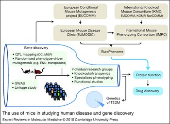
With the increase in sample size and better development of clinical phenotyping protocols, yet more susceptibility loci of smaller effect sizes are likely to emerge. This increases the demand for appropriate mouse models to validate and characterise candidate genes and for subsequent molecular and physiological studies to determine the mechanisms and pathways underlying disease susceptibility – to provide the potential to translate this new knowledge into therapeutic intervention (Fig. 2).
The use of mice in studying human disease and gene discovery. (Bottom left, within bottle representation) So far, most known type 2 diabetes mellitus (T2DM) genes have been discovered by various animal- and population-based analyses. In mice, this includes QTL mapping, with new efforts such as the CC and the MGP to improve resolution and efficiency, as well as various randomised mutagenesis techniques, including the use of chemicals (e.g. ENU) and transposons (e.g. Sleeping Beauty) to screen for interesting phenotypes and identify new genes. In humans, disease loci are being discovered by GWASs and linkage analyses. Based on this information, the classical way employed by individual research groups to study gene functions is by the generation of mouse alleles of interest, and specialised phenotyping. As a result, the past couple of decades have seen great advances in our understanding on the genetics of T2DM using mouse models. However, the generation of transgenic and knockout mice often involves complex genetic manipulation and lengthy breeding procedures and may also be restricted by the lack of easily accessible specialised phenotyping facilities (bottleneck). As a result, the knowledge we have so far explains only a very small proportion of human T2DM cases (pie chart). (Top right) Recently, various mouse projects have been set up with the goal of providing a comprehensive functional annotation of the mouse genome, and to fast-track the process of disease gene identification and the characterisation of protein function. The basic idea is to generate knockout alleles for every known mouse gene, followed by systematic phenotyping of each line. The knockout lines are being generated by the International Knockout Mouse Consortium (IKMC) (consisting of EUCOMM, KOMP and NorCOMM) and are subsequently phenotyped by the International Mouse Phenotyping Consortium (IMPC), which involves various phenotyping centres worldwide. To date, the European Mouse Disease Clinic (EUMODIC) project is serving as a precursor to the IMPC by undertaking a primary phenotype assessment of up to 650 mouse mutant lines generated by EUCOMM. All the phenotyping data generated from the above projects will be available from the EuroPhenome database and will be accessible to the wide research community. At completion, the IMPC project is anticipated to accelerate the process of gene verification and protein characterisation, and may bypass the rate-limiting step of gene identification and protein characterisation for genes that can be systematically characterised through the IKMC/IMPC pipelines. This will allow research efforts to focus on the study of protein function and identification of drug targets, ultimately leading to drug discovery and improvement of current therapy for T2DM. Abbreviations: CC, Collaborative Cross; ENU, N-ethyl-N-nitrosourea; EUCOMM, European Conditional Mouse Mutagenesis project; GWAS, genome-wide association study; KOMP, Knockout Mouse Project; MGP, Mouse Genomes Project; NorCOMM, North American Conditional Mouse Mutagenesis project; QTL, quantitative trait locus.

The development of new mouse resources initiated in the past five years is likely to accelerate the validation of the expanding list of GWAS-mapped susceptibility loci. With the availability of the complete mouse genome sequence, the International Knockout Mouse Consortium (IKMC; http://www.knockoutmouse.org) has been established through global collaboration, with the aim of generating a public resource of null or conditional mutations in each of the ~25 000 genes in the mouse genome. The IKMC includes the European Conditional Mouse Mutagenesis project (EUCOMM), the North American Conditional Mouse Mutagenesis project (NorCOMM) and the Knockout Mouse Project (KOMP). These mouse lines will then be archived in the European Mutant Mouse Archive (EMMA) and will be available to researchers as frozen embryos or sperm. (Website addresses for these projects are given in ‘Further reading, resources and contacts’ near the end of the article.)
The next stage of development would be to determine the phenotype of the knockout mouse lines and elucidate their physiological, developmental and behavioural characteristics. To achieve this, the European Mouse Disease Clinic (EUMODIC; http://www.eumodic.org/) consortium has been established to perform a primary phenotyping assessment of 650 selected mouse lines derived from EUCOMM ES cell lines using standardised operating procedures (SOPs) developed through European Union Mouse Research for Public Health and Industrial Applications (EUMORPHIA; http://www.eumorphia.org). This is being undertaken in four academic phenotyping centres: at GSF (Germany), ICS (France), MRC Harwell (UK) and the Wellcome Trust Sanger Institute (UK). All the data generated from these screens will be publicly available in the EuroPhenome and Mouse Phenome Database (MPD) (Refs Reference Mallon, Blake and Hancock18, Reference Grubb146). The protocols of SOPs for screening these mice are held in the European Mouse Phenotyping Resource of Standardised Screens (EMPReSS; http://empress.har.mrc.ac.uk) database. Depending on the phenotype, interesting mouse lines will go through more specialised phenotyping. Growing out of the EUMODIC project, the International Mouse Phenotyping Consortium (IMPC) aims to apply high-throughput comprehensive phenotyping procedures systematically to all mouse lines generated by the IKMC, to ultimately produce an encyclopaedia of mammalian gene function for every gene in the genome. This will reveal many new genes with a role in glucose homeostasis and will aid in the translation of GWAS loci into new genes and pathways for therapeutic intervention.
Once completed, these projects will provide the largest freely accessible resource for mouse functional genomics research worldwide, which is likely to significantly contribute to our understanding of T2DM and the identification of appropriate targets for therapeutic intervention.
Clinical implications and conclusions
Apart from the administration of insulin by injection, conventional therapeutic agents for treating T2DM include metformin (modulator of hepatic glucose production), sulfonylureas (insulin secretagogues), thiazolidinedione (PPARγ agonists), α-glucosidase inhibitors, analogues of the incretin hormone glucagon-like peptide 1, and dipeptidyl peptidase 4 inhibitors. Some of these agents produce side effects such as hypoglycaemia, weight gain, oedema and gastrointestinal intolerance, as well as having inadequate efficacy and short duration of action (Ref. Reference Colagiuri147). Treatments often become less effective with the progressive deterioration and reduction of β-cell mass that occurs during the natural progression of diabetes. There is therefore a crucial need for the development of new treatments through the identification of alternative drug targets as well as a better understanding of the complex molecular networks that control glucose homeostasis.
Although many of the T2DM mouse models described here share similar phenotypic features with humans, it is important to keep in mind that β-cell dysfunction as well as insulin resistance in mice might be different from that in humans because of fundamental differences in the physiological and pathophysiological mechanisms between the two species in addition to other factors such as social and behavioural influences that might not be replicable in mouse models. Nevertheless, mouse models remain a valuable tool for T2DM because of the ease of genetic manipulation, improved phenotyping protocols that allow the study of genetic, endocrine and metabolic changes, and aetiopathogenic mechanisms, and the option of screening of antidiabetic compounds that would not be possible in humans.
It is anticipated that in the near future, all players (including small- and moderate-effect genes) involved in glucose homeostasis and the development of T2DM will be discovered and characterised in the context of the whole organism through various large-scale mouse projects and human GWASs that are currently under way. Very soon, we will be saturated with vast amounts of data and new knowledge on gene function and possible drug targets in relation to T2DM. This will allow most research efforts to focus on functional studies and identification of suitable drug targets that aim to overcome current limitations in T2DM treatment. The accomplishment of this goal will improve the quality of life for patients with diabetes and fulfil our original justification of animal use in medical research.
Acknowledgements and funding
The authors thank the MRC and the Wellcome Trust for research funding, and the peer reviewers for their constructive and valuable comments.