Introduction
The mineral wadalite (IMA87-045), Ca6Al5Si2O16Cl3, found in skarn xenoliths in two-pyroxene andesite from Tadano, Fukushima Prefecture, Japan, was approved by the International Mineralogical Association Commission on New Minerals and Mineral Names (IMA CNMMN) in 1989. The mineral was named in honour of Dr. Tsunashiro Wada (1856–1920), the first director general of the Geological Survey of Japan. The structure of wadalite from Tadano was studied by Tsukimura et al. (Reference Tsukimura, Kanazawa, Aoki and Bunno1993), but its full description has not been published until now. Recently, a nomenclature for the mayenite supergroup was approved by the IMA Commission on New Minerals, Nomenclature and Classification, and wadalite was defined as a member of the wadalite group, and the wadalite group defined as a subgroup of the mayenite supergroup (Galuskin et al., Reference Galuskin, Gfeller, Galuskina, Armbruster, Bailau and Sharygin2015a). Galuskin et al. (Reference Galuskin, Gfeller, Galuskina, Armbruster, Bailau and Sharygin2015a) also proposed that the general formula of the mayenite supergroup minerals was X 12T 14O32–x(OH)3x[W 6–3x] (X = Ca; T = Al, Fe3+, Mg, Si and Ti; W = □, Cl, F and H2O), and that the ideal formula of wadalite be doubled to Ca12Al10Si4O32Cl6 (the formula originally approved by the IMA CNMMN in 1989 was Ca6Al5Si2O16Cl3).
We have reinvestigated wadalite in type specimens and found that it shows sector zoning and is enclosed by katoite, Ca3Al2(SiO4)3–x(OH)4x (x = 1.5–3). In this paper, we present mineral data for holotype wadalite from Tadano, including a Raman spectrum, although physical and optical properties and powder X-ray diffraction data were taken from the CNMMN proposal (IMA87-045). The type specimens of wadalite are deposited at the Geological Museum, Geological Survey of Japan, AIST, Tsukuba under the registered number GSJ M17112.
Occurrence
The mineral wadalite, was first found in skarn xenoliths (<50 cm in diameter) within Tertiary two-pyroxene andesite in a quarry in Tadano, Fukushima Prefecture, Japan (37°23′38″N, 140°15′21″E). The wadalite-bearing skarn xenolith comprises an anhydrous Ca-silicate-rich core and a hydrous Ca-silicate-bearing rim. The core is composed mainly of wollastonite, grossular, andradite, gehlenite and minerals of the hydroxylapatite–hydroxylellestadite series, whereas the rim consists mainly of wollastonite, calcite, ‘hydrogrossular’, andradite, thaumasite, tobermorite and xonotlite.
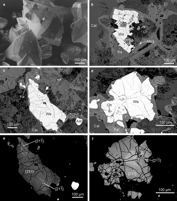
Wadalite occurs in the rim of the xenolith, near the core–rim boundary. Wadalite forms tris-tetrahedral crystals up to 1 mm in size. The dominant form is {211}, and the corners of the crystals are truncated by negative {21¯1} forms (Fig. 1a). The typical internal structure of wadalite is visible in back-scattered-electron (BSE) images (Fig. 1b–d). Along its margins and fractures, the wadalite is partially replaced by katoite. Moreover, wadalite is often replaced by unidentified hydrous minerals that contain Ca, Fe, Mg, Al and Si. In the composite crystals of wadalite, katoite and unidentified hydrous minerals we observed inclusions of gehlenite and magnetite. The inclusions of gehlenite form anhedral to subhedral crystals in the wadalite and the unidentified hydrous minerals, but not in the katoite. In many cases the wadalite crystals are replaced completely by katoite and/or unidentified hydrous minerals, and the aggregates of these minerals form pseudomorphs after wadalite. The pseudomorphs, whose outermost part consists of katoite crystals, preserve a well-defined wadalite form. Pseudomorphs shown in Fig. 1b can be classified as case-like (Glikin, Reference Glikin2009). Galuskina et al. (Reference Galuskina, Galuskin and Sitarz1998) studied ‘actarandite’, a ‘hydrogrossular’ pseudomorph after an unknown mineral with well-preserved primary tris-tetrahedral form and showed that the protophase of ‘actarandite’ was represented by a mineral close to wadalite. The Tadano wadalite replaced by katoite strongly supports their conclusion.
Secondary electron (a) and BSE images (b–f) of wadalite from Tadano: (a) the dominant form tris-tetrahedron {211} with an arrow indicating a negative {21¯1} that truncates the corner of the tris-tetrahedron; (b–d) wadalite replaced by katoite; (e,f) high contrast, BSE images for the {21¯1} and {211} sectors. Abbreviations: Wa = wadalite; Ka = katoite; Gh = gehlenite; Mag = magnetite; Un = unidentified hydrous minerals; Adr = andradite; and Cal = calcite.

Sector zoning of wadalite
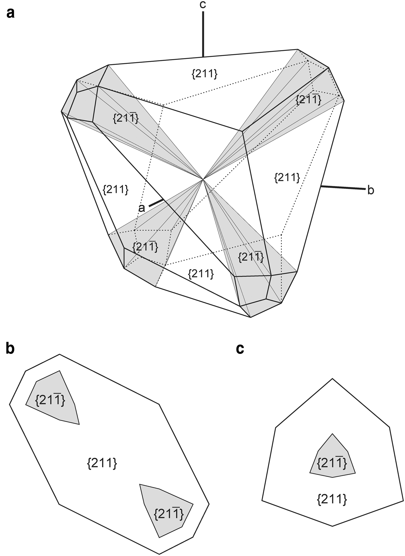
Sector zoning of wadalite in high-contrast, BSE images is shown in Fig. 1e,f. In Fig. 1e, two large brighter parts with polygonal shapes can be observed in the darker part. In Fig. 1f, one large brighter part with a hexagonal shape can be observed in the darker part. An ideal model for the sector-zoned wadalite is shown in Fig. 2, as inferred from the observations described above. Ideal patterns on sections normal to two different directions are shown in Fig. 2b,c, so the pattern shown in Fig. 1e is interpreted as a section almost normal to one of the crystallographic axes but away from the centre (Fig. 2b), and the pattern shown in Fig. 1f is interpreted as a section almost normal to the direction connecting the centre and the corner of the tris-tetrahedron {211}, but away from the centre (Fig. 2c). The brighter and darker parts, shown in Fig. 1e,f, are distinguished as {21¯1} and {211} sectors, respectively.
Ideal model for the sector-zoned wadalite from Tadano: (a) three-dimensional structure; (b) section normal to one of the crystallographic axes but away from the centre; and (c) section normal to the direction connecting the centre and the corner of the tris-tetrahedron {211}, but away from the centre. The drawings were produced using SHAPE by Shape Software.

Physical and optical properties of wadalite
The Tadano wadalite is black to dark grey, and translucent with a vitreous lustre. The streak is white, no cleavage was observed and Mohs hardness is 6.5. Fluorescence was not observed under short- and long-wavelength ultraviolet light. The density calculated from the average chemistry and powder X-ray diffraction data is 3.06 g cm–3. In thin section, the wadalite is transparent and colourless to pale yellowish brown. The wadalite is optically isotropic with n = 1.712(2) (white light). The value of the refractive index for wadalite (this study; Kanazawa et al., Reference Kanazawa, Aoki and Takeda1997; Mihajlović et al., Reference Mihajlović, Lengauer, Ntaflos, Kolitsch and Tillmanns2004) is 1.700(1)–1.712(2), and is smaller than that for eltyubyuite, the Fe3+ analogue of wadalite (n = 1.85 calculated on the basis of the Gladstone–Dale relationship, Galuskin et al., Reference Galuskin, Galuskina, Bailau, Prusik, Gazeev, Zadov, Pertsev, Jeżak, Gurbanov and Dubrovinsky2013). The smaller refractive index of wadalite is due mainly to its relatively low Fe3+ content.
Mineral chemistry
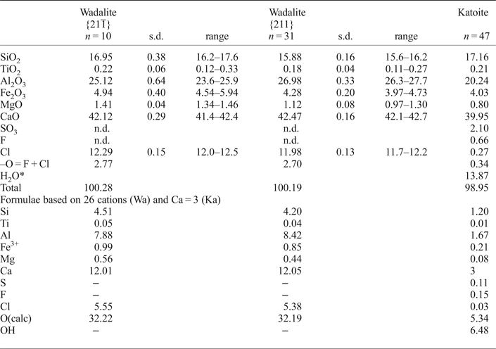
The chemical compositions of the wadalite, katoite, gehlenite and magnetite were determined using a JEOL JXA-8900R electron microprobe analyser (GSJ-Lab, AIST). The accelerating voltage and specimen current were kept at 15 kV and 12 nA on the Faraday cup. A beam diameter of 5 µm was used for the analyses of katoite and 2 µm for other minerals. Synthetic quartz (for Si), rutile (Ti), corundum (Al), Cr2O3 (Cr), MnO (Mn), hematite (Fe), periclase (Mg), wollastonite (Ca), celestine (S), F-phlogopite (F), natural jadeite (Na), adularia (K) and Cl-rich hastingsite (Cl) were used as standard materials. Matrix corrections for silicate minerals and magnetite followed Bence and Albee (Reference Bence and Albee1968) and ZAF methods, respectively. Detection limits for Cl and F were ~0.03 and 0.08 wt.% (3σ level), respectively. Table 1 lists the determined chemical compositions of the wadalite and katoite.
Chemical compositions (wt.%) of wadalite and katoite.

n.d. = not detected.
*H2O calculation based on charge balance and O + OH + F + Cl = 12 apfu.
Wadalite
The chemical formulae of the wadalite sectors were calculated on the basis of 26 cations assuming all iron = Fe3+, as suggested by Mihajlović et al. (Reference Mihajlović, Lengauer, Ntaflos, Kolitsch and Tillmanns2004) and Galuskin et al. (Reference Galuskin, Galuskina, Bailau, Prusik, Gazeev, Zadov, Pertsev, Jeżak, Gurbanov and Dubrovinsky2013). Fluorine and S were sought, but not detected. The empirical formula corresponding to the averaged composition for the {21¯1} sector is Ca12.01(Al7.88 $\hbox{Fe}_{{\rm 0}{\rm. 99}}^{{\rm 3 +}} $Si4.51Mg0.56Ti0.05)Σ13.99O32.22Cl5.55, whereas the formula given by the average composition of the {211} sector is Ca12.05(Al8.42
 $\hbox{Fe}_{{\rm 0}{\rm. 85}}^{{\rm 3 +}} $Si4.20Mg0.44Ti0.04)Σ13.95O32.19Cl5.38. The {21¯1} sector is richer than the {211} sector in Fe3+ [0.90–1.19 atoms per formula unit (apfu)], Si (4.34–4.69 apfu), Mg (0.54–0.58 apfu) and Cl (5.41–5.66 apfu), and the Fe3+, Si, Mg and Cl contents of the {211} sector are 0.79–0.94, 4.13–4.32, 0.38–0.51 and 5.26–5.47 apfu, respectively. The {21¯1} sector is depleted in Al (7.42–8.05 apfu) compared with the {211} sector (8.21–8.58 apfu).
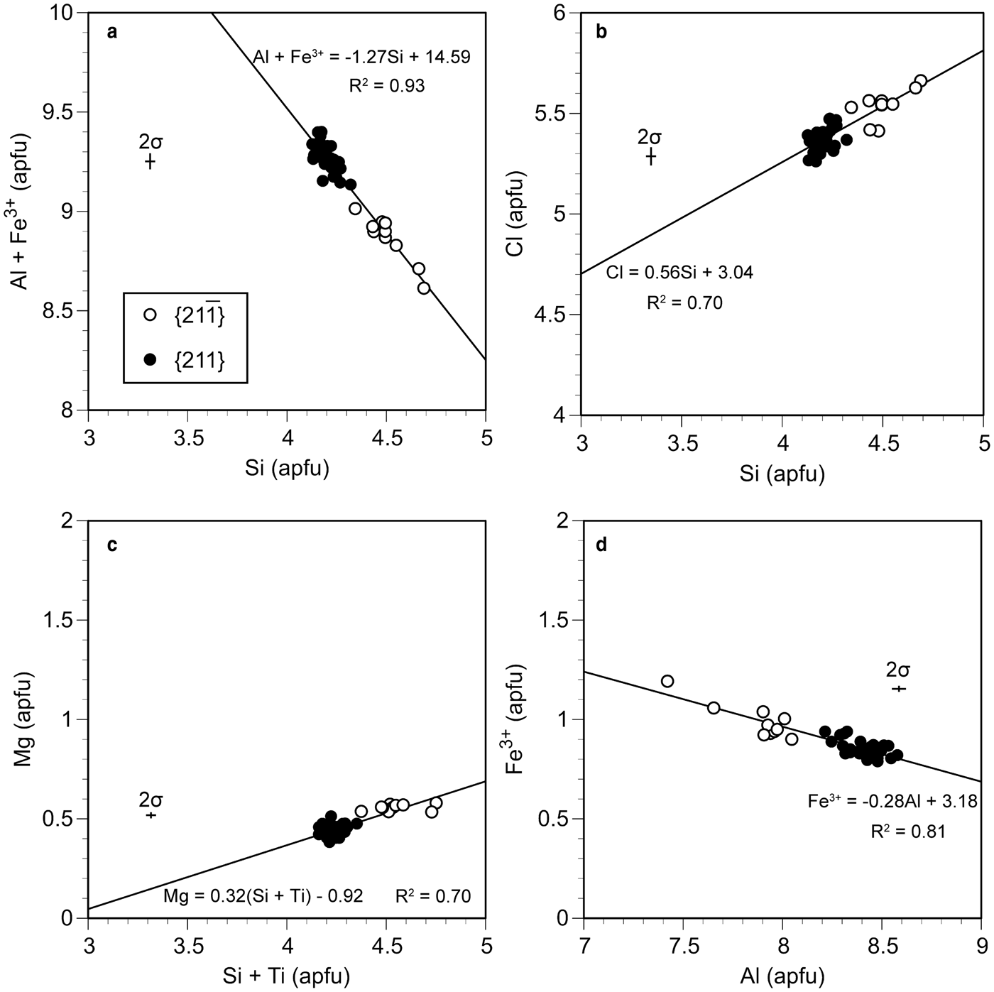
Galuskin et al. (Reference Galuskin, Gfeller, Galuskina, Armbruster, Bailau and Sharygin2015a) summarized the nature of the chemical substitutions in the mayenite-supergroup minerals. They proposed that substitutions displayed by the wadalite-group minerals are as follows: T(Al,Fe3+) + W□ ↔ TSi + W(Cl,F), 2T(Al,Fe3+) ↔ TMg + T(Si,Ti) and TAl ↔ TFe3+. The compositional variations of analysed wadalite in Si vs. (Al + Fe3+) is shown in Fig. 3a, and there is a negative correlation between the two parameters. A positive correlation between Si and Cl is shown in Fig. 3b. These correlations support the substitution: T(Al,Fe3+) + W□ ↔ TSi + W(Cl,F). The narrow range of Mg (Fig. 3c) indicates that the Si contents are controlled mainly by the substitution T(Al,Fe3+) + W□ ↔ TSi + W(Cl,F) and that the effect of the substitution 2T(Al,Fe3+) ↔ TMg + T(Si,Ti) is smaller than that of T(Al,Fe3+) + W□ ↔ TSi + W(Cl,F). The existence of a negative correlation between Al and Fe3+ is shown in Fig. 3d, indicating the substitution TAl ↔ TFe3+.
Plots of the chemical composition of wadalite. (a) Si vs. Al + Fe3+, (b) Si vs. Cl, (c) Si + Ti vs. Mg and (d) Al vs. Fe3+.

Katoite, gehlenite and magnetite
Wadalite is associated closely with katoite, gehlenite and magnetite. The structural formula of katoite was calculated on the basis of Ca = 3 apfu with all Fe cast as Fe3+, as suggested by Galuskin et al. (Reference Galuskin, Gazeev, Lazic, Armbruster, Galuskina, Zadov, Pertsev, Wrzalik, Dzierżanowski, Gurbanov and Bzowska2009). The H2O contents were estimated on the basis of charge balance and O + OH + F + Cl = 12 apfu. The empirical formula corresponding to the averaged composition for katoite is Ca3(Al1.67 $\hbox{Fe}_{{\rm 0}{\rm. 21}}^{{\rm 3 +}} $Mg0.08Ti0.01)Σ1.97(Si1.20S0.11)Σ1.31[(OH)6.48O5.34F0.15Cl0.03]Σ12. The compositional ranges of F, Cl and SO3 are 0.38–0.87, 0.09–0.51 and 1.39–2.99 wt.%, respectively.
The average chemical composition of the gehlenite inclusions is Ca2.01(Al1.79 $\hbox{Fe}_{{\rm 0}{\rm. 06}}^{{\rm 3 +}} $Mg0.07Si1.07)Σ2.99O7. The magnetite contains appreciable amounts of Al2O3 (2.85–7.18 wt.%), Cr2O3 (0.11–9.97 wt.%), MnO (0.69–3.85 wt.%) and MgO (3.28–5.24 wt.%).
X-ray crystallography of the wadalite
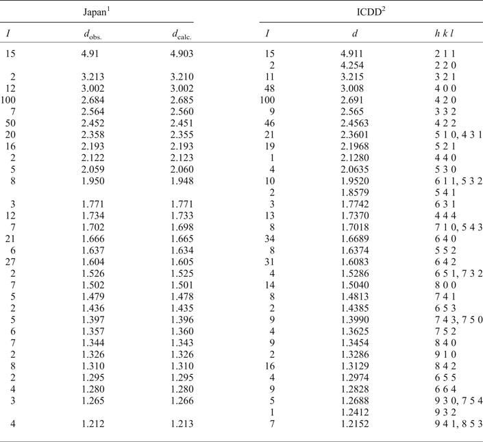
The powder X-ray diffraction data for wadalite were obtained using a diffractometer equipped with a diffracted-beam monochromator, employing CuKα radiation (Table 2). The unit-cell parameters refined from our powder data are a = 12.009(2) Å and V = 1731.8(8) Å3. A single-crystal X-ray study using the type specimen was reported by Tsukimura et al. (Reference Tsukimura, Kanazawa, Aoki and Bunno1993). The results show that wadalite is cubic, I  $ \overline {4}3 $d, with a = 12.001(2) Å, V = 1729(1) Å3 and Z = 2 (changed from Z = 4 based on the doubled formula). The difference between the volumes obtained from the powder data and from the single-crystal data is only ~0.2%. The length of the a axis for wadalite (this study; Tsukimura et al., Reference Tsukimura, Kanazawa, Aoki and Bunno1993; Kanazawa et al., Reference Kanazawa, Aoki and Takeda1997; Mihajlović et al., Reference Mihajlović, Lengauer, Ntaflos, Kolitsch and Tillmanns2004) is 12.001(2)–12.0343(2) Å, and is obviously smaller than that for eltyubyuite, the Fe3+ analogue of wadalite (a = 12.20(3) Å, Galuskin et al., Reference Galuskin, Galuskina, Bailau, Prusik, Gazeev, Zadov, Pertsev, Jeżak, Gurbanov and Dubrovinsky2013; a = 12.2150(2) Å, Gfeller et al., Reference Gfeller, Środek, Kusz, Dulski, Gazeev, Galuskina, Galuskin and Armbruster2015). In tetrahedral sites, the effective ionic radius of Al3+ (0.39 Å) is smaller than that of Fe3+ (0.49 Å) (Shannon, Reference Shannon1976), and thus the smaller unit-cell volume of wadalite is due mainly to its relatively low Fe content.
Powder X-ray diffraction data for wadalite.

1 Wadalite from Tadano, Fukushima, Japan. a = 12.009(2) Å
2 Wadalite, ICDD 49-1856. a = 12.034(1) Å
Raman spectroscopy
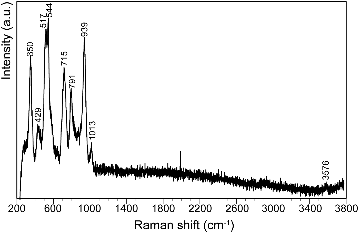
Micro-Raman spectra of the wadalite from Tadano were collected using a JASCO NRS-5100 spectrophotometer with a 532 nm laser, collecting 2 scans over a range of 55–3775 cm–1 with a resolution of 0.42 cm–1. The output power of the laser beam was 6.3 mW and the diameter of the focal spot on the sample was ~1 µm. A spectrum of the wadalite crystal in a polished thin section is shown in Fig. 4. The following main bands are assigned tentatively by referring to the data of Galuskin et al. (Reference Galuskin, Gfeller, Armbruster, Galuskina, Vapnik, Dulski, Murashko, Dzierżanowski, Sharygin, Krivovichev and Wirth2015b) and Gfeller et al. (Reference Gfeller, Środek, Kusz, Dulski, Gazeev, Galuskina, Galuskin and Armbruster2015): 350 cm–1 (ν2 SiO4), 517 and 544 cm–1 (ν4 AlO4), 715 and 791 cm–1 (ν1 AlO4) and 939 cm–1 (SiO4). A very weak band at ~3576 cm–1 can be attributed to OH groups substituting F and/or Cl at the W site of chlormayenite (Galuskin et al., Reference Galuskin, Kusz, Armbruster, Bailau, Galuskina, Ternes and Murashko2012), implying that the Tadano wadalite contains very small amounts of OH ions in the W site.
The Raman spectrum of wadalite from Tadano.

Formation of wadalite
The wadalite-bearing skarn xenolith consists of an anhydrous Ca-silicate-rich core and a hydrous Ca-silicate-bearing rim, and the core and rim parts correspond respectively to the relics of an earlier skarn and the alteration of the skarn during a later stage of hydrothermal activity. Wadalite occurs in the hydrous Ca-silicate-bearing rim and is not present in the anhydrous Ca-silicate-rich core. The presence of anhedral to subhedral gehlenite inclusions in the wadalite implies that wadalite formed at the expense of gehlenite. Feng et al. (Reference Feng, Glasser, Howie and Lachowski1988) synthesized wadalite by heating CaO, SiO2 and Al2O3 in the presence of excess CaCl2 at 850°C in open crucibles. This experiment confirmed that wadalite is stable at high temperatures and with high chlorine concentrations in the system, as suggested by Kanazawa et al. (Reference Kanazawa, Aoki and Takeda1997). Thus, the existence of wadalite in the Tadano xenolith suggests that there was a high concentration of chlorine in the hydrothermal solution from which the wadalite was formed.
We were able to recognize sector zoning in the wadalite by using BSE images and electron probe microanalyses. Sector-zoning typically develops during relatively rapid non-equilibrium crystallization (e.g. Carpenter, Reference Carpenter1980; Ranløv and Dymek, Reference Ranløv and Dymek1991), and the zoning therefore suggests that wadalite grew during a period of rapid non-equilibrium crystallization. The wadalite was replaced by katoite along its margins and along fractures, and this suggests that the katoite formed as a secondary product during retrogressive hydrothermal alteration of the wadalite. Passaglia and Rinaldi (Reference Passaglia and Rinaldi1984) concluded that the crystallization temperature of katoite is ~300 °C, and this conclusion and the experiment by Feng et al. (Reference Feng, Glasser, Howie and Lachowski1988) indicate that the katoite formed at a lower temperature than the wadalite.
Acknowledgements
We thank T. Sato and E. Hirabayashi for making the polished thin sections used in this study. Constructive reviews by I. Galuskina, P. Leverett and C.L. Lengauer improved the manuscript. Thanks are also due to H. Mukai for his assistance in the measurement of Raman spectra.