Introduction
Transcatheter device closure is an established treatment option for perimembranous ventricular septal defects at present. It is done generally by either a retrograde transarterial approach or an upfront antegrade transvenous approach. In case of difficulties with either approach, arteriovenous loop is performed by crossing the defect from the left ventricular side and snaring the tip of the crossing 0.035’’ glide-coated guide wire through the femoral vein. The delivery sheath is now advanced from the right ventricle to the ascending aorta through the left ventricle across the defect. Now the device is taken into the delivery sheath and partially releasing the device, the whole assembly is pulled to align the device across the defect and subsequently released the device. In our case, all these maneuvres either failed or were unsuitable. We report a maneuvre of advancing the delivery sheath towards the left ventricular apex with the help of a cut pigtail.
Case report
A 12-year-old girl (25 kg) came to us for transcatheter device closure of a large (16.5 mm) perimembranous ventricular septal defect with left-to-right shunt, moderate pulmonary arterial hypertension, and evidence of left ventricular volume overload. Heart team consultation was done, and a decision was taken for at least one attempt for device closure. Informed consent was obtained from the parents before attempting device closure, given that the parents and the patient were not willing to undergo open heart surgery.
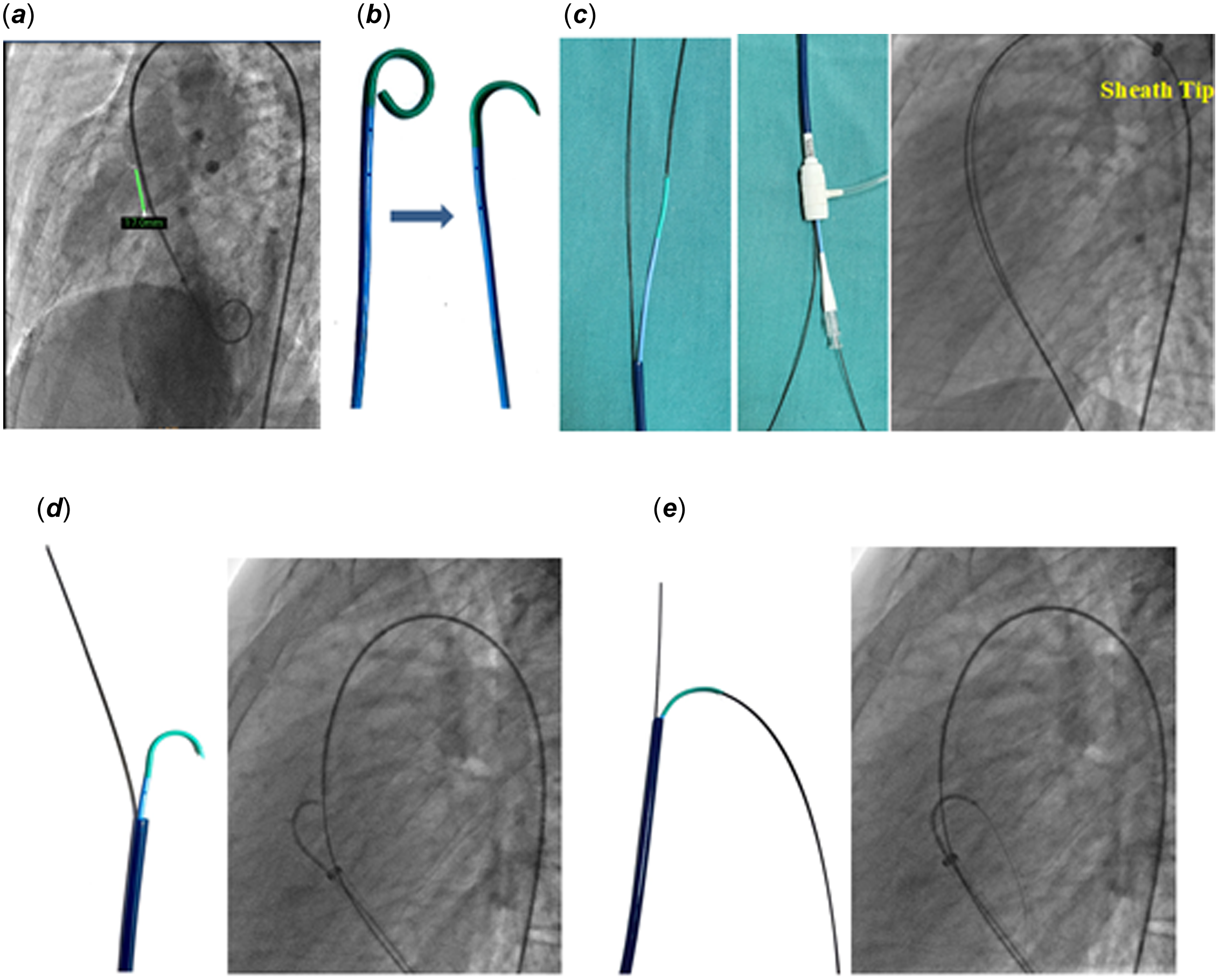
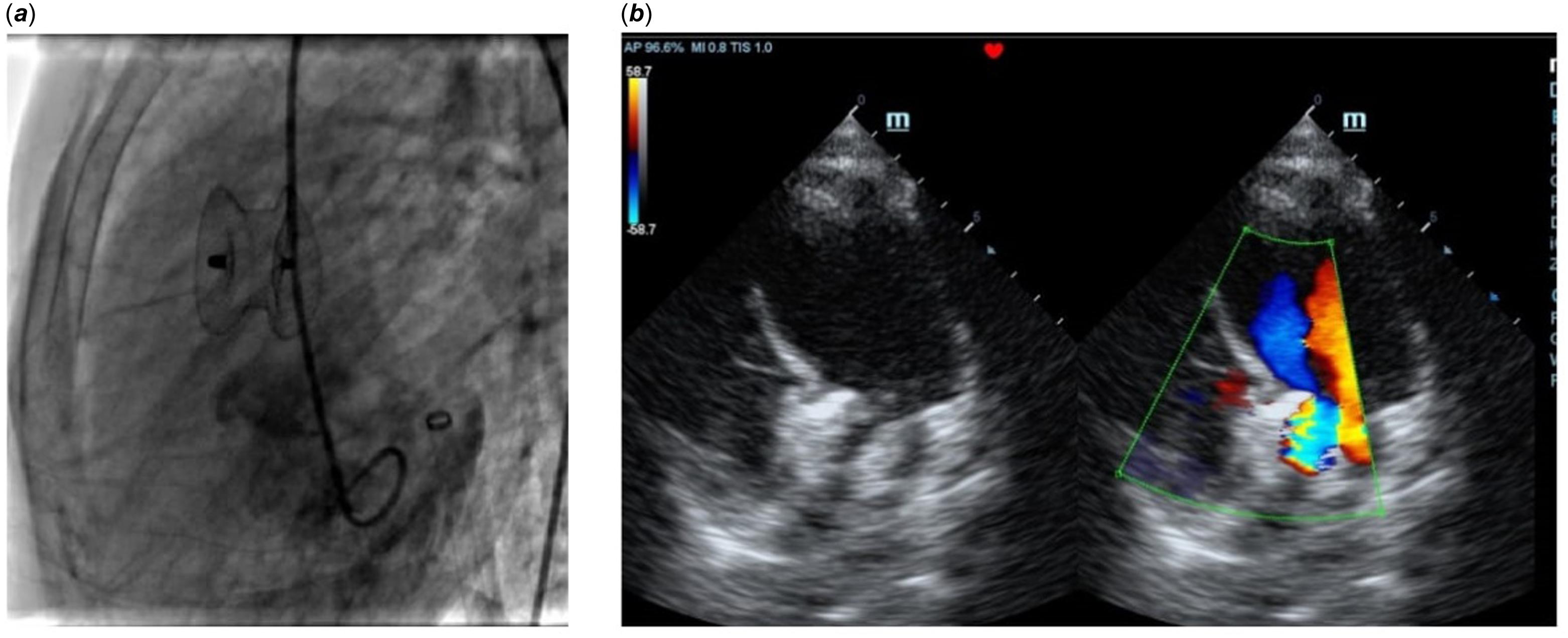
She was taken for device closure under conscious sedation. 6F right femoral arterial and 7F right femoral venous access were taken. Unfractionated heparin was administered intravenously at a dose of 100 units/kg. Left ventricular angiogram was taken in 60 degree left anterior oblique view with 30 degree cranial tilt revealed large defect of 17 mm with left-to-right shunt (Figure 1 a ). Antegrade transvenous crossing of the ventricular septal defect was attempted with a 5F internal mammary catheter and a 0.035” straight tip glide-coated guide wire (Terumo Medical Corporation, Japan). However, it failed even after several attempts. Then retrograde crossing and formation of the arteriovenous loop were done easily. A 12F delivery sheath with dilator was advanced into the descending thoracic aorta, and the dilator and the guide wire were removed. After deairing, a 22 mm muscular septal device (Lifetech, China) was advanced. The sheath was pulled back to the ascending aorta, and the left ventricular disc was released. Now the whole assembly was pulled back slowly. As the device was bulky, the left ventricular disc was getting caught at the aortic valve, and with slightest pull, it was repeatedly coming back to the right ventricle, and the device was not getting properly aligned. So again the arteriovenous loop was formed, and the delivery sheath was advanced into the descending thoracic aorta. We took a 6F cut pigtail catheter (Figure 1 b ), and it was advanced into the ascending aorta over another 0.035" J tipped glide-coated guide wire by the side of the guide wire of the arteriovenous loop (Figure 1 c ). Now, the delivery sheath was slowly pulled back to the right ventricle and positioned just below the ventricular septal defect. The cut pigtail was allowed to direct towards the left ventricular apex across the defect by pulling the guide wire back and rotating the catheter. The guide wire was now advanced to the left ventricular apex (Figure 1 d–e ). The glide-coated guide wire was exchanged for a 0.035" extra-stiff wire, and the arteriovenous loop was broken. The delivery sheath was advanced towards the left ventricular apex. After removing the pigtail with its wire, the same 22 mm muscular device was taken into the delivery sheath and was positioned across the defect with proper device alignment. The device was released with minimal residual flow through the device with mild aortic regurgitation (Figure 2 a–b ). Proper hemostasis was achieved. She was discharged on the 3rd postoperative day in a stable condition.
Device closure of the ventricular septal defect. ( a ) Size of the defect was 17 mm. ( b ) The tip of a 6F pigtail catheter was cut to make a desired curvature. ( c ) Tip of the delivery sheath showing the 0.035’’ glide-coated guide wire of the arteriovenous loop and the second guide wire through the cut pigtail and the proximal end of the delivery sheath showing the wire of the arteriovenous loop and alongside the hub of the cut pigtail through which the second wire was introduced. ( d ) The delivery sheath was pulled back to the right ventricle just below the defect, and the pigtail was prolapsed towards the left ventricular apex. ( e ) The 0.035’’ glide-coated guide wire through the cut pigtail was advanced towards the left ventricular apex (Video 1).

Post-device closure device alignment in the left ventricle. ( a ) Left ventricular angiogram in left anterior oblique 60 degree with 30 degree cranial tilt view after release of the device. ( b ) Echocardiogram in apical five-chamber view showing device in position with proper alignment with minimal residual flow through the device with minimal aortic regurgitation.

Discussion
Transcatheter device closure is an established way of treating ventricular septal defect in various locations either by antegrade transvenous or retrograde transarterial route. Reference Yang, Yang and Yu1–Reference Ratnayaka, Raman and Faranesh4 However, large perimembranous defects, as in our case, may sometimes pose technical challenges for transcatheter device closure. Reference Buntharikpornpun, Roymanee and Promphan5 As a large defect requires a large device and a compatible large delivery sheath, the retrograde transarterial approach may not be a good option in small children. In our patient, we failed to close the defect by antegrade transvenous approach. So, only option was to form an arteriovenous loop and approach the ventricular septal defect from the venous side with taking the device to the ascending aorta and releasing partially (only left ventricular disc) followed by gently pulling the whole assembly across the aortic valve into the left ventricle and then aligning the device properly across the defect followed by release of the right ventricular disc. Reference Alshahrani, Linnane and McCrossan6 However, unfortunately, in our patient, this maneuvre was ineffective. So, we performed the above-described maneuvre to direct the delivery sheath towards the left ventricular apex, and it was successful.
Perimembranous ventricular septal defects are generally closed with an Amplatz duct occluder II (Abbott, USA) or a Konar multifunctional occluder (Lifetech, China) device. Reference Kuswiyanto, Gunawijaya and Djer8,Reference Lee, Song and Choi9 For such a large defect, the highest available size of either of the devices was too small. So, the options for closing the ventricular septal defects were to use either a duct occluder I (Abbott, USA) or a muscular ventricular septal device. Reference Lee, Song and Choi9 Because of the anatomy of the defect, we had chosen a muscular ventricular septal device; otherwise, the device could have caused the right ventricular outflow tract obstruction.
Conclusion
Transcatheter device closure of a large perimembranous ventricular septal defect may sometimes be technically challenging where conventional techniques fail. We have described a method of directing the large diameter delivery sheath towards the left ventricular apex by using a cut pigtail along the side of a preexisting arteriovenous loop. This technique may be helpful for other selected cases where the conventional methods are ineffective.