INTRODUCTION AND BACKGROUND
We present two cases of cellulitis developing as a sequela of radiotherapy tattooing following implant-based breast reconstructions. Without early and aggressive treatment of such infections, the reconstruction is adversely compromised.
CASE 1
A 54-year-old patient presented to the Cambridge Breast Unit with a left sided 20 mm grade III invasive ductal carcinoma with lymphovascular invasion and positive axillary lymph nodes. The multidisciplinary team (MDT) recommended a left mastectomy, axillary clearance and adjuvant radiotherapy. The patient requested a simultaneous right prophylactic mastectomy. Bilateral immediate breast reconstructions were carried out with latissimus dorsi myocutaneous flaps and McGhan style 150 permanent expanders (Allergan Ltd, Marlow, Buckinghamshire, UK). The patient made an unremarkable recovery and was discharged on day 10.
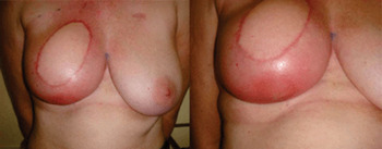
At radiotherapy planning 6 weeks postoperatively, black dye was tattooed over her xiphisternum and the lateral aspect of the right breast. Approximately 36 hours after tattooing, the patient presented with a swollen and painful right breast. She was pyrexial (37.8 °C) with two areas of cellulitis, both corresponding to the tattoo sites (Figure 1). Investigations revealed a white cell of 10 × 10−9/l, a C-reactive protein of 18 mg/l and negative blood cultures. High doses of intravenous (IV) flucloxacillin, benzylpenicillin and metronidazole were commenced. Breast ultrasound revealed no fluid collection.
Cellulitis on the medial aspect of the right breast just lateral to the blue midline tattoo mark.

As no improvement was observed over the next 12 hours, microbiology advice was sought. The patient was started on IV tazocin 4.5 g eight hourly. The tenderness improved dramatically, whilst the cellulitis subsided gradually. After 6 days of tazocin, she was discharged on a 7 days course of oral co-amoxiclav 625 mg eight hourly.
The radiotherapy treatment was delayed by 1 week to allow the tissue oedema to settle. She successfully underwent a three and a half week course of adjuvant radiotherapy (40 grays in 15 fractions) with no recurrence of infection. Her implant-based reconstructions remain soft and intact.
CASE 2
A 41-year-old patient presented to the Cambridge Breast Unit with an 8 mm nodule on the right breast and an underlying 30 mm grade II invasive ductal carcinoma with abundant lymphovascular invasion and positive axillary lymph nodes. The MDT recommended neo-adjuvant chemotherapy, right mastectomy and axillary clearance. The patient selected an immediate right breast reconstruction with a latissimus dorsi myocutaneous flap and McGhan style 150 permanent expander. She made a good recovery and was discharged 1 week postoperatively.
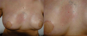
Post discharge the patient was seen weekly in clinic, where her expanders were inflated. Four weeks postoperatively skin tattooing for radiotherapy was performed. Six days later, she developed pain, discomfort and discolouration of the breast near the tattoo. Clinically there was a small area of cellulitis around the tattoo site and a peau d’orange appearance of the right breast (Figure 2). Oral co-amoxiclav 625 mg eight hourly was commenced and gradual improvement was seen.
The area of cellulitis on the right breast shown in close up to be related to the radiotherapy tattoo mark.

Upon complete resolution of the cellulitis, adjuvant radiotherapy (40 grays in 15 fractions) was started. Two weeks into radiotherapy, the patient was advised by the radiographers to return to the surgeons, as she had developed an erythematous and warm area on the inferior aspect of the reconstructed breast. An empirical week course of oral co-amoxiclav was re-started. The breast remained warm and swollen after 6 days. Purulent fluid aspirated from the swollen dependent area of the breast revealed a gram-positive cocci. The patient was admitted to hospital for IV antibiotics comprising metronidazole and tazocin. Having showed no improvement after 48 hours, an emergency exploration of the right breast was performed. This revealed a copious peri-implant fibrinous exudate which necessitated implant removal.
Postoperatively the erythema of the mastectomy skin settled rapidly. Histology showed features in keeping with infection and radiation damage.
Further expandable implant reconstruction was successfully undertaken 12 months later.
DISCUSSION
Whilst absolute causality cannot be proven, the onset of infection following the time of the tattoos and the ink’s location at the centre of the areas of cellulitis suggest that the tattooing process was in some way a trigger for the infection. The simplest mechanism would be direct introduction of micro-organisms by the needle or the dye. A literature search using Pubmed identified cases of reactions to tattoo inks,Reference Sewak, Graham and Nankervis1–Reference Durmazlar, Tatlican and Eskioglu3 however, there are no reports of iatrogenic infections as a result of radiotherapy tattooing.
Our radiographers were informed and enquired about previous similar episodes. This was the first incidence of infection post-tattooing in the radiotherapy department. The dye used is standard black printing ink which is not sterile. The skin is cleaned with alcohol wipes, the dye is applied to the area and a needle stick is made to introduce the ink into the skin. In our unit an aseptic technique is not used, however, a UK based survey would be appropriate to enquire about other units and also identify infections that may have occurred but not reported. Involvement of the radiographers with the first case prompted their rapid referral of the second patient.
It remains to be seen if the first patient develops capsular contracture in her right breast or indeed in the left ‘radiotherapised’ breast. This can be anticipated because, although she has had radiotherapy only to the contralateral breast, dye reactions have been reported to cause capsular contracture.Reference Singh-Ranger and Mokbel4 Singh-Ranger et al. reported a case of capsular contracture following methylene blue dye used in sentinel node biopsy. They postulated that this could have been caused by a localized tissue reaction initiated by or involving the dye. This may represent another cause of capsular contracture in patients undergoing postoperative radiotherapy, as radiotherapy itself is a known risk factor for capsular contracture.Reference Whitfield, Horan, Irwin, Malata, Wishart and Wilson5 Tattoos may cause capsular contracture by an ‘allergic reaction’ or by inducing a peri-implant infection. Using an aseptic technique for tattooing would minimize infection risk.
We conclude that radiotherapy tattooing is a possible iatrogenic cause of tissue reactions and infection. Any suggestion of cellulitis following tattooing should be treated early and aggressively to prevent a catastrophic reconstructive outcome with loss of the implant due to infection. Radiologists and radiographers should be aware of this complication so that they can undertake the tattooing in aseptic conditions. Patients should also be warned of this risk.