Introduction
Increasing number of patients with unilateral or bilateral hip prostheses (b-HP) are being referred for radical radiotherapy for prostate cancer. In 2017, there were 91,698 primary hip replacements performed in England, Wales and Northern Ireland: 40·2% were in men and with a median age of 69 years, largely due to osteoarthritis.1 An aging population together with an increased incidence of prostate cancer will mean this is likely to become a more common problem for radiotherapy departments.
Prosthetic hips, particularly when bilateral, cause a number of challenges to deliver prostate radiotherapy. Materials of high-atomic numbers used for hip prostheses cause much greater photon attenuation than surrounding human tissue and this leads to beam hardening, photon starvation and increased noise. These phenomena usually manifest themselves as bright and dark streaks, or areas with no signal, which can cause severe image quality degradation. Various methods of metal-induced artefacts reduction have been employed by different equipment manufacturers, usually in the form of post-processing algorithms. Philips® (Amsterdam, the Netherlands) scanners use the O-MAR software (Orthopaedic Metal Artefact Reduction) which reduces metal-induced artefacts on CT planning for patients with hip prostheses.Reference Li, Noel and Chen2 O-MAR-processed images improve visualisation of the structures in the affected areas by removing artefacts; however, contrast and spatial resolution often remain affected, particularly for patients with b-HP. Loss of contrast between different density structures in the affected area is due to Hounsfield unit interpolation which is part of the O-MAR image processing. Therefore, even with metal artefact reduction algorithms, larger margins may still need to be used to allow for ongoing uncertainty.Reference Li, Noel and Chen2,Reference Hagen, Kretschmer and Wurschmidt3
The prosthesis-related artefact can also affect the planning process by affecting the ability of treatment planning systems to determine accurate electron densities for modelling dose. Intensity-modulated radiotherapy (IMRT) is the standard treatment technique for such patients, but with consensus to uphold several principles if possible during planning: avoiding the prosthesis, using more arcs, turning off inhomogeneity corrections during treatment planning, estimating dose perturbations and measuring exit doses during treatment delivery.Reference Zaorsky, Showalter and Ezzell4 Artefact can also affect treatment verification, particularly when soft-tissue cone-beam CT is being used.
In this report, we describe the routine use of transperineal ultrasound (TPUS) in the workflow for three prostate radiotherapy cases with b-HP to both aid volume delineation and daily inter- and intra-fractional image guidance.
Methods
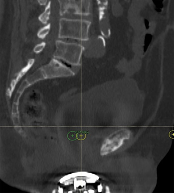
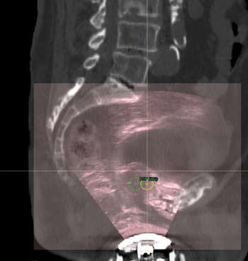
Planning CT scans and co-registered Clarity® (Elekta, Stockholm, Sweden) TPUS images of three cases of men with b-HP who were due to undergo external beam radiotherapy for localised prostate cancer were retrospectively reviewed. Both CT and TPUS scans are acquired within a few minutes of each other, on the same couch and in the same position, in order to minimise the differences in organ position. The TPUS images are not affected by the presence of the hip prostheses (which are outside the field of view) and allow good visualisation of structures in areas obscured by artefact on the O-MAR reconstructed CT images (Figures 1 and 2).
Sagittal CT planning scan without TPUS fusion with transperineal US probe in situ.

Sagittal CT planning scan with TPUS fusion with prostate and penile bulb well visualised.

Two radiation oncologists marked the prostate and seminal vesicles CTV firstly using:
-
(i) the CT planning images alone
and then,
-
(ii) the CT planning images together with the fused 3D TPUS images.
Contouring in both instances was with the aid of non-fused diagnostic prostate MRI images, and neither oncologist had access to the other’s delineated volumes. The sets of contours were compared for each case and are reported here.
All men underwent CT planning and treatment as per our standard departmental workflow. Following standardised bladder and bowel preparation, 2-mm thickness CT scans were performed with patient’s supine with knee and footstock immobilisation on a Philips® Big Bore 16 slice scanner. The Clarity® US probe was in position and at the same time, a reference three dimensional (3D) TPUS image was acquired and fused with the planning CT scan using the Automated Fusion and Contouring Workstation. A metal artefact reduction algorithm was utilised to try to improve the visualisation of anatomical structures on the CT.
A dose of 60 Gy in 20 fractions over 4 weeks was prescribed to PTVp_6000, and plans were generated with a photon beam energy of 10 MV. Treatment was on an Elekta (Stockholm, Sweden) Versa HD machine: two patients were treated with fixed field IMRT and one with dual arc volumetric-modulated arc therapy (VMAT). Patients underwent daily image-guided radiation therapy (IGRT) using TPUS as per our departmental standard, including intra-fractional monitoring.Reference Hilman, Smith and Masson5
Results
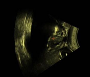
The images of the prostate, bladder and penile bulb produced with Clarity TPUS were clear and, compared to a transabdominal approach, have the advantage of no large bladder fill preparation (Figure 3).
Sagittal TPUS image of prostate, bladder and penile bulb in a patient with b-HP.

In patient 1, the CTV marked by the two radiation oncologists was smaller when the fused TPUS images were utilised (−15·3 and −14·5%). In patient 2, the CTV was consistently larger (+54·2 and +35·8%) when the fused TPUS images were utilised. The same was true for patient 3 (+14·2 and +13·4%). Most of the added volume was due to extra contouring at the superior part of the CTV, which was at the level of the hip replacements and the TPUS images enabled better visualisation of the prostate at this site.
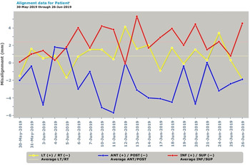
Within our department, IGRT inter-fractional audits have shown a 95% agreement between offline ‘experts’ and online matching by radiographers to within <3 mm, and this was the case for these three patients with b-HP (Figure 4).
Inter-fractional monitoring data for one of the patients with b-HP.

Using TPUS for intra-fractional monitoring using the standard Clarity imaging protocol was feasible. None of the patients were outliers with respect to anterior/posterior; inferior/superior or left/right motion. Intra-fractional motion beyond correction thresholds was observed in 20–40% of fractions and was more common in the IMRT cases than the VMAT case. While some motion was corrected online, a motion was often brief and did not require online position correction. The anterior/posterior direction excursion was most marked as expected often due to a common posterior ‘drift’ of the prostate.Reference Richardson and Jacobs6
Discussion
Target delineation in patients with bilateral hip prostheses (b-HP) is challenging and illustrated by greater levels of inter-observer variability than in patients without prostheses. The use of additional imaging modalities, such as MRI or TPUS has the potential to improve the reliability of outlining.
Here, we describe three cases where the use of co-registered TPUS images (taken at the time of CT planning) improved the confidence of prostate clinical target volume (CTV) delineation and led to a consistent change in volumes across two observers when compared with delineated volumes done on the CT planning scan alone (that was subjected to a metal artefact reduction algorithm).
To our knowledge, our report is the first in the literature to demonstrate the utility of TPUS for this group of patients. Boda-Heggemann et al. described a case of a patient with b-HP in whom trans-abdominal ultrasound (the predecessor to TPUS) along with a planning MRI were used to aid CTV delineation.Reference Boda-Heggemann, Haneder and Ehmann7 They suggested that the CTV could be more reliably defined using the US images than the MRI images (due to differences in the bladder and rectal filling and metal-related image distortion in the latter). Similarly, Dube et al. evaluated four cases with b-HP and concluded that the prostate was clearly visible on trans-abdominal ultrasound and the co-registered images could be used to aid CTV delineation in this group of patients.Reference Dube, Mahadevan and Sheldon8
Since the vast majority of modern orthopaedic implants are constructed from non-ferromagnetic materials, co-registration of MRI images also has the potential to aid CTV delineation in cases with b-HP. However, due to different couch type on both scanners (curved versus flat), as well as the significant time difference between MRI and CT scan image acquisition, relative positions and extents of the organs of interest cannot be guaranteed.Reference Rosewall, Kong and Vesprini9,Reference Charnley, Morgan and Thomas10 In addition, image degradation from metal artefacts can occur in MRI imaging. Rosewall et al. evaluated seven patients with b-HP, and found consistently smaller CTV volumes when MRI was used and a trend towards reduced inter-observer variability.Reference Rosewall, Kong and Vesprini9 Charnley et al. (2005) evaluated four patients with b-HP and concluded that CT-MRI co-registration was feasible and led to the clearer delineation of the CTV.Reference Charnley, Morgan and Thomas10
The patients in this report were also treated using TPUS for daily image-guidance. TPUS can be used for inter- and intra-fraction verification as part of a routine prostate radiotherapy workflow.Reference Camps, Fontanarosa, de With, Verhaegen and Vanneste11 In patients with b-HP, TPUS has a potential advantage over cone-beam CT verification, which shows image degradation due to metal artefact. Image-guidance with intra-prostatic fiducial markers (with electronic portal imaging or kilovoltage image pairs) and radiofrequency transponders.Reference Bittner, Butler, Kurko and Merrick12 both have a similar advantage over CBCT but neither is a useful aid for CTV delineation.
This will be part of our protocol for treating such patients, and we aim to look at a larger series in the future.
Conclusion
The use of co-registered TPUS images can aid target volume delineation in patients with b-HP and has potential advantages over MRI in this situation. In addition, TPUS can be used for daily image guidance in this group of patients in the same way as it is used in patients with non-prosthetic hips, including for intra-fractional monitoring.
Acknowledgements
None.
Financial Support
We received no specific grant from any funding agency, commercial or not-for-profit sectors.
Conflicts of Interest
Petra Jacobs has received financial support from Elekta for attending courses.