Introduction
The interventional closure of patent ductus arteriosus was first introduced by Porstman et al. in 1971. Reference Porstmann, Wierny and Warnke1 Advances in technology have significantly improved the accessibility and safety of interventional cardiology, increasing success rates while reducing complication risks. Although there is no absolute lower weight limit for catheter-based patent ductus arteriosus closure, most studies have been conducted on patient groups weighing more than 4 kg. Reference Dimas, Takao and Ing2 Recent case series suggest that catheter-based patent ductus arteriosus closure is feasible in infants weighing less than 4 kg; however, data on post-procedural complications in this subgroup remain insufficient. Reference Zahn, Nevin, Simmons and Garg3
Here, we report the case of a 12-day-old preterm infant who underwent surgical intervention due to the right external iliac artery injury following catheter-based patent ductus arteriosus closure.
Case presentation
The patient, born at 33 weeks of gestation as a twin, was admitted to the neonatal ICU for monitoring. The infant, with a birth weight of 2045 g, was unable to be weaned from nasal synchronised intermittent mechanical ventilation and presented with a continuous cardiac murmur. A paediatric cardiology consultation was requested. Transthoracic echocardiography identified a 4 mm patent ductus arteriosus. Medical closure therapy with two doses of ibuprofen was administered. Post-treatment transthoracic echocardiography revealed persistence of the patent ductus arteriosus with a left atrium-to-aorta ratio of 2.2, prompting interventional closure.
Following the procedure, the patient developed circulatory impairment in the right lower extremity and abdominal distension. Doppler ultrasonography revealed a haematoma approximately 2 cm in diameter at the level of the right external iliac artery. Due to haemodynamic instability, the patient underwent emergency surgery by the cardiovascular surgery team.
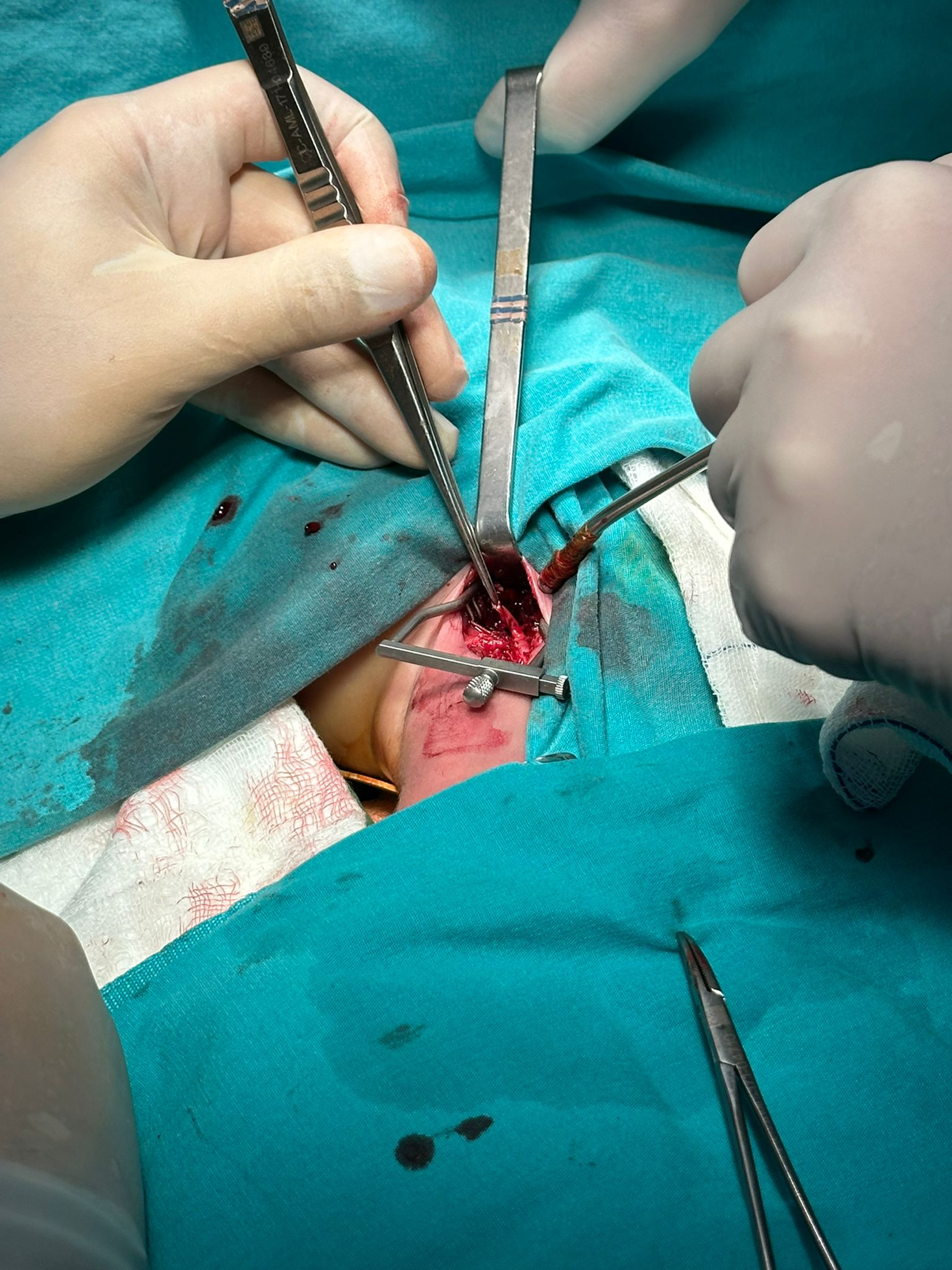
During the procedure, a femoral incision was made, but no pulse was detected in the femoral artery. The femoral artery was followed proximally, and the inguinal ligament was dissected to access the retroperitoneum. The haematoma identified on Doppler ultrasonography was evacuated. It was observed that the external iliac artery was transected at the level of the common iliac artery bifurcation (Figure 1). Although re-anastomosis of the external iliac artery was considered, it was deemed unfeasible due to the inability to reach its original origin and massive bleeding. The proximal external iliac artery was ligated using 7/0 prolene suture. Retrograde blood flow was observed in the femoral artery, leading to the ligation of the distal external artery. The surgical incision was closed, and the patient was transferred back to the neonatal ICU for monitoring.
Transected part of the external iliac artery.

The patient remained haemodynamically stable with no signs of bleeding and was started on subcutaneous enoxaparin at the daily dose of 100 units/kg. Daily Doppler ultrasonography evaluations demonstrated normal flow patterns in the right lower extremity. Ischaemic findings regressed after surgery, and no motor or sensory deficits were observed.
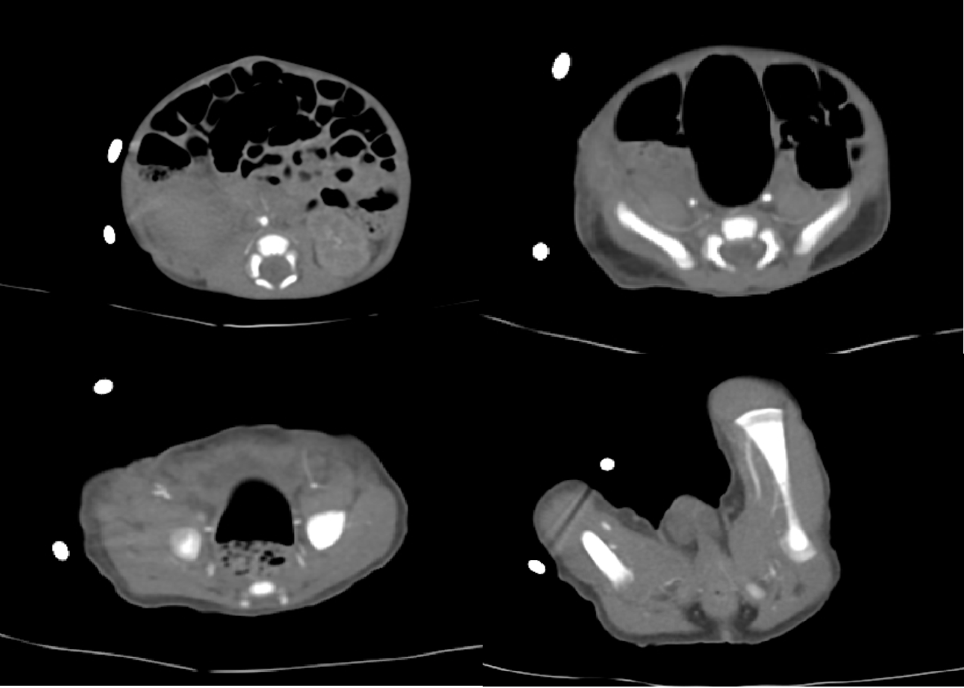
Before discharge, CT angiography showed the absence of the right external iliac artery, with collateral circulation from the internal iliac artery maintaining the continuity of the common femoral artery and its distal branches (Figure 2). Doppler ultrasonography was performed after CT confirmed triphasic flow in the right main femoral artery and its distal portion. The patient was discharged in good health on the 60th postoperative day. At the one-year follow-up, the right lower extremity perfusion was normal, and no developmental delay was observed.
CT angiography of collateral circulation from the internal iliac artery maintaining the continuity of the common femoral artery and its distal branches.

Discussion
Due to complications associated with medical treatment and surgical patent ductus arteriosus ligation, there has been a growing interest in percutaneous patent ductus arteriosus closure in preterm infants. Reference Natarajan, Chawla and Aggarwal4 While percutaneous patent ductus arteriosus closure reduces the risks compared to surgery, studies in low birth weight and preterm infants indicate an increased incidence of catheter-related vascular injuries.
Brotschi et al. identified younger age and low birth weight as significant risk factors for vascular thrombosis. To minimise the risk of arterial injury and reduce vascular thrombosis, studies have recommended retrograde access using a venous approach. Reference Brotschi, Hug and Kretschmar5
In adult patients, vascular injury is one of the most common complications of cardiac catheterisation. Reference Filis, Arhontovasilis and Theodorou6 However, most reported vascular injuries are minor and do not require surgical intervention. In rare cases, emergency surgical intervention is necessary to restore circulation and control bleeding. Reference Babu, Piccorelli and Shah7 While vascular injuries and their management following cardiac catheterisation in adult patients are well-established, the occurrence of such injuries in low birth weight infants is less frequently reported and remains an area of ongoing research.
Vitiello et al. reported a single case of retroperitoneal haemorrhage due to iliac artery injury in a large patient population. Reference Vitiello, McCrindle and Nykanen8 Retroperitoneal haemorrhage is particularly concerning due to its insidious progression and lack of early symptoms, which can lead to high mortality rates. Therefore, early diagnosis and timely intervention are crucial.
Although no specific study has investigated retroperitoneal haemorrhage following catheterisation in low birth weight infants, Amaro et al. described a case of left main iliac artery transection after cardiac catheterisation. In that case, revascularisation was achieved using a saphenous vein graft, allowing successful limb preservation and patient discharge without extremity loss. Reference Amaro and Stephen9
In cases involving aortic and iliac artery bleeding, priority should be given to haemorrhage control, followed by monitoring for lower limb ischaemia. The management of limb ischaemia in paediatric patients is particularly challenging due to ongoing growth. Discrepancies in limb length compared to the contralateral side may occur, as documented in some literature reports. Reference Taylor, Troutman and Feliciano10 Additionally, the selection of surgical materials presents another challenge, as synthetic grafts may necessitate future reoperations due to growth-related complications. Therefore, autologous grafts are generally preferred when possible. Close monitoring and careful decision-making following haemorrhage control are essential.
Conclusion
With the increasing prevalence of catheterisation procedure in paediatric patients, the number of arterial interventions is expected to rise. The timing and intraoperative decision-making for surgical interventions in this context will be challenging. Therefore, selecting the least risky techniques and being well-prepared for potential complications are crucial in managing these cases effectively.