Coarctation of the abdominal aorta, also known as middle aortic syndrome or mid-aortic dysplastic syndrome, is characterised by segmental or diffuse narrowing of the distal descending or abdominal aorta. It may be a congenital anomaly or acquired from a number of conditions. Abdominal coarctation resulting in secondary dilated cardiomyopathy and heart failure is a rare phenomenon, with only one previously reported case.
Case report
A 4-month-old female with newly diagnosed dilated cardiomyopathy was transferred to our institution after an echocardiography performed for a new-onset murmur at her 4-month-old well examination demonstrated a dilated left ventricle (left ventricular diastolic dimension 3.6 cm, z score +3.5 [normal 1.94–2.68 cm]; z score of −2 to +2) and severe left ventricular dysfunction with a fractional shortening of 16% (normal 32–45%). There was moderate mitral regurgitation. Although the abdominal aorta was severely hypoplastic, there was no evidence of coarctation of the proximal descending thoracic aorta. On clinical examination, she was hypertensive in the upper extremities (140/80 mmHg), there was a blood pressure gradient of 70 mmHg between the upper and lower extremities, and her pulses in the lower extremities were diminished. She was started on low-dose calcium channel blockers with the aim of only mildly decreasing her blood pressure in order to avoid renal hypoperfusion. A cardiac CT was performed, which confirmed the diagnosis of middle aortic syndrome.
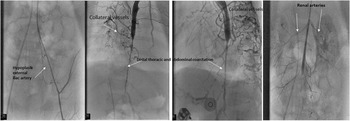
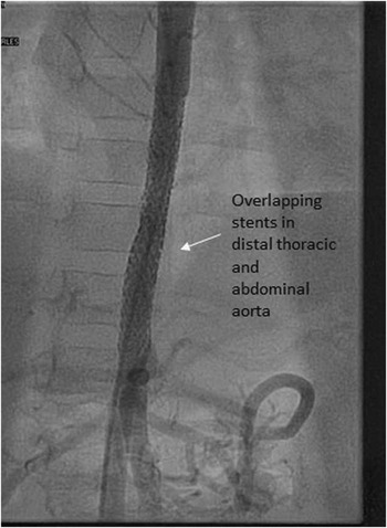
She was presented for a cardiac catheterisation. As hypoplasia of her abdominal and femoral vessels made access and intervention with a 5-F sheath challenging (Fig 1a), a carotid cut-down was performed. A 69-mmHg gradient was measured between the ascending and the abdominal aorta. The distal thoracic and abdominal aorta was diffusely hypoplastic, spanning ~50 mm in length (Fig 1b) and measuring ~1.2 mm at its widest (Fig 1c). Although there was no discrete renal artery stenosis, the renal arteries were hypoplastic (Fig 1d). Prominent collateral circulation via the internal mammary arteries and the intercostal arteries was noted. We placed two VeriFLEX coronary stents (Veriflex stents, Boston Scientific, Massachusetts, United States of America) of around 4×32 and 4×28 mm in an overlapping manner to cover the area of the coarctation (Fig 2). Following stenting, there was no residual gradient between the upper and the lower extremities or by direct measurement, and she was discharged home 2 days later. Clinically, she continued to be hypertensive (120/60 mmHg) and was managed on a dual antihypertensive regimen of calcium channel blockers and enalapril. Notably, a complete evaluation for vasculitic illnesses was performed – and we found no abnormalities to suggest acquired middle aortic syndrome from a vasculitic process.
( a ): Angiogram showing hypoplasia of the external iliac artery. ( b and c ) Angiograms in the antero-posterior view and lateral view show severe hypoplasia of the distal thoracic and abdominal aorta measuring around 5 cm in length and 1.2 mm in width. ( d ) Angiogram showing diffuse hypoplasia of renal arteries and distal abdominal aorta below the renal arteries.

Angiogram showing no residual narrowing after placement of two overlapping stents in the distal thoracic and abdominal aorta.

Repeat catheterisations occurred at 6 months of age for re-stenosis, with acutely favourable responses to balloon dilatation and placement of a 5×28-mm coronary stent and at the age of 2 years with placement of two 8×29-mm Omnilink stents (Abbott Vascular, United States of America). At present, at 2 years of age, her left ventricular dilatation has resolved and her left ventricular systolic function is normal (fractional shortening of 44%); her mitral regurgitation is trivial. Her blood pressure is adequately controlled on low-dose enalapril (100/50 mmHg).
Discussion
Coarctation of the abdominal aorta is rare condition and accounts for less than 0.5–2% of all cases of aortic coarctation. Middle aortic syndrome is a rare disorder caused by a discrete narrowing of the thoracic and abdominal aorta, occurring either as a congenital anomaly or in association with a number of vasculitic diseases including Takayasu syndrome, neurofibromatosis, fibro-muscular dysplasia, systemic lupus erythematosus, and mucopolysaccharidosis.Reference Daghero, Bueno, Peirone, Ochoa, Fao Torres and Ganame 1 , Reference Das, Recto, Shoemaker, Mitchell and Austin 2 A rare disorder, middle aortic syndrome has a prevalence that is difficult to estimate. Most cases are not diagnosed until adolescence or early adulthood. Abdominal coarctation can present with a variety of signs and symptoms, with the predominant manifestations being intractable arterial hypertension and lower extremity arterial insufficiency.Reference Das, Recto, Shoemaker, Mitchell and Austin 2 , Reference Parent, Bendaly and Hurwitz 3 Rarely has it been associated with heart failure at presentation.Reference Zeltser, Parness, Ko, Holzman and Kamenir 4 Only a handful of cases have ever been reported in infants. When symptomatic in newborns and infants, MAS has been reported to be fatal.Reference Zeltser, Parness, Ko, Holzman and Kamenir 4 In adults, the commonest reported co-morbidity is Takayasu arteritis. Vasculitic processes have also been reported to be associated in paediatric series,Reference Parent, Bendaly and Hurwitz 3 with Takayasu arteritis and systemic lupus erythematosus being the most common. The pathogenesis of idiopathic mid-aortic syndrome is unknown.Reference Sethna, Kaplan, Cahill, Velazquez and Meyers 5 When idiopathic, the location of the aortic lesion is intra-renal in 19–52% of cases, supra-renal in 11–40%, infra-renal in 19–25%, and diffuse in 12%.Reference Sethna, Kaplan, Cahill, Velazquez and Meyers 5
Owing to the rarity of the disease, surgical and catheter-based interventions are the only available therapies to date for these patients.Reference Parent, Bendaly and Hurwitz 3 , Reference Tummolo, Marks and Stadermann 6 , Reference Pilati, Pongiglione and Gagliardi 7 In older patients, systemic vasculitic diseases must be excluded before initiation of these interventions.Reference Parent, Bendaly and Hurwitz 3 In the only large case series of children,Reference Das, Recto, Shoemaker, Mitchell and Austin 2 both surgical and catheter-based interventions were used. Those initially treated with catheterisation required repeat interventions to accommodate somatic growth. Given the rarity of the disease and lack of large comparative studies, it is unclear at this time which approach offers better outcomes for these patients.
For infants, patch aortoplasty would theoretically be ideal, given its growth potential. In our patient, however, surgical intervention would have been very risky, as the extensive long-segment narrowing involving the distal thoracic and abdominal aorta necessitated an extensive incision. Although an aortic bypass graft may have been an alternative option, the patient’s age along with the size of her proximal aorta would have necessitated multiple surgical re-interventions in the future to upsize the graft. Arguably stent placement in our patient is not without its complications, as it does set her up for future catheter-based interventions. To accommodate somatic growth, stent re-dilation and additional stent placement will likely be necessary. Although self-expanding stents could have been used, the patient’s young age placed her at high risk for in-stent stenosis, making this approach less than ideal.
Our case highlights the rare presentation of cardiomyopathy resulting from severe mid-aortic syndrome in infancy. A trial of medical management is usually the first-line approach in patients presenting with hypertension.Reference Sethna, Kaplan, Cahill, Velazquez and Meyers 5 , Reference Tummolo, Marks and Stadermann 6 In our patient, however, the presence of severe cardiomyopathy compelled more immediate and invasive therapy. The addition of catheter-based interventional management, performed in such a young infant to avoid the high risk associated with surgical patch homograft aortoplasty needed to address severe hypoplasia of the distal thoracic and abdominal aorta, makes our case particularly unique. Although repeat cardiac catheterisation will be necessary to accommodate somatic growth, the morbidity and mortality associated with an extensive thoraco-abdominal surgery were avoided.
Acknowledgements
None.
Financial Support
This research received no specific grant from any funding agency, commercial, or not-for-profit sectors.
Conflicts of Interest
None.