Case report
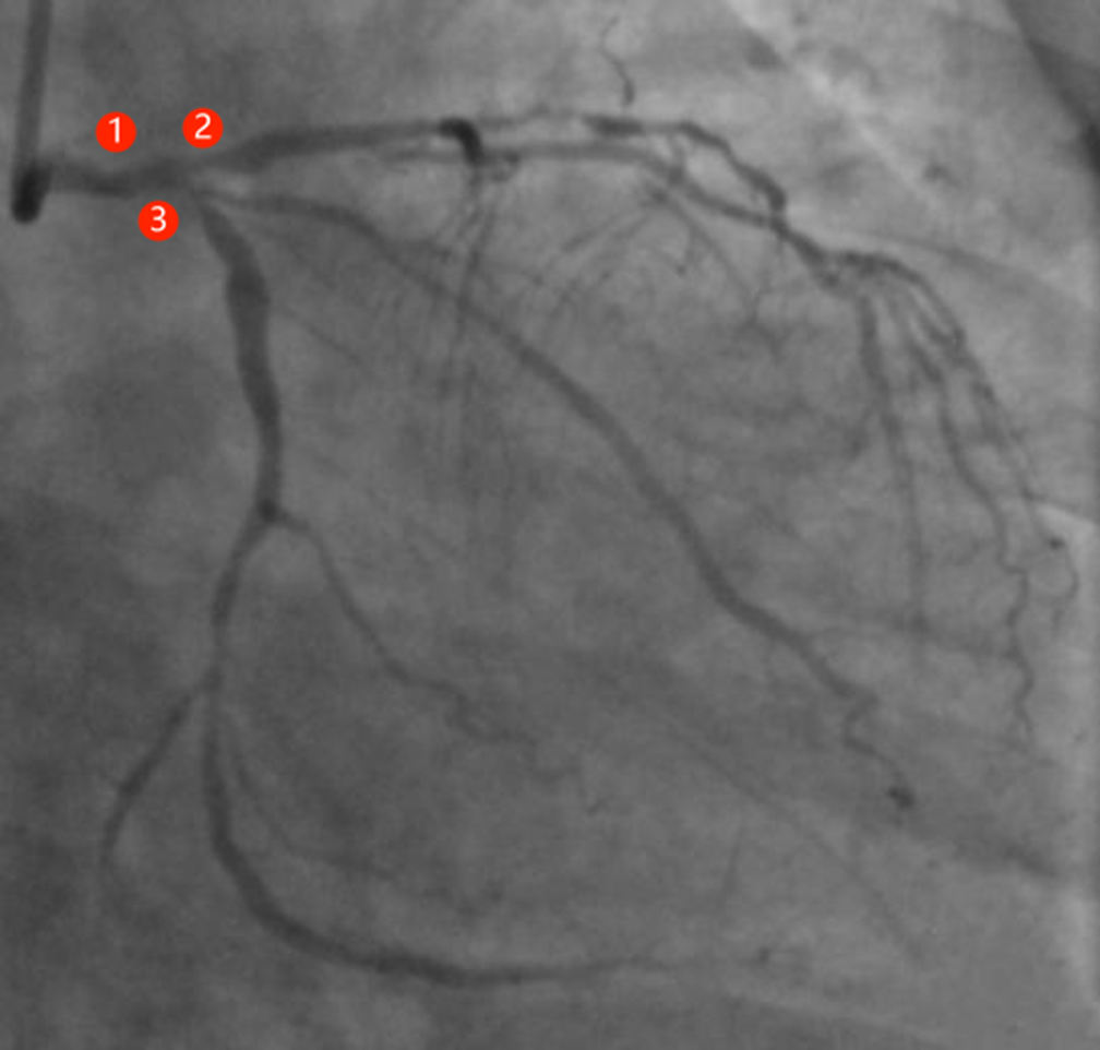
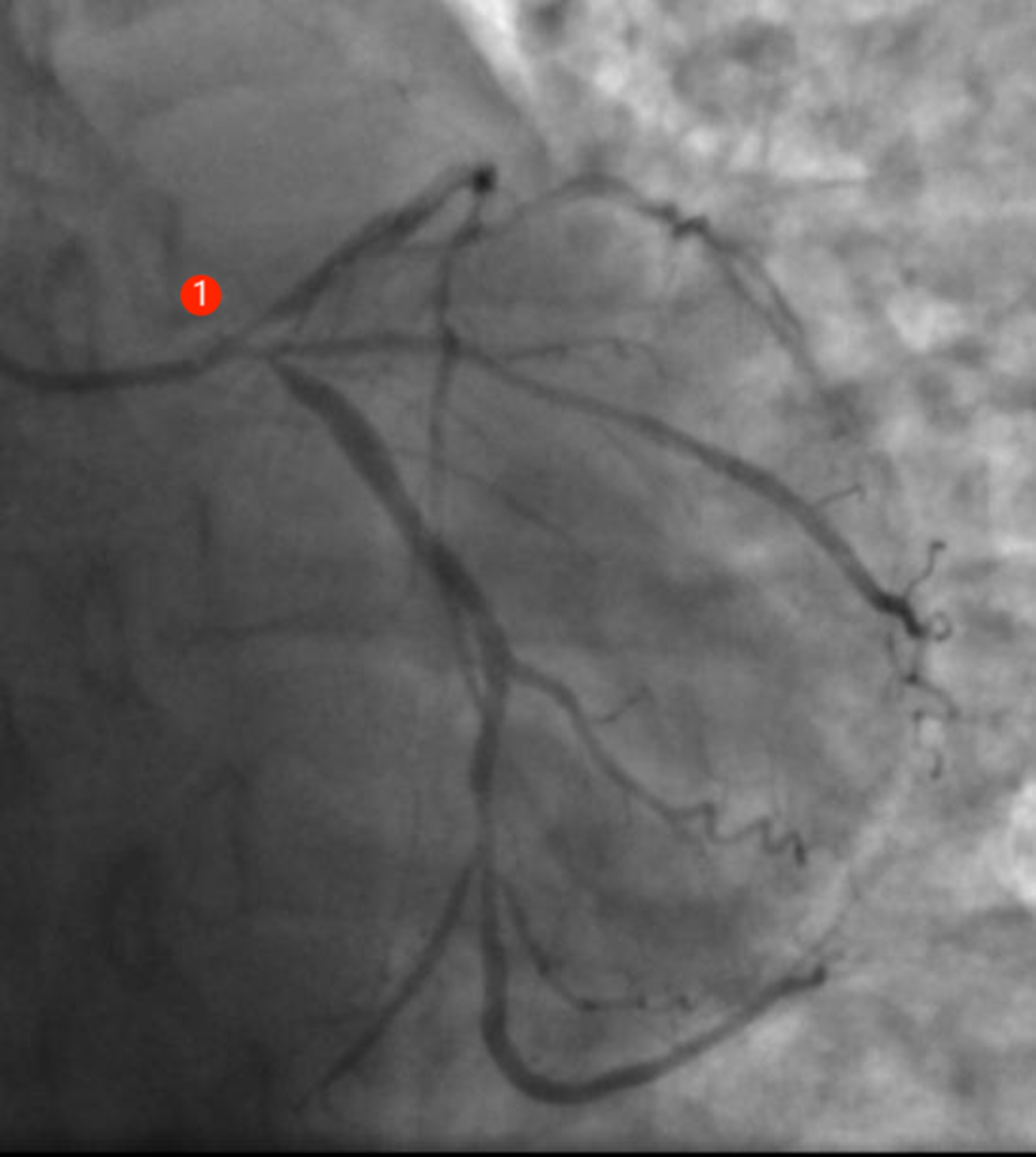
A 41-year-old female without traditional risk factors for coronary artery disease presented with a 1 year history chest discomfort and chest pain after activity which had worsened in the last 5 months and was admitted to the hospital. She was hospitalised elsewhere 5 years before for mild claudication. No classic risk factors for CAD were present. The aorta CT angiography showed the stenosis of the inferior mesenteric artery. Clinical diagnosis of Takayasu arteritis was made based on 1990 American College of Rheumatology criteria (she met three out of six criteria). Physical examination showed normal pulsation of the radial and brachial arteries bilaterally. At the onset of symptoms, the electrocardiogram showed a horizontal shift of >0.1 mV in the anterior wall leads of the heart. Myocardial enzymes were normal. Routine biochemical and haematological tests were normal, and serological tests for C-reactive protein was negative. The arterial ultrasound results showed moderate stenosis of the ostium of the left external carotid artery. Echocardiography results showed a left ventricular end-diastolic volume of 50 mm, EF of 60%, and decreased ventricular wall motion in the lower half of the anterior septum and apical segment of posterior septum. The first coronary angiography (Fig 1) revealed 40% stenosis of the ostium of the left main coronary artery. There were multiple segmental lesions in the left anterior descending artery, with 75% stenosis of the proximal segment and 90% stenosis of the middle segment of the left anterior descending artery. There was 80% stenosis of the proximal segment of the left circumflex artery. There was 90% stenosis of the ostium right coronary artery and long lesions in the proximal-middle segment with stenosis reaching 60%. SYNTAX scores were 40. Optical coherence tomography examination was performed after angiography. Diffuse concentric fibrous hyperplasia was observed from distal to proximal sections of the right coronary artery and the left anterior descending. After discussing the options of coronary artery bypass grafting and percutaneous coronary stenting, the patient declined surgery and opted for percutaneous coronary stenting. A Zotarolimus-Eluting stent (Resolute, 3.5 * 12 mm, Medtronic) was implanted in the right coronary artery ostium, and a Zotarolimus-Eluting stent (Resolute, 2.25 * 18 mm, Medtronic) was implanted in the middle section of the left anterior descending artery. The percutaneous coronary stenting procedure was successful, and the patient was advised to take oral corticosteroids and dual antiplatelet therapy with aspirin and clopidogrel. Nine months later, the chest discomfort symptoms recurred, and she underwent coronary angiography again. After the catheter entered the left main coronary artery ostium, blood pressure dropped to 80/50 mmHg, (Fig 2) showing diffuse lesions in the left main coronary artery, 75% stenosis in the left main coronary artery ostium. The original stent in the middle of the left anterior descending artery was unobstructed. The SYNTAX scores of the left coronary artery were 34. Immediately after the withdrawal of the left coronographic tube, the patient developed chest pain, ST segment depression seen on ECG monitoring, blood pressure increased to 160/80 mmHg. Her symptoms were not relieved with sublingual administration nitroglycerin, and she developed dyspnoea, which was relieved after 10 minutes of oxygen inhalation. Nitroglycerin was given intravenously, morphine intramuscular injection was administered, and the patient was positioned in high occipital supine position. Emergency coronary artery bypass grafting was recommended but still refused by the patient. The results of the 5-year follow-up showed that the patient was continuously treated with a combination of Chinese and Western medicines for Takayasu arteritis with multiple coronary involvement after discharge. She had intermittent angina pectoris that could be alleviated by oral medication. She could engage in general activities of life without need for repeat coronary artery bypass grafting or percutaneous coronary stenting. Regular monitoring of C-reactive protein and ESR showed no increase, and no long-term oral steroid or immunosuppressive therapy was given for Takayasu arteritis.
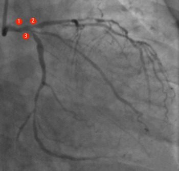
Coronaryangiogram (initial): 40% stenosis of the ostium of the left main coronary artery (LMCA①).There were multiple segmental lesions in the left anterior descending artery (LAD②), with75% stenosis of the proximal segment and 90% stenosis of the middle segment of the LAD. There was 80% stenosis of the proximal segment of the left circumflexartery (LCX③).
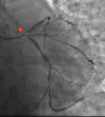
Coronary angiogram (nine months later): Compared with the initial angiography, the progression of de novo lesionsLM from 40% stenosisto 75%(①).
Discussion
Takayasu arteritis refers to chronic, granulomatous full-thickness arteritis involving the aorta and its primary branches. The aetiology is poorly understood, but it is believed that infectious sources, genetic factors, and autoimmune disease play a major role. Coronary artery involvement is found in 7–29% of patients with Takayasu arteritis. In the versus majority of cases, the proximal and ostium parts of the coronary arteries are affected.
However, the optimal treatment strategy for Takayasu arteritis with multiple coronary involvement is still unclear. The treatment options to improve prognosis include drugs (steroids, immunosuppressants), percutaneous transluminal coronary angioplasty, percutaneous coronary stenting and coronary artery bypass grafting. There have been many reports on the application of drug-eluting stents in coronary invasive therapy for Takayasu arteritis. Reference Kang, Han, Ahn and Shin1,Reference Sakai, Oyama, Kishimoto, Takahashi, Urasawa and Tsutsui2 In the patient reported by Yokota et al Reference Yokota, Shimpo and Iwata3 repeated stent restenosis appeared 5 months after Cypher stent implantation. Finally, stent restenosis was prevented by adding oral steroid therapy, and the patient was asymptomatic at the 4-year follow-up. However, the patient we reported regularly take steroid and other related drugs, and the symptoms recurred 9 months after percutaneous coronary stenting. The original left anterior descending artery stent was unobstructed by coronary artery angiography, which may be related to the application of the second-generation stent. Using the new generation of drug-eluting stents that is expected to further reduce the inflammatory response, the incidence of restenosis may be significantly reduced.
Drug-coated balloons are semi-compliant angioplasty balloons covered with an antiproliferative drug that is rapidly released from a lipophilic matrix upon contact with the vessel wall. The advantage of drug-coated balloons over stents is that it allows a “leaving nothing behind” strategy, no metal implants with biocompatible coatings or biodegradable coatings, which minimise the long-term inflammatory stimulation of foreign bodies to coronary artery. The principle of action is applicable to the pathological characteristics of Takayasu arteritis with endointimal hyperplasia of coronary artery lesions. Reference Chiew and Lim4 A patient with active Takayasu arteritis presenting with acute coronary syndrome was recently reported to have been treated with drug-coated balloon under optical coherence tomography guidance in the critical left anterior descending artery lesion and remained asymptomatic for more than 3 years with long-term follow-up.
However, the reexamination of angiography in our case showed the progression of de novo lesions in the left main coronary artery. Unfortunately, the acute condition of the patient after occlusion of the angiography catheter at that time made the plan of reexamination of optical coherence tomography impossible to perform, so the pathological features of the lesions with left main coronary artery progression could not be observed. However, the progression of de novo left main coronary artery lesions from 40% stenosis to 75% after 9 months suggests the possibility of local inflammatory reaction and hyperplasia caused by the previous invasive manipulation of percutaneous coronary stenting. In our case, the SYNTAX score of the left coronary artery was 34 points based on the calculation of the second coronary angioplasthy, and IA was recommended according to the guidelines, Reference Neumann, Sousauva and Ahlsson5 but the guidelines were based on research into atherosclerotic heart disease. Some cases have reported coronary artery bypass grafting results in patients with Takayasu arteritis that are not satisfactory. Although internal mammary artery transplantation has an excellent patency rate in atherosclerotic coronary heart disease, Takayasu arteritis patients have a greater risk of developing subclavian artery stenosis. Reference Haridas, Kumar, Rajesh, Kumar and Pannekal6 Therefore, unlike most coronary artery bypass graftinggrafts, saphenous vein grafts are commonly used in patients with Takayasu arteritis. The 10-year survival rate after saphenous vein grafts-left anterior descending artery transplantation was 80%, and the event-free survival rate was 77%. Reference de Soeiro, Almeida M.C. and Torres7 Some retrospective studies Reference Wang, Zhang, Shen, Fang, Liu and Zhang8,Reference Huang, Zhang, Wang, Yang, Qiao and Hu9 showed that neither percutaneous coronary stenting nor coronary artery bypass grafting showed a reduction in MACE and mortality in patients with Takayasu arteritis complicated with coronary stenosis when compared with drug therapy alone. Percutaneous coronary stenting was independently associated with a higher incidence of MACE and revascularization than coronary artery bypass grafting. For patients with revascularization indications, coronary artery bypass grafting may be the preferred method of choice. Macedo et al reported a case of Takayasu arteritis with multiple coronary involvement. Reference Macedo, Lima, Junior and Bannon10 Steroid and immunosuppressant treatment was started immediately after coronary artery bypass grafting treatment. However, 8 months after the operation, a 70% stenosis of the venous graft to the posterior descending artery occurred while the arterial graft remained patent. The patient has been asymptomatic for a long time after placing DES in the venous graft to the psterior descending artery. This shows the complexity of the treatment of these patients.
Conclusion
However, the case we reported was only treated with drug therapy after percutaneous coronary stenting. The results of 5 years of follow-up showed that the patient could still engage in general activities of life under the condition of drug maintenance and stable control of inflammatory response monitoring. This case review makes us inclined to the following strategies for the treatment of Takayasu arteritis with multiple coronary involvement: adequate and aggressive drug therapy, regular monitoring of inflammatory indicators to maintain low levels, invasive intervention strategies for next-generation stent implantation, or coronary bypass grafting should be considered when symptoms are not controlled with medication or when coronary artery lesions are unstable. However, the feasibility of this strategy needs to be clarified by accumulating more cases, increasing the sample size and designing prospective randomized controlled studies. Interventional cardiologists should also cooperate with multi-disciplinary doctors including rheumatology and cardiothoracic surgery, so as to jointly study and develop an optimal strategy for Takayasu arteritis with multiple coronary involvement, in order to improve their quality of life and life expectancy.
Acknowledgements
Thanks to all the teachers who gave me support in the course of experiment and thesis writing!
Conflicts of interest
None.
Financial support
This research received no specific grant from any funding agency, commercial, or not-for-profit sectors.