

Hemifacial spasm (HFS) is characterised by irregular, involuntary and recurrent tonic and clonic contractions of muscles innervated by the ipsilateral facial nerve. HFS is usually attributed to compression of the facial nerve at the root entry zone by compressive or non-compressive lesions (Reference Jankovic1).
Homocysteine, an amino acid important in metabolism, causes endothelial cell injury and cell detachment that initiates the development of arteriosclerosis. A markedly elevated homocysteine level has been associated with a higher risk of forming blood clots and vascular disease. However, mild hyperhomocysteinaemia has also been linked to these complications (Reference Wierzbicki2).
A 59-year-old man complained of a 5-year history of intermittent, involuntary muscle spasms involving the right lower and mid portions of his facial region. Neurological examination was consistent with right HFS. He also reported intermittent short-lasting episodes of dizziness and gait instability during the last years, without objective findings of brainstem dysfunction on examination. Taste was normal. Tendon reflexes were increased in all four limbs with flexors plantar reflexes. The rest of the examination was normal. His past medical history was significant for mild well-controlled hypertension and prostate hypertrophy. There was no family history for cardiovascular diseases.
Blood counts, general biochemistry, collagen, rheumatic, venereal disease and tumour markers were either normal or negative. Vitamin B12 and folate levels were within normal limits. Likewise, coagulation studies including protein S, protein C, antithrombin III, lupus anticoagulant and anticardiolipin antibodies were unremarkable, with the exception of serum homocysteine, which was mildly elevated (22.1 µmol/l with normal values ranging between 5 and 15 µmol/l).
The heart ultrasound and the carotid and vertebral arteries triplex examination were normal. Late-onset demyelination of the central nervous system was thoroughly excluded by appropriate examinations, including visual evoked potentials and oligoclonal bands testing in cerebrospinal fluid, which were either normal or negative.
Right facial nerve conduction study and repetitive stimulation study in the orbicularis oculi (OOc) muscle were normal. Needle electromyography of the OOc and orbicularis oris muscles at rest showed a continuous activity of irregularly firing low-amplitude motor unit potentials. The interference pattern was incomplete.
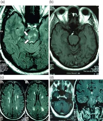
The magnetic resonance imaging of the brain revealed multiple, confluent high-intensity lesions at the level of the pons on the right and fewer similar lesions on the left. At right, the high-intensity lesions involved the ipsilateral medial cerebellar peduncle and the facial colliculus (Fig. 1a). No contrast-enhancing lesions were noticed after gadolinium administration (Fig. 1b). Diffuse high-intensity white matter lesions were also depicted in Fluid Attenuated Inversion Recovery (FLAIR) images (Fig. 1c and d). There was no evidence of mass lesion at the cerebellopontine angle both before and after the administration of contrast media (Fig. 1d). Treatment with combined folate and B-vitamin supplementation was initiated.
No contrast-enhancing confluent high-intensity lesions at the level of the pons on the right involving the ipsilateral medial cerebellar peduncle and the facial colliculus (arrows). (a and b) Fewer similar lesions on the left. (c) Diffuse high-intensity white matter lesions on FLAIR images. (d) Contrast T1W image and non-contrast axial FLAIR image depicting no evidence of mass lesion at the cerebellopontine angle.

The pathogenesis of HFS is not completely understood. Most instances of HFS are thought to be caused by vascular compression from aberrant blood vessels or cerebellopontine angle masses compressing the facial nerve within the cerebellopontine angle (Reference Papapetropoulos, Argyriou, Guevara, Sengun, Mitsi and Singer3). Other causes of HFS include stroke, multiple sclerosis plaques and tumour (Reference Jankovic1). In any case, causes other than mass lesions or vascular loops are rarely reported in the medical literature (Reference Han, Chang, Chang, Huh and Chung4).
We are confident that there is no evidence of facial nerve compression at its root entry zone in cerebellopontine angle in our case, as T1-weighted (T1W) sequences both before and after the administration of contrast media have ruled out this possibility. T1W imaging is still considered the optimal screening tool to detect even small tumours surrounded by cerebrospinal fluid at the cerebellopontine angle, because it is able to reveal with high sensitivity a contrast between physiological structures and pathological processes. Other modern techniques, such as the constructive interference in steady-state sequences, might also be used but as a complementary investigation and not as a primary screening tool (Reference Goebell, Ries and Kucinski5).
The facial motor nucleus is located in the ventrolateral portion of the caudal pons. Its fibres form the facial colliculus in the floor of the fourth ventricle and then exit from the lower body of the pons through the pontine regmentum. The facial colliculus is formed by the internal genu of the facial nerve and the abducens nucleus around which the facial fibres curve (Reference Lacombe6). To our knowledge, our case is the fist to describe the occurrence of hyperhomocysteinaemia-associated cerebral small vessel disease that was clinically manifested with HFS due to an ischaemic lesion at the facial colliculus. Apart from HFS, hyperhomocysteinaemia was the prominent factor that gave rise to the genesis of multiple silent cerebral infarcts.
The manifestation of HFS in our case is attributed to the interruption of the ipsilateral facial nerve fibres at the genu as they arch behind the abducens nerve nucleus and form the colliculus. Despite that the blink reflex pathway was not studied, we suggest that the signs of HFS might also be caused by hyperexcitability of the facial nucleus secondary to either compression of the root entry zone or loss of inhibitory impulses from the brainstem, due to the vascular lesions at the pons (Reference Moller7).
As compared to the general population, persons with hyperhomocysteinaemia have increased the risk of manifesting peripheral arterial disease, stroke, cerebral vascular disease and heart disease. The risk of serum hyperhomocysteinaemia for the presence of the above-mentioned conditions may be as significant as the risk associated with hypertension, dyslipidaemia or smoking history (Reference Ford, Smith, Stroup, Steinberg, Mueller and Thacker8).
Our case further highlights the close relation between serum hyperhomocysteinaemia and development of cerebral small vessel disease, as reflected by the effects on both subclinical and overt disease. Even mild serum hyperhomocysteinaemia represents a significant risk factor for the formation of cerebral small vessel disease with subsequent manifestation of atypical clinical symptomatology such as HFS. In our case, we suggest a causal association between hyperhomocysteinaemia, cerebral small vessel disease and HFS. However, the combined occurrence of hyperhomocysteinaemia with mild hypertension should not be disregarded as it might possibly have increased the risk for the development of cerebral small vessel disease in the patient reported herein.
Acknowledgement
No funding source had a role in the preparation of this paper or in the decision to submit it for publication. We have no conflicts of interest to declare.