I. INTRODUCTION
Morphology-selective synthesis of lanthanide orthovanadate nanomaterials is important due to their promising application in optical,Reference Wang and Li1 electrical,Reference Picardi, Varsano, Decker, Opara-Krasovec, Surca and Orel2 magneticReference Yan and Yan3 and catalytic fields.Reference Fang, Hong, Zhou, Dai, Weng and Wan4 Therefore, great effort has been devoted to the synthesis of lanthanide orthovanadates of various morphologies, including rods,Reference Fan, Song, Bu, Sun and Zhao5 wires,Reference Nguyen, Dinh, Nguyen and Do6 spheres,Reference Nguyen, Dinh and Do7 plates,Reference Deng, Yang, Xiao, Gong and Wang8 cube-likeReference Sunand and Xia9 and persimmons-like.Reference Qian, Zhu, Chen, Gui, Gong and Yuan10 In addition, quite a few methods have been developed for preparing these compounds, such as sol–gel, microwave-assisted chemical synthesis, Czochralski method and solid-state reaction.Reference Wang, Zhuang, Peng and Li11–Reference Yu, Cheng, Zhang, Wang, Cheng and Xia16 However, most of the above methods need severe reaction conditions such as high temperature.Reference Errandonea, Lacomba-Perales, Ruiz-Fuertes, Segura, Achary and Tyagi15, Reference Yan and Su17 Compared with the synthetic techniques listed above, the hydrothermal method is very mild (the reaction temperature is often below 200 °C). Additionally, the hydrothermal strategy is simple, convenient, safe and scalable, which can affect the crystallization of single particles and allow the control of structures and morphologies of the final product.
Organic complexing agents are often used to prepare inorganic materials with uniform and special morphologies for different applications. For instance, Ferrari et al.Reference Ferrari, Parreira, Pires, Lima and Davolos18 obtained neodymium ion-doped gadolinium (III) oxide (Gd2O3:Nd3+) with different particle size by increasing the amount of ethylene diamine tetraacetic acid (EDTA). Fu et al.Reference Fu, Tian, Chen, Zhou, Pan and Dong19 reported the controllable preparation of hollow-structured bismuth molybdate (Bi2MoO6) spheres as high-performance visible-light-driven photocatalysts with the aid of ethylene glycol. Besides, our group recently reported that erythrocyte-like holmium vanadate (HoVO4) and lutetium vanadate (LuVO4) nanocrystals could be successfully fabricated via the complexing agent-assisted hydrothermal method.Reference He, Zhang, Zhu, Zheng and Fang20, Reference Zhang, He, Zhu and Zheng21
Although the synthesis and properties of rare earth orthovanadate nanomaterials have been well documented, most studies are focused on yttrium orthovanadate (YVO4) and lanthanum orthovanadate (LaVO4). As one of the important lanthanide-group elements, the electronic structure of ytterbium ion (Yb3+) is composed of only two states, the 2F7/2 ground state and the 2F5/2 excited state, which bestows on the Yb3+ ion no excited-state absorption, no upconversation and negligible nonradiative relaxation rate between the two states.Reference Yu, Cheng, Zhang, Wang, Cheng and Xia16, Reference Cheng, Zhang, Zhang, Xin, Yang and Xu22 Due to the above features, ytterbium (Yb) is commonly used as a vanadate laser host. However, most previous approaches for preparing those erbium ion-doped ytterbium orthovanadate (YbVO4:Er3+) compounds needed high temperature and severe reaction conditions. The hydrothermal method has its own advantages. It not only affects the crystallization of single particles, but also allows the controls of structures and morphologies of the resulting materials. Moreover, the hydrothermal system still possesses many other merits, such as simplicity, safety and convenience.
In this study, a mild complexing-agent-assisted hydrothermal strategy was used for the synthesis of YbVO4 nanocrystals with well-defined morphology. Trisodium citrate (Na3Cit) was selected as the complexing agent for synthesis. The influence of the reaction temperature, the reaction time and the molar ratio of Na3Cit to Yb3+ on the formation of erythrocyte-shaped YbVO4 nanocrystals were systemically investigated. In addition, the YbVO4:Er3+ crystals synthesized using different complexing agents exhibited interesting morphology-dependent optical properties.
II. EXPERIMENTAL SECTION
A. Materials
All reagents [ytterbium oxide (Yb2O3), erbium oxide (Er2O3), vanadium pentoxide (V2O5), Na3Cit, ethylene diamine tetra acetic acid (EDTA), ethylene diamine (EDA)] were of analytical purity, obtained from Shanghai Chemical Reagent Ltd. Co. (Shanghai, China) and used without further purification.
B. Synthesis of nanoerythrocyte-shaped YbVO4 and YbVO4:Er3+
The YbVO4 nanocrystals were prepared by a simple and mild complexing agent-assisted hydrothermal method. In a typical procedure, ytterbium nitrate [Yb(NO3)3] crystal was prepared by dissolving 0.28 mmol diytterbium trioxide [Yb2O3] in 1 mol/L nitric acid and drying at elevated temperature. Afterward, 30 mL 0.073 mol/L Na3Cit aqueous solution was added to form a clear solution under magnetic stirring. Then 0.56 mmol sodium orthovanadate [Na3VO4] obtained by adding 0.28 mmol V2O5 into the dilute sodium hydroxide (NaOH) solution was introduced drop wise to the clear solution. After agitation for 10 min, the as-obtained mixture was transferred into a 50 mL Teflon liner and some distilled water was added up to 40 mL. The system was sealed and heated at 180 °C for 24 h. The obtained precipitate was collected at room temperature after centrifugal separation at 3500 rpm for 15 min, washed with distilled water and then dried at 80 °C in air.
A similar process was used to prepare YbVO4:Er3+ powder by adding a proper amount of erbium nitrate [Er(NO3)3] into the Yb(NO3)3 solution at the initial stage as described above. All the doping 3%, 6%, 9% Er3+ were the molar ratios of the reactants Er3+:Yb3+ in our experiments.
C. Characterization
Powder x-ray diffraction was carried out with an x-ray diffractometer (XRD; Rigaku model No. D/max rA, Tokyo, Japan) equipped with graphite-monochromatized high-intensity Cu Kα radiation (λ = 0.15478 nm). The scanning rate was 0.05°/s in the 2θ range from 10 to 70°. The x-ray photoelectron spectroscopy analysis was performed on a x-ray photoelectron spectrometer (XPS; model No. ESCALAB MK II, East Sussex, UK), using Mg Kr radiation as the excitation source. The high-resolution transmission electron microscopy images were carried out on a transmission electron microscope working at 200 kV (HRTEM; Hitachi model H-800, Tokyo, Japan). The field-emission scanning electron microscopy images were obtained on a field-emission scanning electron microscope with an accelerating voltage of 15 kV (FESEM. model No. FEI SIRION-200, OR).The photoluminescence was carried out using a steady-state/lifetime spectrofluorometer at room temperature (PL; Jobin Yvon model No. Fluorolog-3-TAU, Paris, France).
III. RESULTS AND DISCUSSION
A. Characterization, morphology and the growth mechanism of YbVO4 powder
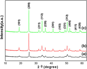
The overall crystallinity and phase purity of the samples obtained with Na3Cit at different reaction temperatures after 24 h were characterized are shown in Fig. 1. The XRD patterns of the samples indicate all of the reflections can be indexed to pure tetragonal phase YbVO4 (space group:I41/amd) with lattice constants a = b = 0.70435 nm and c = 0.62470 nm [Joint Committee on Power Diffraction Standards (JCPDS) card 017-0338]. No XRD peaks corresponding to metal oxide of V2O5 or Yb2O3 are detected, indicating that pure tetragonal YbVO4 crystals were obtained. The well-resolved diffraction peaks show the high crystallinity of the products, and the tetragonal phase remains stable up to 220 °C.
XRD patterns of the samples obtained at different reaction temperatures for 24 h with Na3Cit: (a) 140 °C; (b) 180 °C; (c) 220 °C.

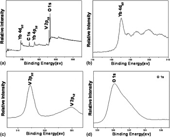
The product obtained at 180 °C for 24 h with the molar ratio of Na3Cit to Yb3+ of 2:1 was also characterized by XPS for the evaluation of its composition and metal oxidation states on the particle surface (Fig. 2). The binding energies obtained in the XPS analysis are corrected for specimen charging by referencing the C 1s orbital to 284.17 eV. The Yb 4d5/2 peak at 185.10 eV, and the V 2p3/2, V 2p 1/2 peaks at 517.2–524.9 eV are attributed to Yb3+ and V5+, respectively [Figs. 2(b) and 2(c)]. Reference Uwamino, Ishizuka and Yamatera23–Reference Kruczek, Talik, Sakowska, Gala and Swirkowicz26 No characteristic peaks of other metals are detected. The O 1s peak at 530.11 eV is attributed to both Yb–O and V–O bonds in the tetragonal YbVO4 and another shoulder peak at 531–532 eV is attributed to the presence of hydroxyl species or adsorbed water on the nanocrystal surface.Reference Nguyen, Dinh and Do7, Reference Salvi, Decker, Varsano and Speranza27
XPS spectra of the as-synthesized product obtained at 180 °C for 24 h with the molar ratio of Na3Cit to Yb3+ of 2:1: (a) survey spectrum; (b) Yb 4d region; (c) V 2p region; (d) O 1s region.

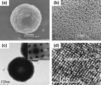
The high magnification FESEM images illustrate the YbVO4 is spherical and biconcave with the diameter around 500 nm [Figs. 3(a) and 3(b)]. It can be seen that the erythrocyte-like YbVO4 nanocrystal is self-assembled by many smaller nanoplates through face-by-face attachment and the yield of the erythrocyte-like YbVO4 in the product obtained at 180 °C for 24 h is 100% [Figs. 3(a) and 3(b)]. The assembled YbVO4 nanocrystal has a well-defined single-crystalline structure, which is confirmed by the selected-area electron diffraction (SAED) pattern [inset of Fig. 3(c)]. The HRTEM image shows that the nanocrystals are structurally uniform with an interplanar spacing of about 0.352 nm, which corresponds to the (200) lattice spacing of tetragonal phase YbVO4. The (200) and (020) directions are parallel to the view erythrocyte-like YbVO4 in Fig. 3(c), showing that the growth of the architecture occurs along the [100] and [010] directions.Reference Liu and Li12
As-synthesized YbVO4 obtained at 180 °C for 24 h with the molar ratio of Na3Cit to Yb3+ of 2:1 (a and b) FESEM images; (c) TEM image of an individual nanoerythrocyte-shaped YbVO4 and its SAED(inset); (d) HRTEM image.

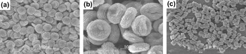
It is well known that the crystal morphology has important effect on its physical and chemical properties. The morphology of the product is affected by the reaction temperature, reaction time and the addition of complexing agents.Reference Luo, Jia, Song, You and Yan28, Reference Tao, Wang, Yao, Cai and Li29 The FESEM images of the samples obtained at different reaction temperatures are shown in Fig. 4. Many obvious changes were observed with the increase of reaction temperature when fixing the molar ratio of Na3Cit to Yb3+ and reaction time. When the reaction temperature was 140 °C, the obtained product consisted of many thin nanodisk particles [Fig. 4(a)]. As the reaction temperature was increased to 180 °C, the uniform erythrocyte-like YbVO4 nanoparticles were obtained in the product [Fig. 4(b)]. It was noted that the diameter of the particles increased with the changes of the temperature from 140 to 180 °C. The length of the aggregated pillar extended when the reaction temperature was further increased to 220 °C [Fig. 4(c)].
FESEM images of YbVO4 obtained at different temperatures for 24 h with the molar ratio of Na3Cit to Yb3+ of 2:1: (a) 140 °C; (b) 180 °C; (c) 220 °C.

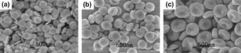
For understanding the formation mechanism, the detailed growth process of YbVO4 was carefully studied by time-dependent experiments. The time-dependent experiments were carried out at 180 °C with the molar ratio of Na3Cit to Yb3+ of 2:1, and indicated that the reaction time had a great role on the morphology of the nanoerythrocyte-shaped YbVO4 (Fig. 5). At short periods of time, many thin nanodisk particles were found in the product [Figs. 5(a) and 5(b)]. The regular and uniform nanoerythrocyte-shaped YbVO4 architectures were created when the reaction time was prolonged to 24 h [Fig. 5(c)]. Meanwhile, the diameter and thickness of the particle increased when the reaction time increased from 12 to 24 h. These images obviously demonstrated that the nanoerythrocyte-shaped YbVO4 was developed from thick nanodisk particles.
The FESEM images of YbVO4 obtained at 180 °C with the molar ratio of Na3Cit to Yb3+ of 2:1 for different reaction times: (a) 12 h; (b) 18 h; (c) 24 h.

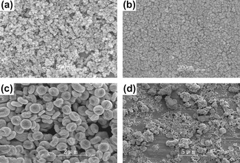
The mophologies of the products obtained at different dosages of Na3Cit when the other conditions were unchanged are shown in Fig. 6. It is found Na3Cit is crucial for the formation of nanoerythrocyte-shaped YbVO4. The experiments showed that only YbVO4 nanoparticles of 10–40 nm diameter were obtained with the molar ratio of Na3Cit to Yb3+ 0:1 or 1:1 and the average diameter of the latter was smaller than that of the former [Figs. 6(a) and 6(b)]. However, when the molar ratio was increased to 2:1, the nanoerythrocyte-shaped YbVO4 could be observed clearly. It was determined that the nanoerythrocyte-shaped YbVO4 particles were obtained from the assembly of nanoplates [Fig. 6(c)]. As the molar ratio was increased to 3:1, stable nanoerythrocyte-shaped YbVO4 architectures were not formed [Fig. 6(d)].
The FESEM images of YbVO4 obtained at 180 °C for 24 h with different molar ratio of Na3Cit to Yb3+: (a) 0:1; (b) 1:1; (c) 2:1; (d) 3:1.

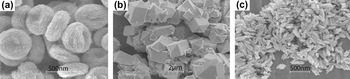
To further investigate the influence of complexing agents on the YbVO4 morphologies, different complexing agents were used. Generally, Na3Cit, EDTA and EDA have similar properties because they are regarded as complexing agents, which can slow down the nucleation rate and prevent further aggregation of the particles.Reference Tao, Wang, Yao, Cai and Li29 Nanoerythrocyte-shaped, dodecahedron and spindle-like YbVO4 were obtained with Na3Cit, EDTA or EDA respectively (Fig. 7), which establishes that the complexing agents have great influence on the morphology evolution of the products. These differences have some connection with their different chelating constants and coordination modes with Yb3+.Reference Xu, Kang, Li, Hou, Zhang and Yang30–Reference Sun, Chen, Tian, Yu, Kong, Zhao and Zhang32
The FESEM images of YbVO4 obtained with different complexing agents for 24 h: (a) Na3Cit(180 °C); (b) EDTA(260 °C); (c) EDA(260 °C).

The reaction process for the formation of nanoerythrocyte-shaped YbVO4 could be described as follows:
 $${\rm{Yb}}^{{\rm{3 + }}} {\rm{ + 2Cit}}^{{\rm{3 - }}} ⇌\,\,{\rm{Yb(Cit)}}_{\rm{2}} ^{{\rm{3 - }}} \quad,$$
 $${\rm{Yb}}^{{\rm{3 + }}} {\rm{ + VO}}_{\rm{4}} ^{{\rm{3 - }}} ⇌\,\,{\rm{YbVO}}_{\rm{4}} \downarrow {\rm{\quad. }}$$
B. Photoluminescence properties of YbVO4:Er3+
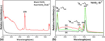
The excitation spectrum of YbVO4:Er3+ sample consisted of a strong absorption band with a maximum at 379 nm and several weak bands monitoring with 553 nm, which was obviously different from that of the YbVO4 [Fig. 8(a)]. It could be found that Er3+ had been doped in the YbVO4 powder because of the close ionic radii (Er3+ = 0.089 nm, Yb3+ = 0.087 nm) from Fig. 8(b). The room-temperature emissions of YbVO4:3% Er3+, YbVO4:6% Er3+, YbVO4:9% Er3+ are centered at 408, 524 and 553(544) nm under 379 nm excitation, which were associated with the typical transitions from 2H9/2, 4H11/2 and 4S3/2 levels to 4I15/2 level of Er3+ ions respectively .Reference Golab, Solarz, Dominiak-Dzik, Lukasiewicz and Ryba-Romanowski33 Among them the hypersensitive emission transition 4S3/2→4I15/2 at 553 nm was the most prominent. No emission from VO43− groups was detected, indicating the energy transfer from VO43− groups to Er3+ is effective. By increasing Er3+ doping concentration from 3 to 9 mol%, the PL intensity of the YbVO4:6% Er3+ and YbVO4:9% Er3+ is higher than that of YbVO4:3% Er3+ sample. However, there is no obvious enhancement between the YbVO4:6% Er3+ and YbVO4:9% Er3+ samples.
(a) Excitation spectrum of YbVO4:3% Er3+ and YbVO4, monitoring the emission at 553 nm; (b) Emission spectrum of YbVO4:Er3+ (a:YbVO4:3% Er3+ b:YbVO4:6% Er3+, c:YbVO4:9% Er3+).

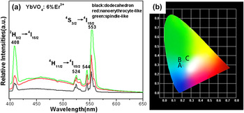
In addition, the luminescent properties of the YbVO4:6% Er3+ with different morphologies by changing the complexing agents were also investigated. It can be seen clearly that the emission spectra of the three samples are similar in shape and different only in the relative intensity under identical measurement conditions [Fig. 9(a)] namely, the relative emission intensity of the dodecahedron YbVO4:6% Er3+ (green line) is the highest, while that of the spindle-like (black line) is the lowest. The influence of the complexing agents on the product morphologies had been described above. In general, the photoluminescence emission intensity of materials has a significant correlation with their particle sizes. In the studies covered by our article, the size of the dodecahedron particle is the largest and that of the spindle-like particle is the smallest. It is well known that the surface area of materials increases along with the decrease of their particle size. The larger the surface area of materials, the more the defects, which are disadvantageous to the PL intensity for the phosphors, as they provide nonradiative recombination routes for electrons and holes. If the surface area were greatly reduced, a phosphor with fewer defects would show greater improvement in the PL intensity.Reference Sun, Chen, Tian, Yu, Kong, Zhao and Zhang32 This may be the reason why the relative emission intensity of the dodecahedron YbVO4:6% Er3+ is the strongest and the spindle-like YbVO4:6%Er3+ shows the lowest intensity. As illustrated in the inset of Fig. 9(b), the emission color of the dodecahedron, nanoerythrocyte-shaped and spindle-like YbVO4:Er3+ samples could be expressed by the Commission internationale de I’eclairage (CIE) coordinates, which represented blue–green light and their chromaticity coordinates were x = 0.227, y = 0.344; x = 0.212,y = 0.334; x = 0.207, y = 0.294, respectively.
(a) Emission spectrum of YbVO4:6% Er3+ with different morphologies; (b) Position of the synthesized spindle-like (A), nanoerythrocyte-shaped (B) and dodecahedron YbVO4:6% Er3+(C) emissions in the chromaticity diagram.

IV. CONCLUSIONS
In summary, a convenient and facile complexing agent-assisted hydrothermal method has been utilized for the preparation of nanoerythrocyte-shaped, dodecahedron and spindle-like YbVO4. The condition experimental results show that 180 °C, 24 h and the molar ratio of Na3Cit to Yb3+ of 2:1 are the preferable experiment parameters to synthesize the nanoerythrocyte-shaped YbVO4. The YbVO4:Er3+ exhibits the typical blue–green emissions at 408, 524 and 553 nm under 379 nm excitation, which can be considered as a promising material for light phosphor powders. The YbVO4:Er3+ samples obtained using different complexing agents reveal the interesting morphology-dependent optical properties. Furthermore, the facile method could be extended to prepare other lanthanide orthovanadates with nanoerythrocyte-shaped morphology.
ACKNOWLEDGMENTS
This research is supported by the Structure Research Laboratory of CAS. We thank S. Q. Fu (University of Science and Technology of China) for the helpful discussion.