Case report
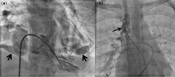
A 14-year-old girl was admitted for surgical correction of tetralogy of Fallot. Preoperative chest radiograph showed ill-defined opacities in bilateral lower lung zones. Cardiac catheterisation proved them to be large bilateral pulmonary arteriovenous malformations (Figure 1A). Significant aortopulmonary collateral to the right lung was also evident (Figure 1B). Computed tomography angiogram ruled out additional arteriovenous malformations in liver or brain. She neither had bleeding manifestations nor cutaneous telangiectasias. A history of liver disease, recurrent infections, metastatic carcinoma, other systemic disorders, surgical intervention, or chest trauma was absent. None of her first-degree relatives suffered from a similar illness. In view of large arteriovenous malformations, she would be at high-risk for life-threatening haemoptysis, systemic embolism, or brain abscess. She was anticipated to have persistent hypoxemia and cyanosis despite surgical correction of tetralogy of Fallot and, thereby, a prolonged ventilator weaning postoperatively. Hence, she was planned for preoperative percutaneous closure of arteriovenous malformations and aortopulmonary collateral.
a. Angiographic imaging demonstrating bilateral large pulmonary arterivenous malformations (arrows). b. Angiographic imaging showing a large aortopulmonary collateral to the right lung (arrow).

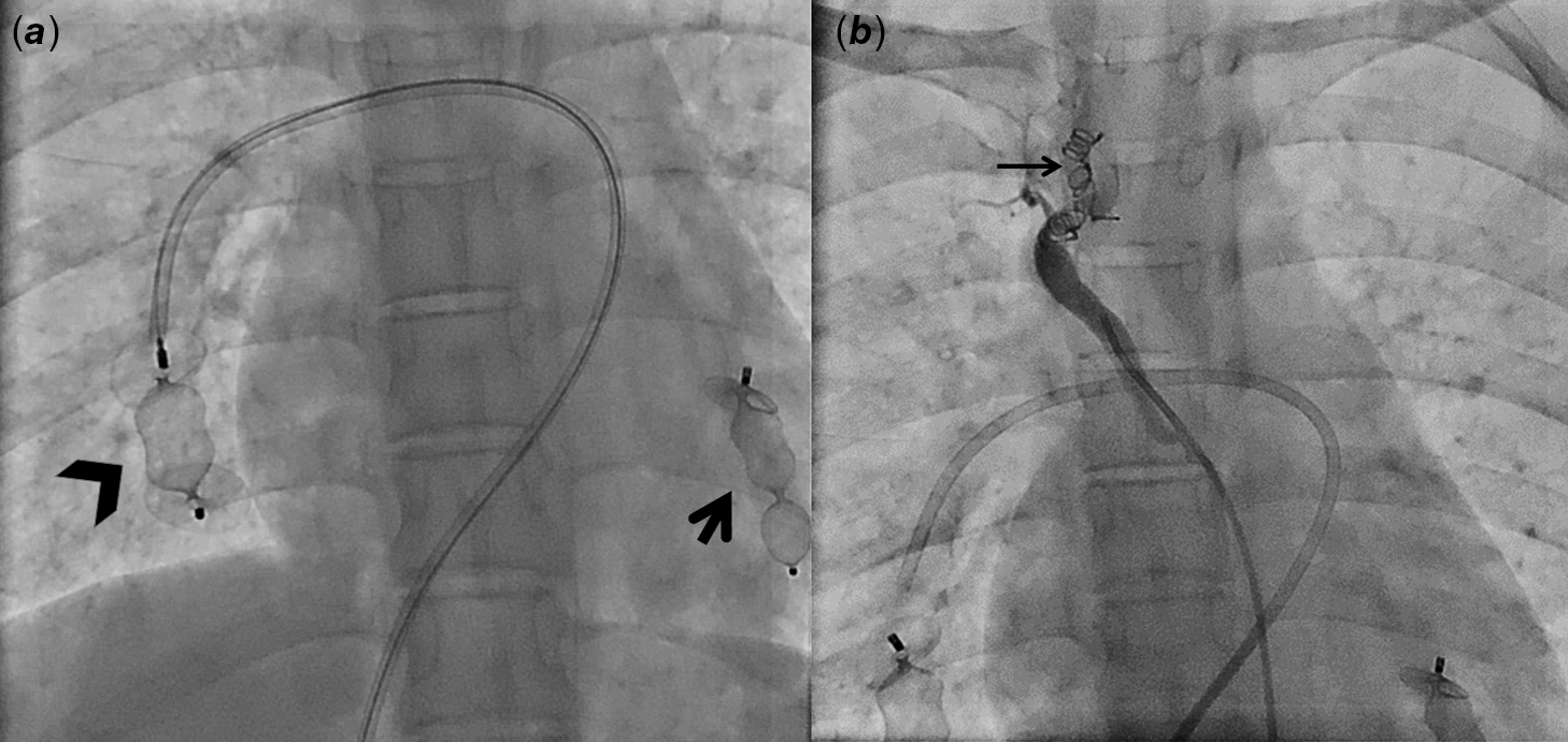
Through a right femoral venous approach, the left pulmonary arteriovenous malformation was engaged using an 8 Fr Judkins Right 3.5 guide catheter (Boston Scientific, Natick, Massachusetts, United States of America) and a 12-mm Amplatzer vascular plug II (AVP II) (AGA Medical, Golden Valley, Minnesota, United States of America) was then deployed successfully proximal to the arteriovenous malformation (Figure 2A). Appropriate plug positioning was confirmed fluoroscopically, with minimal contrast flowing through the plug. Similarly, a 16-mm Amplatzer vascular plug II was deployed in the right pulmonary arteriovenous malformation (the diameter at the site of intended device deployment was 12 mm) (Figure 2A). A check angiogram showed a residual left pulmonary arteriovenous malformation with a 6-mm diameter at the site of intended device deployment. Another 10-mm Amplatzer vascular plug II was deployed. Subsequently, through a right femoral arterial approach, the aortopulmonary collateral to the right lung was hooked with a cobra catheter. It was embolised using multiple coils (two coils sized 5 × 5 mm, one 4 × 4 mm, and another 6 × 5 mm) (Figure 2B). Postprocedure chest radiograph demonstrated multiple plugs and coils in both lungs.
a. Postinterventional angiographic imaging shows an Amplatzer vascular plug II occluding the left pulmonary arterivenous malformation (arrow) and another one occluding the right arterivenous malformation (arrowhead). b. Angiographic imaging demonstrating multiple coils occluding the aortopulmonary collateral to the right lung.

The systemic arterial oxygen saturation improved from 69 to 89% suggesting significant contribution of arteriovenous malformations to her cyanosis. Reference Meredith1 The patient underwent total correction for tetralogy of Fallot with an uneventful postoperative course and doing well on follow-up.
Discussion
Pulmonary arteriovenous malformations can occur in a variety of clinical situations including liver diseases, infections, metastatic carcinomas, systemic disorders, and after the palliation of congenital heart diseases. Reference Marianeschi, McElhinney and Reddy2 In addition to being a vascular mass lesion, they are a source of right-to-left shunt and affected patients may suffer haemoptysis, dyspnea, cyanosis, paradoxical emboli, and brain abscess.
Pulmonary arteriovenous malformations are rarely described in association with tetralogy of Fallot. To the best of our knowledge, only one case of an infant with tetralogy of Fallot and pulmonary atresia with pulmonary arteriovenous malformation exists in the literature. Reference Kim, Park and Park3 The pulmonary arteriovenous malformation in that infant was detected postoperatively when ventilator weaning failed. This seems to be an expected complication with this association as pulmonary arteriovenous malformations, being a right-to-left shunt, would worsen the cyanosis in a tetralogy of Fallot patient. Though an extremely rare association, a high index of suspicion is needed to diagnose these arteriovenous malformations preoperatively in a tetralogy of Fallot patient with unusual lung shadows or disproportionate cyanosis. A timely diagnosis is essential to avoid disastrous postoperative course and known complications related to the pulmonary arteriovenous malformations. Percutaneous closure definitely prevented early postoperative complications and helped in timely weaning of our patient from the ventilator.
Financial support
This research received no specific grant from any funding agency, commercial or not-for-profit sectors.
Conflicts of interest
None.
Ethical standards
Observational case report not revealing identity of the patient.