Introduction
In vitro embryo production (IVP) in cattle is hampered by the comparatively low rate of successful development of oocytes: only about 30% of immature oocytes develop to the blastocyst stage in culture. In addition, variations in physiological condition among cows make it difficult to achieve a reliable rate of embryo production; mastitis (Lonergan et al., Reference Lonergan, Monaghan, Rizos, Boland and Gordon1994; Roth et al., Reference Roth, Dvir, Kalo, Lavon, Krifucks, Wolfenson and Leitner2013), postpartum period (Leroy et al., Reference Leroy, Van Soom, Opsomer, Goovaerts and Bols2008), liver abnormality (Borjigin et al., Reference Borjigin, Ogata, Taguchi, Kato and Nagao2013), and heat stress (Ai-Katanani et al., Reference Ai-Katanani, Et, Paula-Lopes and Hansen2002) have been found to influence oocyte quality and subsequent embryo development. Various aspects of folliculogenesis, such as follicle size (Lonergan et al., Reference Lonergan, Monaghan, Rizos, Boland and Gordon1994), follicle wave (Vassena et al., Reference Vassena, Mapletoft, Allodi, Singh and Adams2003), and hormone regulation (Nivet et al., Reference Nivet, Bunel, Labrecque, Belanger, Vigneault, Blondin and Sirard2012), also influence oocyte quality.
In vitro-matured oocytes have a lower developmental competence compared with those that mature in vivo (Rizos et al., Reference Rizos, Ward, Duffy, Boland and Lonergan2002). The use of different supplements (such as growth factors and hormones) during in vitro maturation (IVM), and the adoption of appropriate morphological criteria to select oocytes (such as evaluation of oocyte cytoplasm and of cumulus cell compaction) can improve the rate of successful IVP (Lonergan et al., Reference Lonergan, Rizos, Gutierrez-Adan and Boland2003). Nevertheless, there are considerable differences in the reported rates of successful IVP (Ward et al., Reference Ward, Enright, Rizos, Boland and Lonergan2002). The accumulation of maternal mRNA during the final phase of folliculogenesis is essential for the development of the embryo until activation of the embryonic genome (Memili and First, Reference Memili and First2000). A microarray study identified several differentially expressed genes in oocytes assessed as having different developmental competences on the basis of different levels of G6PDH activity (Torner et al., Reference Torner, Ghanem, Ambros, Holker, Tomek, Phatsara, Alm, Sirard, Kanitz, Schellander and Tesfaye2008). Previously, Corcoran et al. (Reference Corcoran, Fair, Park, Rizos, Patel, Smith, Coussens, Ireland, Boland, Evans and Lonergan2006) showed that there were many differences in the patterns of mRNA expression between in vitro and in vivo derived bovine blastocysts. Therefore, the origin of the oocytes is the main factor affecting blastocyst yield and molecular signature is likely to be a valuable selection criterion for improving IVP results. Establishment of a non-invasive assessment of oocyte quality with respect to potential further development will be important for the elucidation of the mechanisms of developmental competence and for application to animal reproduction.
Cumulus cells closely surround the oocyte and bidirectional communication between the cumulus cells and the oocyte maintains meiotic arrest and supports cytoplasmic maturation in the germ cell (Tanghe et al., Reference Tanghe, Van Soom, Nauwynck, Coryn and De Kruif2002). The removal of cumulus cells during IVM results in developmental arrest at an early embryo stage (First et al., Reference First, Leibfried-Rutledge and Sirard1988). Similarly to the findings in oocytes, large-scale analysis of gene expression identified different levels of mRNA between cumulus cells associated with oocytes of different developmental competence, such as different follicle size (≥ 8.0 mm vs 1–3 mm; Melo et al., Reference Melo, Cordeiro, Pellegrino, Wei, Daye, Nishimura and Dode2017) or age of the cow (adult vs prepubertal, Bettegowda et al., Reference Bettegowda, Patel, Lee, Park, Salem, Yao, Ireland and Smith2008). Moreover, in cows treated with follicle-stimulating hormone (FSH), significantly different levels of mRNA expression were found in cumulus cells at different times of recovery of immature oocytes (Nivet et al., Reference Nivet, Vigneault, Blondin and Sirard2013). Therefore, cumulus cells provide a promising route for the non-invasive assessment of oocyte quality (Kussano et al., Reference Kussano, Leme, Guimaraes, Franco and Dode2016). Although global transcript profiling can lead to the identification of hundreds of differentially expressed transcripts, it is difficult to identify the best potential candidate biomarkers for assessment of the developmental ability of the oocytes. One practical difficulty may be the choice of in vitro fertilization (IVF) protocol: IVM, IVF or in vitro culture (IVC). Tracking single oocytes in culture from maturation to the blastocyst stage and analysis of cumulus cell gene expression at the different stages may be effective for identifying suitable markers of developmental competence.
Cumulus cells and granulosa cells develop from preantral granulosa cells in response to FSH during antral follicle formation. Cumulus cells play a supporting role in oocyte development, whereas granulosa cells principally fulfil an endocrine role by producing steroid hormones and other ligands (Su et al., Reference Su, Sugiura and Eppig2009). Nevertheless, cumulus cells have a higher level of FSHR mRNA compared with granulosa cells. It has been reported that the microsurgical removal of the oocyte from the cumulus–oocyte complexes (COCs) inhibits both FSH-induced proliferation of cumulus cells and production of progesterone in cumulus cells (Li et al., Reference Li, Norman, Armstrong and Gilchrist2000). Moreover, the addition of denuded oocytes suppresses expression of the FSH-induced LH receptor (LHR), CYP19a1, and CYP11a1, which are well known markers of differentiation, in cumulus cells (Baumgarten et al., Reference Baumgarten, Convissar, Fierro, Winston, Scoccia and Stocco2014; Diaz et al., Reference Diaz, Wigglesworth and Eppig2007). It is unclear whether the differentiation state of the cumulus cells reflects oocyte developmental competence.
The present study aims to determine whether the differentiation state of cumulus cells can be used as a suitable marker of the developmental potential of the COCs. We cultured individual COCs to be able to effectively monitor development from the immature oocyte to the blastocyst stage.
Materials and methods
Collection of bovine ovaries
Bovine ovaries were obtained from a slaughterhouse, placed in a saline solution supplemented with antibiotics and antimycotics (AB; No. 15240–062, Gibco, Grand Island; NY, USA) at 15–20°C, and transported to the laboratory within 2 h. The ovaries were then placed in saline supplemented with 0.3 mM reduced glutathione (GSH; G4251, Sigma-Aldrich, St. Louis, MO, USA) and 1% AB at 15°C for 20 h with shaking.
COCs collection and IVM
COCs were aspirated from follicles 2–6 mm in diameter using a 5 ml syringe equipped with a 20-gauge needle. Oocytes with the intact layers of cumulus cells and evenly granulated cytoplasm were selected as good quality for further study. Selected oocytes were washed and cultured individually in 20-µl drops of maturation medium covered with mineral oil (M8410, Sigma-Aldrich) in a culture dish (Falcon 351007, Becton, Dickinson and Company, Franklin Lakes, NJ, USA) under 5% CO2, 5% O2 and 90% N2 at 39°C with high humidity. A maturation medium consisting of medium 199 (No. 12340, Invitrogen) supplemented with 0.1% (w/v) polyvinyl alcohol (P8136, Sigma-Aldrich), 0.5 mM sodium pyruvate (Nacalai Tesque, Kyoto, Japan), 1% AB, 0.02 AU/ml FSH (Antrin, Kyoritsu Seiyaku, Tokyo, Japan) and 1 µl/ml estradiol-17β (E2758, Sigma-Aldrich) was used. A small number of cumulus cells (approximately 100 cells) was obtained from each COC before IVM by gentle pipetting using a glass pipette; these cells were frozen at −196°C, and then stored at −80°C until RNA analysis.
IVF of mature oocytes
IVF was carried out as previously described (Nagao et al., Reference Nagao, Iijima and Saeki2008) using the modified medium drop size suggested by Carolan et al. (Reference Carolan, Lonergan, Khatir and Mermillod1996). Briefly, after IVM, each COC was individually transferred to a 10-µl drop of fertilization medium (Brackett and Oliphant medium supplemented with 10 µg/ml heparin, Sigma-Aldrich; m-BO). Frozen–thawed semen from a single bull was washed with a discontinuous Percoll solution and 10 µl were added to a fertilization droplet at a final concentration of 2 × 10⁶ spermatozoa/ml. All culture drops were covered with mineral oil in the culture dish at 39°C under 5% CO2 in air at high humidity for 18 h.
IVC of presumptive zygotes
IVC was carried out essentially as described by Nagao et al. (Reference Nagao, Saeki, Hoshi, Takahashi and Kanagawa1995) using the culture dish system of Sugimura et al. (Reference Sugimura, Akai, Hashiyada, Aikawa, Ohtake, Matsuda, Kobayashi, Kobayashi, Konishi and Imai2013). Briefly, a 100-µl drop of protein-free synthetic oviductal fluid containing the amino acid component (m-SOF) was placed in a microwell group culture dish (LinKID micro25, Dai Nippon Printing, Tokyo, Japan) and overlaid with mineral oil. Presumptive zygotes were denuded by gentle pipetting, washed in m-SOF medium, transferred to the microwells and cultured under 5% CO2, 5% O2 and 90% N2 at 39°C with high humidity. Oocyte developmental competence was evaluated by screening for cleavage (≥2-cell) and development to the blastocyst stage under a stereomicroscope (×60 magnification) at 7 days after insemination.
Gene expression analysis
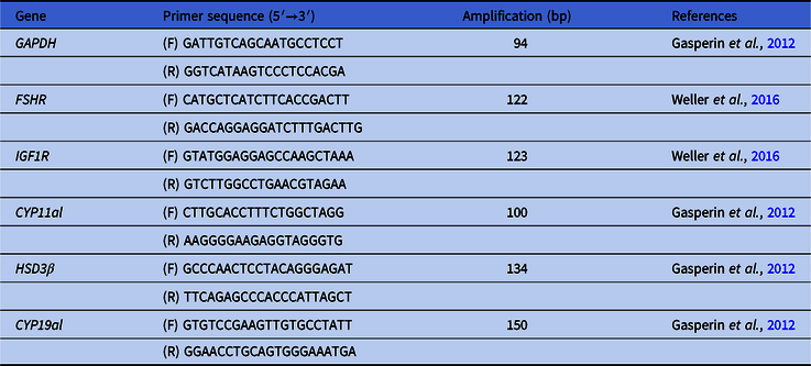
Cumulus cell samples were discarded when the corresponding oocytes did not cleave after IVF. Total RNA was isolated using a PicoPure RNA Isolation Kit (KIT0204, Thermo Fisher Scientific) following the manufacturer’s instructions. The concentration and quality of each RNA sample was assessed using a NanoVue system (GE Healthcare, Little Chalfont, UK). Purified RNA samples were incubated in gDNA Wipeout Buffer at 42°C for 2 min to remove contaminating genomic DNA, and then mRNA was used for reverse transcription with a QuantiTect Reverse Transcription Kit (No. 205311, QIAGEN, Valencia, CA, USA) in accordance with the manufacturer’s instructions. Real-time PCR amplifications were performed in a LightCycler 96 instrument (Roche, Basel, Switzerland) using a QuantiTect SYBR Green PCR kit (No. 204143, Qiagen). Real-time PCR was used to determine the relative expression levels of FSHR, IGF1R, CYP11al, HSD3β and CYP19al under the following conditions: 95°C for 15 min, followed by 45 cycles of 15 s at 95°C for denaturing, 25 s at 55°C for annealing, and 20 s at 72°C for extension. The annealing temperature was optimized for our experimental conditions. Melting curves were examined to confirm the specificity of product amplification. The relative amount of each mRNA was determined by normalization against the level of glyceraldehyde 3-phosphate dehydrogenase (GAPDH) as the internal control. GAPDH was selected as the housekeeping gene control due to its documented performance as a stable control in analyses of cattle granulosa cells and cumulus cells (Melo et al., Reference Melo, Cordeiro, Pellegrino, Wei, Daye, Nishimura and Dode2017). Primer sequences are shown in Table 1.
Primers used for quantitative reverse transcription-polymerase chain reaction (RT-qPCR)

Abbreviations: GAPDH, glyceraldehyde 3-phosphate dehydrogenase; FSHR, follicle-stimulating hormone receptor; IGF1R, insulin-like growth factor 1 receptor; CYP11al, P450 cholesterol side-chain cleavage; HSD3β, 3β-hydroxysteroid dehydrogenase; CYP19al, cytochrome P450 aromatase.
Statistical analysis
The frequencies of cleavage and development to the blastocyst stage calculated for every replicate were summarized as means ± standard error of the mean (SEM). The relative levels of each mRNA were calculated for each COC, which was classified for its embryo development stage after IVP. All data were summarized as means ± SEM, and were tested for normality using the F-test; if the data conformed to a normal distribution, they were analyzed using Student’s t-test. Differences at a P-value < 0.05 were considered to be statistically significant. Correlations between the levels of expression of different genes were analyzed using Pearson’s correlation coefficient with Fisher’s r-to-z transformation.
Results
Ovaries were collected from five cows. The mean number of good quality COCs recovered from individual ovaries for use in the following experiment was 15.0 ± 2.0. The embryo cleavage rate was 88.1 ± 4.6% and the rate of blastocyst formation was 30.0 ± 3.9%. Overall, cumulus cells were sampled from 76 COCs prior to IVM and 30 COCs were selected for subsequent gene analyses.
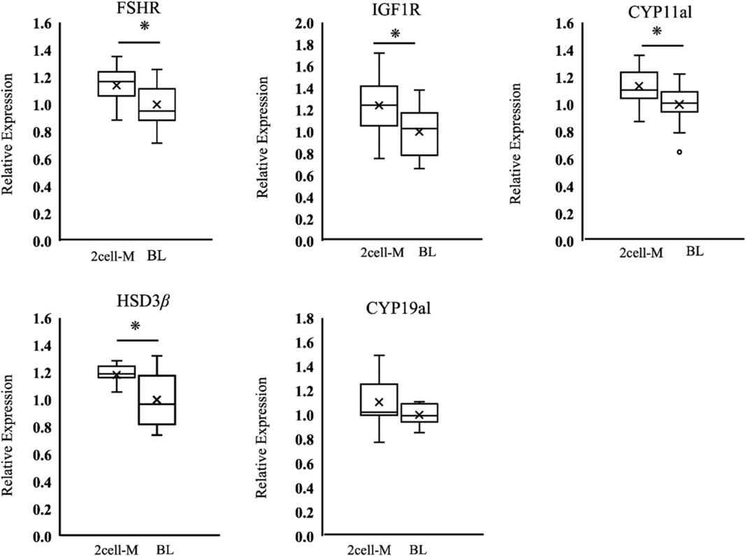
After evaluation of embryo development, gene expression was compared in cumulus cells of COCs that failed to develop to the blastocyst stage with that of COCs that developed into blastocysts. Among the five biomarker candidate genes analyzed, FSHR, IGF1R, CYP11al, and HSD3β mRNAs were present at significantly higher levels in the 2 cell-morula (2cell-M) group than in the blastocyst (BL) group. The level of CYP19al mRNA was no different between the 2cell-M group and the BL group (Fig. 1).
Relative mRNA levels of FSHR, IGF1R, CYP11al, HSD3β and CYP19al in bovine cumulus cells derived from cumulus–oocyte complexes (COCs) that failed to develop to blastocysts (2cell-M) and those that developed to the blastocyst stage (BL). mRNA levels were normalized against GAPDH and expressed relative to the mean value of a blastocyst group, which was set at 1. Boxes reflect two quartiles, the 25th and 75th percentiles, and the interior horizontal line indicates the median. Whiskers indicate the maximum and minimum values within the acceptable range defined by two quartiles. Open circles denote outliers, crosses indicate mean value. Asterisks indicate significant difference at P < 0.05.

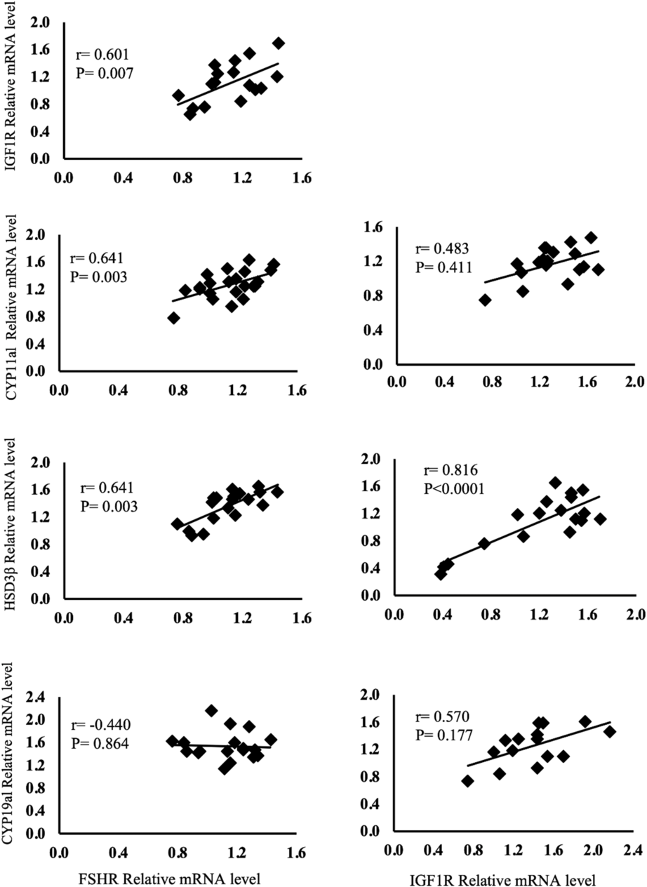
Regardless of COC developmental potential, the level of FSHR in cumulus cells was positively correlated with those of IGF1R, CYP11al, and HSD3β (Fig. 2); the level of IGF1R was positively correlated with that of HSD3β (Fig. 2).
Analysis of relative FSHR mRNA level and relative IGF1R mRNA level relative to mRNA level in steroidogenesis-related enzymes.

Discussion
Oocytes at the maturation or fertilization steps are able to develop to the blastocyst stage in single cell cultures, similarly to cultures of multiple cells (Carolan et al., Reference Carolan, Lonergan, Khatir and Mermillod1996). However, zygotes in single cell culture fail to form blastocysts (Carolan et al., Reference Carolan, Lonergan, Khatir and Mermillod1996; Nagao et al., Reference Nagao, Iijima and Saeki2008). An appropriate density of embryos has been shown to be necessary for stable developmental competence (Nagao et al., Reference Nagao, Iijima and Saeki2008). The use of a well-of-the-well (WOW) system, consisting of microwells in the bottom of the dish, enables tracking of each zygote without any reduction in developmental competence (Sugimura et al., Reference Sugimura, Akai, Hashiyada, Aikawa, Ohtake, Matsuda, Kobayashi, Kobayashi, Konishi and Imai2013). The rate of developmental success obtained from single oocyte culture in the present study was similar to that reported using group culture of 10–25 oocytes or embryos in 50-µl droplets during all procedures (Nagao et al., Reference Nagao, Ohta, Murakami and Kato2010). The single cell culture system was essential to this study.
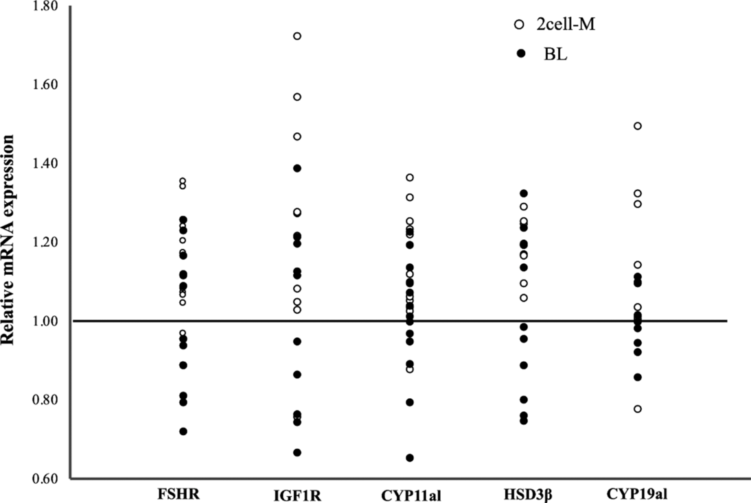
Our analysis here indicated that the levels of FSHR, IGF1R, CYP11al and HSD3β mRNAs in cumulus cells from COCs that failed to develop to blastocysts (2cell-M group) were significantly higher than those in cumulus cells from COCs that developed to the blastocyst stage (BL group) (Fig. 1). The level of CYP19al mRNA in cumulus cells from COCs with low developmental competence tended to be higher than in those with higher competence (Fig. 1). Synthesis of progesterone is mainly regulated by CYP11al, which converts cholesterol to pregnenolone, and by HSD3β, which converts pregnenolone to progesterone; by comparison, oestrogen is produced by conversion of androstenedione by CYP19al. In a previous study, it was reported that FSH-induced progesterone synthesis was the main output of human granulosa cells without luteinization (Oktem et al., Reference Oktem, Akin, Bildik, Yakin, Alper, Balaban and Urman2017). Cumulus cells resemble undifferentiated granulosa cells (Baumgarten et al., Reference Baumgarten, Convissar, Fierro, Winston, Scoccia and Stocco2014) and are less steroidogenically active than granulosa cells (Li et al., Reference Li, Norman, Armstrong and Gilchrist2000). The differentiation process in cumulus cells is regulated by a synergistic action of FSH and IGF1 signals (Zhou et al., Reference Zhou, Baumgarten, Wu, Bennett, Winston, Hirshfeld-Cytron and Stocco2013). The results of the present study are consistent with earlier reports on cumulus cell differentiation: here, we found that the level of FSHR was positively correlated with those of IGF1R, CYP11al, and HSD3β (Fig. 2). The level of IGF1R was found to be positively correlated with that of HSD3β (Fig. 2). Therefore, our results indicated that mRNA expression of these genes varied in cumulus cells and that this difference might be related to the developmental competence of COCs; moreover, the synergistic action of FSHR and IGF1R might contribute to the differentiation of cumulus cells to granulosa cells. To determine whether the levels of FSHR, IGF1R, CYP11al and HSD3β mRNAs could be suitable markers of the developmental competence of COCs, we further analyzed the distribution of relative mRNA levels in each sample. The analysis indicated that most of relative levels of FSHR, IGF1R, CYP11al and HSD3β mRNAs against GAPDH mRNA in COCs with high developmental competence were below the relative value of 1.0 (Fig. 3).
Scatter plots showing the relative mRNA levels against GAPDH mRNA of genes in cumulus cells from cumulus–oocyte complexes (COCs) with different developmental competence. White circles indicate relative mRNA levels in cumulus cells from COCs with low developmental ability (2cell-M). Black circles indicate relative mRNA levels in cumulus cells from COCs that developed to the blastocyst stage (BL).

The relationship between cumulus cell differentiation and oocyte developmental competence has been investigated previously. With regard to steroid hormones, it has been reported that oestrogen stimulates natriuretic peptide precursor type C (NPPC) activity to increase cGMP in cumulus cells, and subsequently inhibits meiotic resumption by maintaining phosphodiesterase 3 (PDE3) activity in the oocyte (Zhang et al., Reference Zhang, Su, Sugiura, Xia and Eppig2010). Progesterone has been shown to have no effect on the maturation of oocytes, although there are conflicting results with respect to blastocyst formation. The addition of progesterone to the maturation medium has been reported to reduce the rate of blastocyst formation; this effect was partially reversed by addition of the anti-progesterone compound mifepristone (Silva and Knight, Reference Silva and Knight2000). However, when progesterone synthesis was inhibited using Trilostane or when signalling of the progesterone receptor was blocked using RU486 during the maturation step, the rate of blastocyst development was dramatically reduced (Aparicio et al., Reference Aparicio, Garcia-Herreros, O’Shea, Hensey, Lonergan and Fair2011). These conflicting results indicated that progesterone may be implicated in cytoplasmic maturation of the oocyte. In addition, the oocyte itself could be a determinate factor in its further development. It was reported that low developmental competence in oocytes from a calf was associated with low expression of growth differentiation factor-9 (GFD9) and bone morphogenetic protein 15 (BMP15); both of these are important oocyte-secreted factors in the intrafollicular environment (Hosoe et al., Reference Hosoe, Kaneyama, Ushizawa, Hayashi and Takahashi2011). Moreover, the addition of oocyte-secreted factors to IVM medium improves oocyte quality and subsequent rates of embryo and fetal development (Hussein et al., Reference Hussein, Thompson and Gilchrist2006). It has been reported that BMP15 inhibits some of the most important events in FSH-dependent granulosa cell differentiation, including the expression of FSHR itself (Otsuka et al., Reference Otsuka, Yamamoto, Erickson and Shimasaki2001) and inhibition of FSH-induced progesterone synthesis (Otsuka et al., Reference Otsuka, Yao, Lee, Yamamoto, Erickson and Shimasaki2000). Our present results are consistent with these reports, and indicate that differentiation of cumulus cells into granulosa cells may be determined by inherent oocyte quality, with no direct relationship to the developmental competence of the COCs. Inherent oocyte quality or the differentiation state of cumulus cells may determine the developmental competence of COCs; further research is needed to determine the relative importance of these factors. In future, it is possible that improvements in maturation culture or in the identification of suitable hormone stimulation treatments may reduce cumulus cell differentiation and improve the developmental competence of COCs with poor developmental ability.
In conclusion, our study showed that the differentiation state of cumulus cells reflects the developmental competence of immature COCs. Screening of COCs for FSHR, IGF1R, CYP11al, and HSD3β RNA levels in cumulus samples may offer a simple, quick, and non-invasive means of identifying the developmental potential of COCs. COCs with high levels of expression of these genes are likely to have poor developmental competence.
Financial Support
This research received no specific grant from any funding agency, commercial or not-for-profit sectors.
Conflict of Interest
None.
Ethical Standards
None.