INTRODUCTION
Neurocysticercosis (NCC) is a zoonotic infection of the central nervous system (CNS) caused by the larval form of Taenia solium. The CNS is the most frequent and severe location of this parasite and NCC is the most common parasitary infection in this site (Flisser, Reference Flisser1991; García and Del Brutto, Reference García and Del Brutto2005). NCC incidence is increasing in developed countries because of travel and immigration from endemic areas and because of the low socioeconomic level of the immigrants. This prospect shows NCC as an emerging disease in countries such as the USA (Flisser et al. Reference Flisser, Sarti, Lightowlers and Schantz2003; Del Brutto and García, Reference Del Brutto and García2012; Del Brutto, Reference Del Brutto2014). NCC is endemic in Latin America, Sub-Saharan Africa, India, China and Southern Asia (Garcia et al. Reference Garcia, Nash and Del Brutto2014a ). In Brazil NCC incidence varies from 1·5% in autopsies to 3% in clinical studies (Takayanagui et al. Reference Takayanagui, Castro e Silva, Santiago, Odashima, Terra and Takayanagui1996; Agapejev, Reference Agapejev2003).
NCC is a pleomorphic disease with no predilections towards gender, race or age. Its clinical manifestations depend on several factors such as morphological type, number, location and stage of evolution of the parasite as well as the in situ and systemic immunopathologic reactions of the host (Sotelo and Marin, Reference Sotelo and Marin1987; Takayanagui, Reference Takayanagui1990; Carabin et al. Reference Carabin, Ndimubanzi, Budke, Nguyen, Qian, Cowan, Stoner, Rainwater and Dickey2011; Garcia et al. Reference Garcia, Nash and Del Brutto2014a ). This multiplicity of factors leads to diverse neurological signs and symptoms including neurological syndromes that vary from the asymptomatic infection to sudden death. The neurological syndrome is influenced by the location of the cysticerci which may be parenchymal, meningeal, medullar, cerebellar and intraventricular (Matushita et al. Reference Matushita, Pinto, Cardeal and Teixeira2011).
The immune response of the human NCC varies from complete absence (asymptomatic) to a severe inflammatory one (symptomatic) (Del Brutto et al. Reference Del Brutto, Santibanez, Idrovo, Rodriguez, Diaz-Calderon, Navas, Gilman, Cuesta, Mosquera, Gonzalez, Tsang and Garcia2005). In most of the cases viable parasites present little inflammation in its surroundings which results in the asymptomatic cases of NCC. However the majority of the symptomatic cases are characterized by an intense inflammatory response in the host tissue (Cardona et al. Reference Cardona, Gonzalez and Teale2003). The inflammatory response may lead to the parasite destruction and is responsible by its pathological effects as well as by its epileptogenic activity (Nash et al. Reference Nash, Del Brutto, Butman, Corona, Delgado-Escueta, Duron, Evans, Gilman, Gonzalez, Loeb, Medina, Pietsch-Escueta, Pretell, Takayanagui, Theodore, Tsang and Garcia2004; Rathore and Radhakrishnan, Reference Rathore and Radhakrishnan2012).
The intraventricular and meningeal location of the cysticerci is less frequent than the parenchymal location. However the clinical manifestations of the extra-parenchymal cysticercosis are more aggressive and happen in about 15–54% of the symptomatic patients (Apuzzo et al. Reference Apuzzo, Dobkin, Zee, Chan, Giannotta and Weiss1984; Zee et al. Reference Zee, Segall, Destian, Ahmadi and Apuzzo1993; Khade et al. Reference Khade, Lemos and Toussaint2013). The parasite reaches the ventricles through the choroid plexus occluding the cerebrospinal fluid flow in a continuum or intermittent manner leading to an obstructive hydrocephaly (Apuzzo et al. Reference Apuzzo, Dobkin, Zee, Chan, Giannotta and Weiss1984; Khade et al. Reference Khade, Lemos and Toussaint2013). Its consequence may be sudden death due to focal compression resulting in mass effect and distension of the ventricle. Besides the hydrocephaly, it may also lead to ependymitis, basal arachnoiditis and periventricular edema which may require draining of the cerebrospinal fluid followed by resection of the cysticerci (Kelesidis and Tsiodras, Reference Kelesidis and Tsiodras2012; Garcia et al. Reference Garcia, Nash and Del Brutto2014a ). Ventriculitis is one of the NCC complications which requires the implant of a shunt and elevates the patient's mortality rates (Colli et al. Reference Colli, Martelli, Assirati, Machado and de Vergueiro1986; Cuetter and Andrews, Reference Cuetter and Andrews2002; Garcia et al. Reference Garcia, Nash and Del Brutto2014a ).
To promote a better understanding of the factors involved in the NCC pathogenesis two experimental models were developed, one using Mesocestoides corti and the other using Taenia crassiceps (Cardona et al. Reference Cardona, Restrepo, Jaramillo and Teale1999; Matos-Silva et al. Reference Matos-Silva, Reciputti, Paula, Oliveira, Moura, Vinaud, Oliveira and Lino2012). According to these models low amounts of parasites induce an initial T helper 1 (Th1) immune response which is able to eliminate the infection (Alvarez et al. Reference Alvarez, Mishra, Gundra, Mishra and Teale2010; Freitas et al. Reference Freitas, Moura, Gonçalves, Rodrigues, Félix, Soares, Irusta, Vinaud, Oliveira and Lino2012). One of the main cells responsible for this inflammatory response is the Tγδ (gamma-delta) lymphocytes from the Th1 profile which plays an important role in the development of the neuropathology and in the severity of the NCC (Cardona et al. Reference Cardona, Restrepo, Jaramillo and Teale1999; Restrepo et al. Reference Restrepo, Alvarez, Castano, Arias, Restrepo, Trujillo, Colegial and Teale2001b ). In the late stages of the disease these cells may lead to a modification of the local macrophages profile inducing the formation of alternatively activated macrophages – AAMϕ (M2). The latter, in turn, present suppressive activity and are considered responsible by the immunoregulation of the inflammatory response (Terrazas, Reference Terrazas2008; Freitas et al. Reference Freitas, Moura, Gonçalves, Rodrigues, Félix, Soares, Irusta, Vinaud, Oliveira and Lino2012). These findings support the hypothesis that the parasites during the initial phase of the infection maintain a balance with the host's immune response which is lost by the switch to the Th2 immune response (Reyes et al. Reference Reyes, Terrazas, Vera-Arias and Terrazas2009).
The NCC pathogenesis is limited by the chronic progression of the disease and also by the treatment's influence and the host's complex immune response. Therefore the aim of this study was the evaluate the in situ and systemic immune response through cytokines quantification (interleukin (IL)-4, IL-10, IL-17 and interferon-γ (IFN-γ)) as well as the local inflammatory response throughout 90 days of the experimental NCC model with T. crassiceps.
METHODS
Maintenance of the parasites
This study obeyed the rules established by the Brazilian Society of Science with Laboratory Animals (SBCAL) and was approved by the Ethics Committee in Animal Use of the Federal University of Goias (CEUA/UFG), protocol n° 010/11.
The maintenance of the Ontario Research Facility (ORF) strain of T. crassiceps was performed through successive intraperitoneal passages with 90 days interval in female Bagg Albino (inbred research mouse strain) (BALB)/c mice of 8–12 weeks old (Vaz et al. Reference Vaz, Nunes, Piazza, Livramento, da Silva, Nakamura and Ferreira1997). This strain is maintained since 2002 in the Animals Facilities of the Tropical Pathology and Public Health Institute of the Federal University of Goias, Brazil.
Experimental NCC infection
For the experimental NCC infection female BALB/c mice of 8–12 weeks old were used (n = 40). After the removal of the T. crassiceps cysticerci from a mice with intraperitoneal infection of parasite maintenance, the cysticerci were washed twice with phosphate buffer solution (PBS), placed into petri dishes and separated according to their stage of evolution (Vinaud et al. Reference Vinaud, Lino and Bezerra2007). Only the initial stage (no buds with translucent fluid and vesicular membrane) cysticerci were selected to be inoculated into the CNS of the mice. The control group received an inoculation of 10 µL of physiologic solution.
After the anaesthetics (0·01 mL g−1 of Xilazine 2% and Ketamine 10%) the trichotomy and anti-sepsy were performed with iodated alcohol. A longitudinal incision in the skin and subcutaneous tissue was performed as to expose the cranial surface. To allow the visualization of the bone junctions and the bregma point 30% hydrogen peroxide was used. The bregma point was used to standardize the posterior region of the bone junctions in the cranial surface as the point of inoculation. The cranial perforation was performed using a micromotor (LB100–Beltec) and diamond drill (44·5 × 2 mm2). The inoculation occurred through a 1 mL syringe – containing 3 initial stage cysticerci – and a 25 G adapted needle (2 mm long) which enabled the reach of the encephalic ventricles. After the surgical procedures the incisions were closed with dental acrylic resin and instantaneous glue. The control group mice received 10 µL of physiological solution (Matos-Silva et al. Reference Matos-Silva, Reciputti, Paula, Oliveira, Moura, Vinaud, Oliveira and Lino2012). Five animals per cage (20 × 30 cm2) were maintained under controlled illumination (12/12 h), temperature of 25 °C and free access to sterilized/acidified water and sterilized ration. After the inoculation the animals were observed in 1 h periods during the first day and daily afterwards until the euthanasia day. Five infected animals and five control ones were used at each experimental day – 7, 30, 60 and 90 days after the inoculation (DAI).
Encephala removal
After the euthanasia through cervical dislocation the encephala were removed for the histopathologic analysis as well as the in situ cytokines quantification through enzyme-linked imunosorbent assay (ELISA).
In situ cytokines quantification
After the removal, the encephala were frozen at −80 °C. After defrosting, fragments of 100 mg of brain tissue were homogenized into 1 mL of cytokines and chemokines extraction solution supplemented with 0·4 M of NaCl, 0·05% of Tween 20, 5% of albumin, 0·1 mm of benzethonium chloride, 10 mm ethylenediamine tetraacetic acid (EDTA) and 20 Kallikrein Inhibitor Unit (KIU) of aprotinin. A rate of 1 mg of tissue to 100 mL of buffer was used. After the maceration the sample was centrifuged at 10 000 g , 10 min, 4 °C. The supernatant was removed for the cytokines detection.
Histopathological analysis for in situ evaluation of tissue injury
The encephala were removed and fixed into 10% tamponated formaldehyde for 24 h, transferred to 70% ethanol for paraffin inclusion and block confection, 5 µm sections were stained with hematoxylin and eosin.
The presence of cysticerci, ventriculomegaly and inflammation were microscopically analysed in the host tissue at 7, 30, 60 and 90 DAI (Lino Jr. et al. Reference Lino, Ribeiro, Antonelli, Faleiros, Terra, Reis and Teixeira2002a).
The ventriculomegaly was analysed in a semi-quantitative – absence (score 0 – no ventricular increase when compared with the contralateral non-infected ventricle), discrete (score 1 – up to 50% of ventricular increase), moderate (score 2–50 to 100% of ventricular increase) and accentuated (score 3 – more than 100% of ventricular increase) (Shook et al. Reference Shook, Lennington, Acabchuk, Halling, Sun, Peters, Wu, Mahajan, Fellows and Conover2014).
Spleen cells culture
After the euthanasia the spleen of the animals were removed and transferred into Petri dishes with 2 mL of PBS. The organ was macerated and the cells in suspension were washed twice through centrifugation (300 g , 10 min, 10 °C). The obtained cells were incubated with lysis buffer at 37 °C, 5 min. Afterwards Roswell Park Memorial Institute (RPMI) culture medium supplemented with 10% of fetal bovine serum was added as to block the reaction. Cells were washed again twice with RPMI culture medium and resuspended. A 1/10 Tripan blue dilution was performed as to allow quantification and analysis of the cell viability through a Newbauer chamber. The cell concentration was adjusted to 5 × 10⁶ cells mL−1 in complete RPMI culture medium supplemented with fetal bovine serum, 200 mm of L-glutamine, 100 U mL−1 of penicillin, 100 mg mL−1 of streptomicin and 50 mm of 2- mercaptoethanol. Spleen cells suspension of 1 mL was added into each well of a 24 well plate (CORNING-EUA), incubated with 5 µg mL−1 of Concanavalin-A, 36 °C at 5% of CO2 for 48 h. After this period the supernatant was collected and used for the quantification of IFN-γ, IL-10, IL-4 and IL-17 through ELISA.
ELISA
The IL-4, IL-17, IFN-γ and IL-10 cytokines were quantified through ELISA sandwich according to the manufacturer instructions (BD OptEIA™). A capture monoclonal antibody, anti-Mouse IL-4 (Lote: 26389), anti-Mouse IL-10 (Lot: 20169) Set BD OptEIA™ (BD Biosciences, San Diego, CA), anti-Mouse IL-17A (Lot: B167940) ELISA MAX™ (BioLegend) and IFN-γ (5 µg mL−1 of clone XMG 1,2) were used. These antibodies were fixed into 96 well plate and incubated according to the manufacturer instructions. After this period the plates were washed with washing buffer and blocked with PBS/bovine serum albumin 1% buffer. After one washing cycle the samples and serial dilutions of the standard curve were added and incubated according to instructions. Afterwards the biotin conjugated anti-cytokines antibodies were added and incubated again. After a washing cycle the streptoavidin-horseradish peroxidase (1:1000) (BD Pharmingen, EUA) was added for a 30 min period at room temperature. After another washing cycle the Tetramethylbenzidine (3,3′,5,5′-Tetramethylbenzidine) was added and followed until colour formation. The colorimetric reaction was interrupted with a 2N sulphuric acid solution. The optic density was detected in a Thermo Labsystems microplate reader using two filters at 450 and 620 nm (Oliveira et al. Reference Oliveira, Santiago, Lisboa, Ceravollo, Trinchieri, Gazinelli and Vieira2000)
The cytokines concentrations were determined through a regression analysis comparing the absorbance of the samples to the ones in the standard curve. The results were expressed in ng mL−1.
Statistical analysis
The results of each parameter were analysed through the analysis of variance test followed by the Bonferrone post-test. The differences were considered significant when P < 0·05.
RESULTS
Histopathological description and cytokines quantification in situ
The mice encephala were removed and the initial stage (no buds with translucent fluid and vesicular membrane) cysticerci were found in 100% of the infected ventricles. In the initial phase of the infection (7 DAI) it was possible to observe the influx of inflammatory cells composed both by polymorphonuclear cells and by mononuclear cells in the inoculation site whereas in the ventricles there was no inflammation. At 30 DAI it was possible to observe inflammatory infiltration of polymorphonuclear and mononuclear cells. At 60 DAI there were polymorphonuclear cells with predominance of mononuclear cells in the inflammatory infiltration. At 90 DAI there was a predominance of mononuclear cells in the inflammatory infiltration. In addition the cysticerci found were classified as initial stage (no buds with translucent fluid and vesicular membrane) and larval stage (presenting buds with translucent fluid and vesicular membrane), with no degeneration signs (Fig. 1).
Photomicrograph of BALB/c mice encephalic ventricle infected with Taenia crassiceps cysticerci, stained with HE. (A) Absence of inflammation in the inoculated animals at 7 DAI, (*) choroid plexus; (B) initial stage (no buds with translucent fluid and vesicular membrane) cysticerci (arrow) in the lateral ventricle and discrete ependymitis (arrow head) at 30 DAI; (C) initial stage cysticerci (arrow) in the lateral ventricle and discrete gliosis (†) at 60 DAI; (D) initial stage cysticerci (arrow) and discrete ependymitis (arrow head) at 90 DAI. Scale bar – 100 µm. (E) BALB/c mouse encephalon of control group with no infection and no ventriculomegaly at 90 DAI. (F) BALB/c mouse encephalon with T. crassiceps larval stage (presenting buds with translucent fluid and vesicular membrane) cysticercus (arrow) inducing ventriculomegaly in the third dorsal ventricle at 90 DAI. Scale bar – 10 mm. Abbreviations: DAI, days after the inoculation; HE, hematoxylin and eosin.

The control group, which received the inoculation of PBS, did not show ventriculomegaly. The mean of the semi-quantitative analysis of the ventriculomegaly in the infected groups was 30 DAI (2·4 ± 0·9), 60 DAI (1·2 ± 1·3) and 90 DAI (2·0 ± 0·7). At 7 DAI the trauma caused by the surgery of the parasites inoculation did not allow the observation of ventriculomegaly.
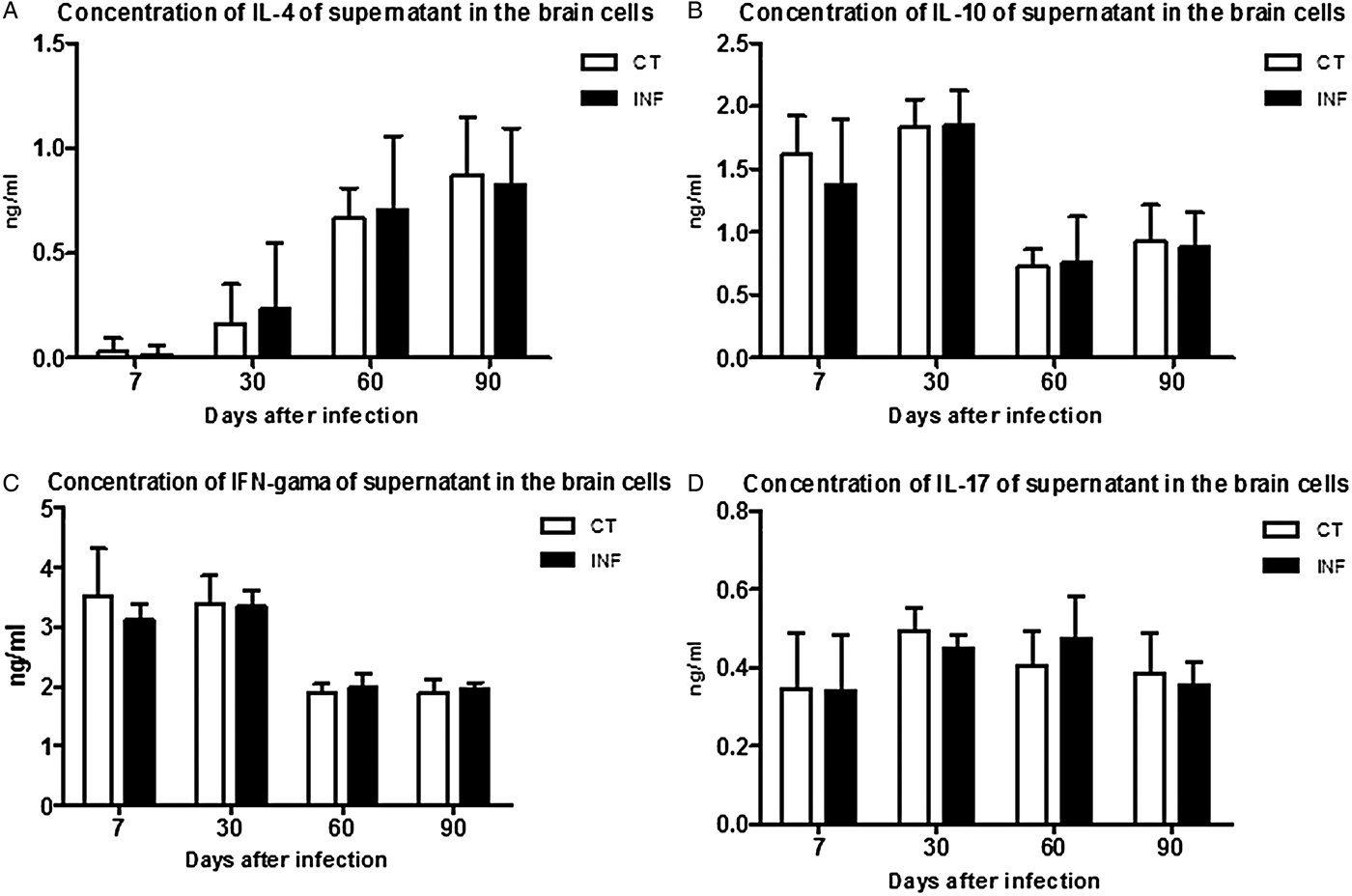
The in situ immune response was evaluated through the levels of cytokines measured in the brain tissue during the experimental days (Fig. 2). The Th2 immune response was quantified through the IL-4 levels throughout the experimental days (Fig. 2A) which showed a gradate increase during the experiment (P > 0·05).
in situ quantification of cytokines in BALB/c mice encephala infected with Taenia crassiceps cysticerci and in the control group. (A) IL-4 levels in different experimental days. (B) IL-10 levels in different experimental days. (C) IFN-γ levels in different experimental days. (D) IL-17 levels in different experimental days. Results expressed in mean ± standard deviation of the cytokines levels at each point. Statistical analysis performed through ANOVA followed by Bonferrone posttest. (n = 5 animals in independent experiments).Abbreviation: ANOVA, analysis of variance.

To evaluate a regulatory immune response the IL-10 levels were quantified (Fig. 2B) as the concentrations were higher at the initial phases of the infection (P > 0·05).
The Th1 immune response was evaluated through the quantification of IFN-γ (Fig. 2C) which was higher at the initial phases of the infection and reduced its levels toward the late phase of infection (P > 0·05).
The Th17 immune response was quantified through the levels of IL-17 (Fig. 2D) which did not present statistical difference throughout the experimental days (P > 0·05).
Systemic cytokines quantification
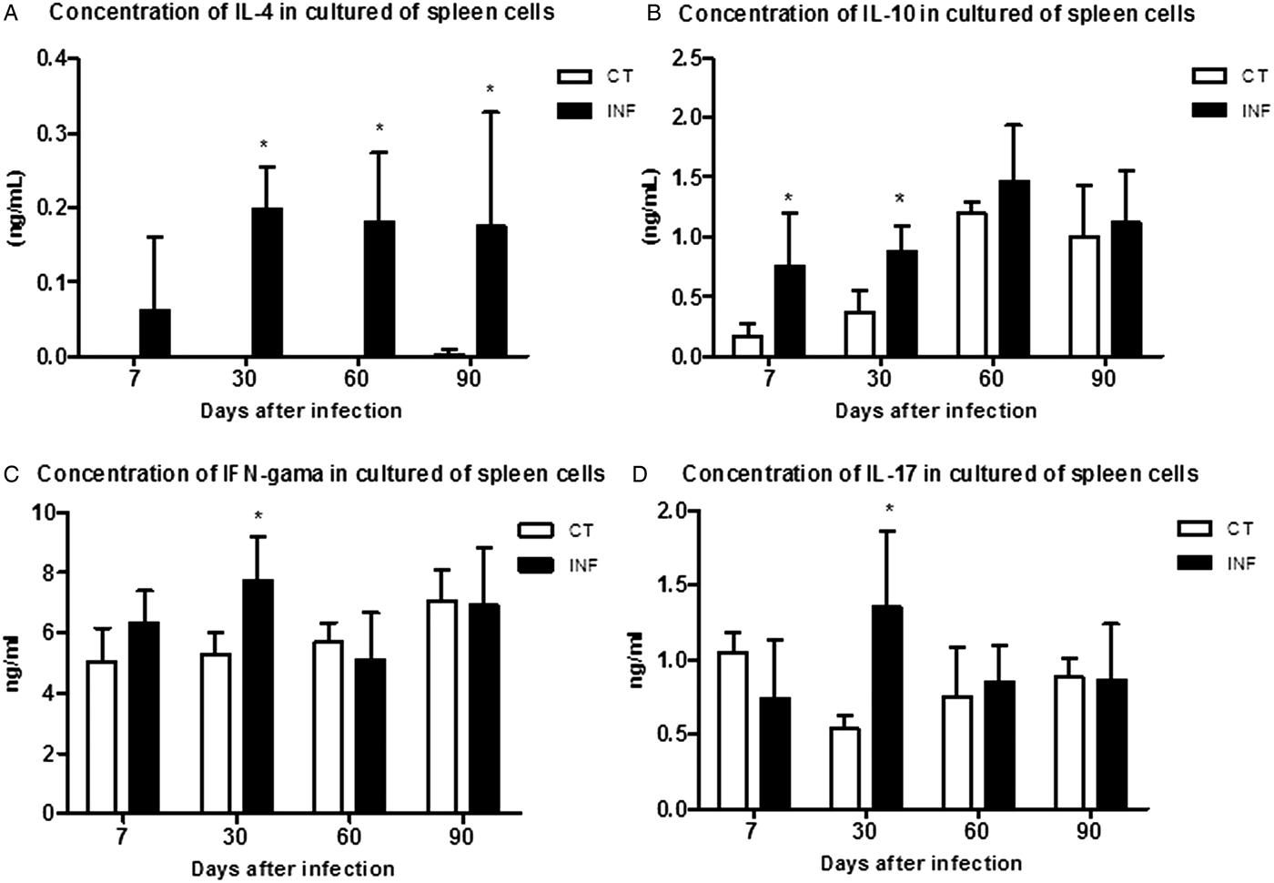
To evaluate the systemic immune response the cytokines levels were quantified in spleen cells cultures during the experimental infection (Fig. 3).
Systemic analysis of cytokines from spleen cells culture from BALB/c infected mice and control group. (A) IL-4 increasing levels throughout the infection. (B) IL-10 higher levels at the initial phase of the infection. (C) IFN-γ levels with statistical difference at 30 DAI. (D) IL-17 levels with statistical difference at 30 DAI. Results expressed in mean ± standard deviation at each point. *P < 0·05 (ANOVA followed by Bonferrone posttest. (n = 5 animals in independent experiments). Abbreviation: ANOVA, analysis of variance.

It was possible to observe a gradate increase in the IL-4 levels in the infected group when compared with the control group. From 30 DAI (0·067 ± 0·10 ng mL−1) until the end of the experimental infection there was a significant difference in this cytokine levels which was higher (0·17 ± 0·01 ng mL−1) at 90 DAI (P < 0·05) (Fig. 3A). It was not possible to detect IL-4 in the control group throughout the experimental period.
The regulatory immune profile was determined through the IL-10 levels quantification. In the infected groups at 7 and 30 DAI the IL-10 levels were 0·72 ± 0·24 ng mL−1 and 0·87 ± 0·13 ng mL−1, respectively. These results were statistically higher than the ones detected in the control group (P < 0·05) (Fig. 3B). The IL-10 levels continued high at 60 and 90 DAI in spite of not presenting a statistical significance when compared with the control groups (P > 0·05).
It was possible to detect IFN-γ throughout all the experimental days with levels varying from 5·78 ± 0·91 ng mL−1 to 7·02 ± 1·52 ng mL−1 (Fig. 3C). Only at 30 DAI there was a significant difference when compared with the control group (P < 0·05).
The IL-17 levels presented a similar behaviour as the one observed in the IFN-γ levels. It was detected throughout the infection, varying from 0·71 ± 0·39 ng mL−1 to 1·34 ± 0·45 ng mL−1 (Fig. 3D). Also it was significantly higher than the control group at 30 DAI (P < 0·05).
The mice did not present behavioral alterations during the experimental days.
DISCUSSION
This is the first report of the in situ and systemic immune response of the experimental NCC caused by T. crassiceps. In this study 100% of the cysticerci were recovered from intraventricular location. This cysticercus location is a frequent cause of intracranial hypertension due to an obstruction of the cerebrospinal fluid and leading to edema in different degrees (Apuzzo et al. Reference Apuzzo, Dobkin, Zee, Chan, Giannotta and Weiss1984; Khade et al. Reference Khade, Lemos and Toussaint2013). It was possible to detect in situ cytokines however there was no significant difference when compared with the non-infected control group. This fact may be explained by the small amount of inflammatory cells and the analysis was performed within the brain macerate which diluted the amount of cytokines produced. Furthermore the better form to dose cytokines produced in an intraventricular NCC would be from the cerebrospinal fluid, which in mice are in a very low volume.
The destruction of the cysticerci induces an intense inflammatory process which is associated to the NCC clinical symptoms and leading to the release of great amounts of cytokines (Sotelo and Marin, Reference Sotelo and Marin1987; Garcia et al. Reference Garcia, Rodriguez and Friedland2014b ). If the cysticerci are still viable, similar to the ones found in this study, they are still capable of modulating the immune and inflammatory response from the host and resulting in the absence of symptoms in the great majority of cases (Laclette et al. Reference Laclette, Merchant, Damian and Willms1990; White et al. Reference White, Robinson and Kuhn1997; Arechavaleta et al. Reference Arechavaleta, Molinari and Tato1998; Gomez-Garcia et al. Reference Gomez-Garcia, Rivera-Montoya, Rodriguez-Sosa and Terrazas2006). Another relevant factor that explains the non-observation of in situ immune response is that the brain is considered an immunological privileged organ where the immune and inflammatory reactions are minimized avoiding accentuated tissue damage (Sotelo and Marin, Reference Sotelo and Marin1987; Garcia et al. Reference Garcia, Rodriguez and Friedland2014b ).
In spite of not presenting an in situ immune response the cysticerci were able to induce ventriculomegaly and ependymitis. Human meningoencephalic NCC presents a similar inflammatory process than the one found in our study leading to the symptoms observed in the acute phase of the infection (Matushita et al. Reference Matushita, Pinto, Cardeal and Teixeira2011). Usually the parenchyma infections lead to epilepsy and a better prognosis than the extra parenchymatous infections in which the main symptom is intracranial hypertension (Khade et al. Reference Khade, Lemos and Toussaint2013; Garcia et al. Reference Garcia, Nash and Del Brutto2014a ). In the cases of intraventricular symptomatic NCC obstructive phenomena are observed or the symptoms begin when the cysticerci changes their stage of evolution and begin to be attacked by the immune system. The obstructive processes may lead to an intense inflammatory process of the ependyma and consequent ependymitis, fibrosis and secondary stenosis of the cerebrospinal fluid leading to hydrocephaly and ventriculomegaly (Sotelo and Marin, Reference Sotelo and Marin1987; Kelesidis and Tsiodras, Reference Kelesidis and Tsiodras2012; Khade et al. Reference Khade, Lemos and Toussaint2013).
Several studies have been performed aiming the characterization of the immune and inflammatory responses and their variations according to the stage of evolution and morphology of the parasite (Cardona et al. Reference Cardona, Restrepo, Jaramillo and Teale1999; Cardona and Teale, Reference Cardona and Teale2002; Khade et al. Reference Khade, Lemos and Toussaint2013; Garcia et al. Reference Garcia, Nash and Del Brutto2014a ). The immune response behaves differently when the in situ and systemic responses regarding peripheric cells are evaluated (Restrepo et al. Reference Restrepo, Aguilar, Melby and Teale2001a ; Sáenz et al. Reference Sáenz, Fleury, Chavarría, Hernández, Crispin, Vargas-Rojas, Fragoso and Sciutto2012). This is in accordance to the findings of this study in which it was possible to detect differences among the in situ and systemic responses. Other studies also observed an increase in cytokines levels such as IL-5, IL-6 and IL-10 in NCC patients with active symptomatology in comparison with NCC asymptomatic patients or cerebrospinal fluid controls (Evans et al. Reference Evans, Garcia, Hartnell, Gilman, Jose, Martinez, Remick, Williams and Friedland1998; Rodrigues et al. Reference Rodrigues, De-Mello, Magalhaes, Ribeiro and Marquez2000; Aguilar-Rebolledo et al. Reference Aguilar-Rebolledo, Cedillo-Rivera, Llaguno-Violante, Torres-López, Muñoz-Hernández and Enciso-Moreno2001; Chavarria et al. Reference Chavarria, Fleury, Bobes, Morales, Fragoso and Sciutto2006). In the systemic evaluation of the immune response the supernatant of peripheral blood mononuclear cells stimulated with antigens from cysticerci removed from NCC asymptomatic patients showed increased levels of Th2 cytokines such as IL-4, IL-5 and IL-13 (Chavarría et al. Reference Chavarría, Fleury, Garcia, Marquez, Fragoso and Sciutto2005; Chavarria et al. Reference Chavarria, Fleury, Bobes, Morales, Fragoso and Sciutto2006; Sáenz et al. Reference Sáenz, Fleury, Chavarría, Hernández, Crispin, Vargas-Rojas, Fragoso and Sciutto2012). These results are in accordance with the findings of this study in which the type of immune response found is a Th2-biased immune profile and reinforcing that this is a good experimental model for NCC immunopathology studies.
In this study it was possible to observe that the intraventricular cysticerci probably induced a rupture of the blood brain barrier allowing the influx of inflammatory cells since the early stages of the infection. The assemblage of inflammatory cells surrounding the cysticerci in extra parenchymatous locations, including meninx and ventricles was reported previously and is in accordance to our study (Cardona et al. Reference Cardona, Restrepo, Jaramillo and Teale1999). These authors also found an initial Th1 immune response in experimental NCC caused by M. corti but, differently from our findings, there was no change in the local immune response probably due to the cysticerci location.
The systemic immune response was initially regulated by the IL-10 presence which is capable of modulating the immune mediators through microglia and resident glial cells leading to the reduction of the release of pro-inflammatory cytokines (Sciutto et al. Reference Sciutto, Chavarria, Fragoso, Fleury and Larralde2007; Terrazas, Reference Terrazas2008). In addition IL-10 is capable of activating T regulatory lymphocytes decreasing the tissue damage (Terrazas, Reference Terrazas2008). At 30 DAI was possible to observe an increase in the immune response with the presence of all the analysed immune profiles. In spite of the presence of IFN-γ and IL-17 the parasite is capable of modulating the immune response in self-benefit favouring its survival through the induction of anti-inflammatory cytokines such as IL-10 and IL-4. The IFN-γ belongs to the Th1 immune profile which is related to severe symptomatology and tissue damage and is regulated by the IL-10 cytokine which modulates the immune response and tissue damage (Terrazas et al. Reference Terrazas, Bojalil, Govezensky and Larralde1998; Rodrigues et al. Reference Rodrigues, De-Mello, Magalhaes, Ribeiro and Marquez2000; Cardona and Teale, Reference Cardona and Teale2002; Chavarría et al. Reference Chavarría, Roger, Fragoso, Tapia, Fleury, Dumas, Dessein, Larralde and Sciutto2003, Reference Chavarría, Fleury, Garcia, Marquez, Fragoso and Sciutto2005; Sciutto et al. Reference Sciutto, Chavarria, Fragoso, Fleury and Larralde2007).
Interestingly the intraperitoneal experimental cysticercosis shows an initial Th1 immune profile through serum cytokines quantification which inhibits a humoral Th2 immune profile (Restrepo et al. Reference Restrepo, Aguilar, Melby and Teale2001a ; Cardona and Teale, Reference Cardona and Teale2002; Rodriguez-Sosa et al. Reference Rodriguez-Sosa, David, Bojalil, Satoskar and Terrazas2002; Toenjes and Kuhn, Reference Toenjes and Kuhn2003) and is associated to the destruction of the parasite (Terrazas et al. Reference Terrazas, Bojalil, Govezensky and Larralde1998; Alonso-Trujillo et al. Reference Alonso-Trujillo, Rivera-Montoya, Rodriguez-Sosa and Terrazas2007). This initial Th1 immune response is capable of inducing classically activated macrophages (CMø or M1) which are responsible for the parasites elimination through the release of nitric oxide (NO). Also NO contributes to the death of several extra cellular pathogens such as intraperitoneal T. crassiceps cysticerci (Alonso-Trujillo et al. Reference Alonso-Trujillo, Rivera-Montoya, Rodriguez-Sosa and Terrazas2007), as well as intracellular pathogens (Gazzinelli et al. Reference Gazzinelli, Talvani, Camargo, Santiago, Oliveira, Vieira, Martins, Aliberti and Silva1998; Gordon, Reference Gordon2003).
Simultaneously it was also possible to detect IL-4 which is a Th2 immune profile cytokine and acts in congruence with IL-10 minimizing the tissue damages but incapable of favouring the parasite's proliferation through the M2 macrophages activation. Also high levels of IL-17 which is a pro-inflammatory cytokine were detected. This cytokine recruits neutrophils to the infection site and is involved in chronic inflammatory diseases. When it is released in excess, it leads to tissue damage and auto-immune diseases. The Th17 cells have been described in the CNS leading to inflammation and tissue damage (Langrish et al. Reference Langrish, Chen, Blumenschein, Mattson, Basham, Sedgwick, McClanahan, Kastelein and Cua2005; Park et al. Reference Park, Li, Yang, Chang, Nurieva, Wang, Wang, Hood, Zhu, Tian and Dong2005). On the other hand the blockage of IL-17 is efficient in preventing bone and cartilage erosion and consequently reducing the clinical symptoms of rheumatoid arthritis (Lubberts et al. Reference Lubberts, Koenders, Oppers-Walgreen, van den Bersselaar, Coenen-de Roo, Joosten and van den Berg2004). IL-17 may act as a therapeutic target in inflammatory diseases of the CNS such as NCC because humanized anti-IL17A antibodies are already being used against inflammatory diseases such as rheumatoid arthritis, psoriasis and uveitis (Genovesse et al. Reference Genovesse, Van Den Bosch, Robertson, Bojin, Ryan and Sloan-Lancaster2010; Hueber et al. Reference Hueber, Patel, Dryja, Wright, Koroleva, Bruin, Antoni, Draelos, Gold, Durez, Tak, Gomez-Reino, Foster, Kim, Samson, Falk, Chu, Callanan, Nguyen, Rose, Haider and Di Padova2010).
In this study it was possible to observe that as the infection progresses there is an immune conversion towards a Th2 immune profile. This change of immune profile favours the parasite's proliferation in chronic infections (Terrazas et al. Reference Terrazas, Bojalil, Govezensky and Larralde1998; Toenjes et al. Reference Toenjes, Spolski, Mooney and Kuhn1999; Rodriguez-Sosa et al. Reference Rodriguez-Sosa, David, Bojalil, Satoskar and Terrazas2002; Toenjes and Kuhn, Reference Toenjes and Kuhn2003). A similar result was reported by Moura et al. (Reference Moura, Silva, Batista, Gomes, Leenen, Lino and Oliveira2013) who evaluated the profile of M2 intraperitoneal macrophages after intraperitoneal experimental cysticercosis and concluded that this response favoured the parasite's proliferation. Also this profile was observed by other authors (Terrazas et al. Reference Terrazas, Bojalil, Govezensky and Larralde1998; Toenjes et al. Reference Toenjes, Spolski, Mooney and Kuhn1999; Rodriguez-Sosa et al. Reference Rodriguez-Sosa, David, Bojalil, Satoskar and Terrazas2002; Toenjes and Kuhn, Reference Toenjes and Kuhn2003).
One of the probable mechanisms responsible for this immune profile change during the experimental NCC is that the parasites have the ability to release glycoproteins which are responsible for the increase in the secretion of IL-4 or IL-13. Another hypothesis is that the parasite's tegument is covered with glycoproteins or glycoconjugates which are highly antigenic and capable of modulating the host's immune response (White et al. Reference White, Robinson and Kuhn1997). Mendlovic et al. (Reference Mendlovic, Cruz-Rivera, Avila, Vaughan and Flisser2015) reported that a calreticulin from T. solium is capable of inducing a Th2-biased immune response in experimental taeniasis in hamsters showing that the parasite is able of secreting immunomodulating agents.
Therefore we conclude that the presence of cysticerci within the ventricles induced ventriculomegaly throughout the experimental period. Also the systemic immune response showed a pro-inflammatory mixed Th1/Th17 profile in the acute phase of the infection accompanied by high levels of IL-10. As the infection progressed there was a predominance of a Th2 immune profile
FINANCIAL SUPPORT
The authors would like to thank CNPq (National Counsel of Technological and Scientific Development) for the financial support. Grant number 471243/2013-2, awarded to R. S. L. J.