Introduction
The onset of neurodegenerative cognitive disorders is strongly related with feeding behaviors and stress that may lead to alteration of energy expenditure or metabolism (Tezapsidis et al., Reference Tezapsidis, Smith and Ashford2009). An association was widely accepted between development and/or progression of dementia and metabolic syndrome parameters including obesity, hypertension, dyslipidemia, and elevated glucose levels (Magalhães et al., Reference Magalhaes, Carvalho, Sousa, Caramelli and Gomes2015). Furthermore, mid-life central adiposity is accounted for the increase in dementia after 32 years (Gustafson et al., Reference Gustafson2012).
There is growing interest of neuropeptides in the literature such as leptin, ghrelin and recently nesfatin-1 those are suggested to be related with learning and memory. In preclinical studies, both leptin and ghrelin have shown to have influences on hippocampal long term potentiation (Diano et al., Reference Diano2006; Oomura et al., Reference Oomura2006) and they are in favor of promoting learning and memory (McNay Reference McNay2007; Signore et al., Reference Signore, Zhang, Weng, Gao and Chen2008) and have been found to mitigate amyloid-β and τ deposition in Alzheimer's disease (AD) transgenic mice (Greco et al., Reference Greco2010). In addition, peripheral leptin levels were detected to be lower in patients with AD (Tezapsidis et al., Reference Tezapsidis, Smith and Ashford2009; Harvey, Reference Harvey2010) and higher circulating level of leptin was shown to be associated with lower risk of AD (Lieb et al., Reference Lieb2009). Nevertheless, in a recently published study has shown that serum leptin level neither altered nor related with cognitive decline in patients with AD (Teunissen et al., Reference Teunissen2015). Nesfatin-1 has been found to be directly targeted by leptin and we have found that it was significantly lower in patients with mild cognitive impairment (MCI) than people with subjective memory complaining (SMC) (unpublished data).
Phoenixin (PNX) is the most recently discovered neuropeptide that abundantly localized in hypothalamic region likewise as nesfatin-1 (Pałasz et al., Reference Pałasz, Rojczyk, Bogus, Worthington and Wiaderkiewicz2015) which has been shown to be distributed in various rat tissues including heart, thymus, stomach, and pituitary (Yosten et al., Reference Yosten2013). In preclinical studies, intracerebroventricular injection of PNX was related with the facilitation of memory formation and retention of prolonged memory (Jiang et al., Reference Jiang2015a,Reference Jiangb) and found to decrease scopolamine induced memory impairment that is an anticholinergic agent (Jiang et al., Reference Jiang2015b). In the last decade GnRH and its receptors were accused of causing memory impairments (Liu et al., Reference Liu, Lee and Herbison2008; Jiang et al., Reference Jiang2015b). Although the receptor of PNX is not detected yet, the improving effect of PNX on memory might be mediated by GnRH receptors (Jiang et al., Reference Jiang2015b). In the literature, there is no study about the contribution of PNX on memory or learning in patients with AD or even in healthy or psychiatric human beings. Here, we hypothesized that (1) people with SMC would have higher level of plasma PNX than patients with MCI; (2) plasma PNX level would be lower in patients with MCI than patients with mild AD. Thus, we aimed to compare the plasma PNX level in people with SMC, patients with MCI, and mild AD via ELISA kit of PNX via cross sectional design.
Methods
Participants
The study was conducted in outpatient clinics of Geriatrics and Geriatric Psychiatry in Medical School of Cerrahpasa between 1st of January and 31st of October in 2015. Two hundred thirty one patients who have prominent subjective memory complaints were screened for matching the including criteria of the study from approximately 4,000 applications to these two policlinics. In screening phase, the patients were assessed with Mini-Mental State Examination, Clinical Dementia Rating scale (CDR), Geriatric Depression Scale (GDS), and Hachinski Ischemic Scale (HIS) after unstructured psychiatric interview according to Diagnostic and Statistical Manual of Mental Disorder, Text Revised (DSM-IV-TR). The inclusion criteria for all participants are as follows: (i) being older than 60 years old and having prominent complaining of memory. (ii) onset of symptoms occurred at least 6 months before the date of the enrolment, (iii) vision and hearing sufficient for compliance with testing procedures, and (iv) presence of caregiver able to help all testing procedures if needed, (v) and ≤4 points in HIS and (vi) scoring ˂6 points in GDS. For early AD: (i) meeting the criteria of probable AD according to DSM-IV-TR, (ii) scoring ≥18 points in MMSE, scoring 1 point in CDR, and (iii) being drug naïve for anti-dementia drugs. For MCI individuals: (i) to be impaired (≥1.5 SD) on at least one cognitive domain compared with age- and education-matched published norms, (ii) no impairment of basic functional activities of daily living, (iii) scoring 0.5 point in CDR, (iv) not fulfill the DSM-IV-TR dementia diagnostic criteria (Pendlebury et al., Reference Pendlebury, Mariz, Bull, Mehta and Rothwell2012). For individuals with SMC: (i) not having met the criteria for AD or MCI, (ii) scoring 0 in CDR, (iii) to be impaired (<1SD) on cognitive domain tests compared with age- and education-matched published norms, (iv) confirmation of cognitive decline by a proxy informant. The exclusion criteria are: (i) hospitalization requirement, (ii) history of head trauma leading to unconsciousness or other neurologic diseases apart from AD; (iii) clinically significant or unstable major medical illnesses (i.e. diabetes mellitus, obstructive pulmonary disease or asthma, hematologic disorders, failure of renal, hepatic, endocrine, or cardiovascular disorders), (iv) dementia other than probable AD, (v) moderate/severe dementia (MMSE ˂18 and CDR ˃1 points).
Sixty-eight patients were excluded from the study because of having met the criteria of depressive disorder or mixed anxiety disorder and having score of more than 6 points according to GDS. Of 231 participants, 27 patients did not take any diagnoses while 22 of them had probable dementia other than AD: vascular dementia (n = 13), frontotemporal dementia (n = 4), dementia due to Parkinson Disease (n = 3), Diffuse Lewy Body Dementia (n = 1), and dementia due to normal pressure hydrocephalus (n = 1). Thirty-two patients were excluded from the study because of incomplete survey (deficient data or drop out). Finally, 92 participants enrolled in the study and they were classified into three groups as follows: (i) SMC, (ii) MCI, and (iii) probable mild AD. All participants were assessed with a neuropsychological battery for further cognitive evaluations.
After the written informed consent, all participants were asked to be evaluated with clinical assessment scales. The local ethics committee of Cerrahpasa Medical School approved the study.
Blood samples for phoenixin (PNX) level
Blood samples were drawn in the morning around 08.00–10.00 h from a forearm vein of the participants at the end of an overnight fasting period for at least 8 h. Tubes with a 5-mL capacity and containing EDTA were used to collect blood. Prior to the centrifuge process, the centrifuge tubes were gently rocked several times to inhibit the activity of proteinases. Between the centrifuge process, which took place under 1,600g rate, at 4°C and for 15 min, and the time of assay plasma was obtained and stored at −80°C. Hemolyzed samples were excluded. There was no time lag between collection and storage. On the day of assay, plasma was thawed for analyses for single time. There were no repeated freeze and thaw cycles.
Measurement of phoenixin (PNX)
The Phoenixin-14 Amide kit (Phoenix Pharmaceuticals, Inc.) uses a double-antibody sandwich enzyme-linked immunosorbent assay (ELISA) to assay the level of Human PNX in samples. As in accordance with instructions, PNX was added to monoclonal antibody enzyme well, which was pre-coated with Human PNX monoclonal antibody. Then, we added PNX antibodies labeled with biotin, and combined with Streptavidin-HRP to form an immune complex. We carried out incubation and washing again to remove the uncombined enzyme. Then we added Chromogen Solution A, B, and the color of the liquid changes into blue and at the effect of acid, the color finally becomes yellow. The chroma of color and the concentration of the Human Substance PNX of the sample were positively correlated. The inter-assay variation coefficient was <12% and intra-assay variation coefficients was <10%.
Neuropsychological screening assessment
Mini mental state examination (MMSE)
We screened the global cognitive impairment by the Turkish version of MMSE, (Güngen et al., Reference Güngen, Ertan, Eker, Yasar and Engin2002) a widely used neurocognitive screening test measuring orientation, verbal memory, attention, language, visuospatial function, and mental control (Folstein et al., Reference Folstein, Folstein and McHugh1975). MMSE score ranges in 11 different items and lower scores mean higher cognitive impairment. Based on MMSE total score, the patients were classified as having mild AD (18–24 points) and MCI or SMC (˃24 points).
Clinical dementia rating scale (CDR)
We used CDR for obtaining cognitive functioning via semi-structured interviewing of patients and informants and investigated six domains of functioning including: memory, orientation, judgment and problem solving, community affairs, home and hobbies, and personal care. According to functioning: 0, no impairment; 0.5, questionable impairment; 1, mild impairment; 2, moderate impairment; and 3, severe impairment (personal care is scored on a 4-point scale without a 0.5 rating available) (Burke et al., Reference Burke1988).
Geriatric depression scale (GDS-15)
To assess depressive symptoms, we used the Turkish version of 15-item GDS-15 in participants with an MMSE score >18 (Ertan et al., Reference Ertan, Bugay and Eker2004). For all items (positive and negative) participants were asked if they agreed with the statement. Participants received zero or one point per item. The total scores ranged from 0 (no complaints) to 15 (severe depressive symptoms). Individuals with six or higher GDS score were considered as having clinical depressive symptomatology (Sheikh and Yesavage, Reference Sheikh and Yesavage1986).
Hachinski ischemic scale (HIS)
It was originally developed to establish a relationship between cerebral blood flow and dementia and was used to differentiate vascular dementia from primary neurodegenerative dementia. Patients scoring ≥7 points are classified as having “multi-infarct dementia,” and patients scoring ≤4 points are considered as having “primary degenerative dementia” (Hachinski et al., Reference Hachinski, Iliff and Zilhka1975). The HIS possesses sufficient psychometric properties and had strong inter-rater reliability (O'Neill et al., Reference O'Neill, Gerrard, Surmon and Wilcock1995).
Neuropsychological testing battery
The tests were chosen from Bilnot battery in which all tests were adapted and standardized for Turkish people (Karakas, Reference Karakas2004). The neuropsychological battery contains some sub-tests of Wechsler Memory Scale-Revised (WMS-R) such as: Digit Span Forward and Digit Span Backward, measuring attention; Immediate and Delayed Logical Memory, measuring episodic memory; visual reproduction test for visuospatial function, (1 = correct, 0 = incorrect) (Wechsler, Reference Wechsler1987), immediate and delayed word-list recall from Rey Auditory Verbal Learning Word List test (RAVLT) (Rey, Reference Rey1964), Stroop Color - Word Interference Test (Golden, Reference Golden1978) for selective attention and interference effect which was consisted of four parts as word naming (Part 1), color naming (Part 2), color interference (Part 3), and word interference (Part 4). The time taken to complete Part 3 was subtracted from that for Part 1 to indicate color interference (STROOP 1), and the time taken to complete Part 4 was subtracted from that for Part 2 to indicate word interference (STROOP 2). We added two tests to this battery as clock drawing test for visuo-constructive ability and executive function (Sunderland et al., Reference Sunderland1989) and SET test for verbal fluency (Isaacs and Akhtar, Reference Isaacs and Akhtar1972).
Statistical analysis
The statistical analyses were performed using Statistical Package for Social Sciences version 22 for Windows. Results are presented as mean ± standard deviation. Kolmogorov–Smirnov test was used to determine whether the parameters were normally distributed. One-way analysis of variance (ANOVA) with Tukey's post hoc analysis was performed for normally distributed parameters. Kruskal–Wallis test for group comparisons of non-parametric values and Mann–Whitney U test for pairwise comparisons and Spearman's correlation tests were used. Significance was set at a p value less than 0.05.
Results
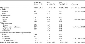
The mean age was significantly different between three groups and patients with AD were significantly older than people with SMC (p = 0.02) while no significances were detected between patients with AD versus patients with MCI (p = 0.149) or SMC versus MCI group (p = 0.252). There was no significant difference between groups according to gender (p = 0.435). Significantly more people with SMC had high school and university degree while being illiterate or having primary school degree was more frequent in AD and MCI groups (p=0.005). There were no significant differences between groups according to having medical comorbidities such as: cardiovascular disease (p = 0.159), diabetes mellitus (p = 0.542), and liver related disorder (p = 0.142) (Table 1).
The socio-demographical features of the participants

aWith one-way ANOVA, bwith chi-square test; MCI, mild cognitive impairment; SMC, subjective memory complaining.
The mean plasma PNX levels are as follows: 292.93 ± 277.30 ng/mL in patients with AD, 230.68 ± 270.00 ng/mL in patients with MCI and 339.52 ± 351.64 ng/mL in people with SMC and the mean PNX level was not significantly different between groups (p = 0.279) and within groups (Table 2). There was no significant correlation between plasma PNX level and metabolic or neuropsychological parameters in patients with AD. The mean plasma PNX level in MCI group was positively correlated with BMI (r = 0.402 and p = 0.028), serum HDL level (r = 0.454 and p = 0.012), blood systolic pressure (r = 0.428 and p = 0.018) and negatively correlated with logical memory (r = −0.335 and p = 0.031). The mean plasma PNX level was positively correlated with immediate recall in SMC groups (r = 0.417 and p = 0.034).
Comparisons of plasma Phoenixin (PNX) level between groups

aKruskal–Wallis test between groups and Mann–Whitney U test was used for within groups, p1, mild AD versus MCI; p2, mild AD versus SMC, p3, MCI versus SMC.
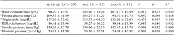
There were no significant differences between three groups according to mean BMI (p = 0.064), mean waist circumference (p = 0.325), mean triglyceride level (p = 0.622), mean HDL level (p = 0.710), mean fasting glucose level (p = 0.105), mean systolic pressure (p = 0.877), and mean diastolic pressure (p = 0.842) (Table 3).
Comparison of the data of metabolic parameters within groups

awith one-way ANOVA post hoc Tukey's test, bwith Kruskal–Wallis test. MCI, mild cognitive impairment; SMC, subjective memory complaining. p1, mild AD versus MCI; p2, mild AD versus SMC, p3, MCI versus SMC.
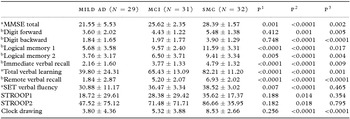
The mean total Mini-Mental State Examination score was significantly different between groups (p < 0.0001). The mean digit forward score and mean digit backward score were significantly different between groups (p=0.001 and p < 0.0001, respectively). Both logical episodic memory subtests were significantly different between groups (p < 0.0001, for both). The mean immediate verbal recall, total verbal learning, remote verbal recall, and SET verbal fluency scores were significantly different between groups (for all, p < 0.0001). There was a trend of difference between groups according to mean time in STROOP 1 (p = 0.061) while mean time in STROOP 2 was insignificant (p = 0.100). The mean clock drawing test score was significantly different between groups (p < 0.0001). The paired comparisons are shown in Table 4.
Comparison of the data of Bilnot neuropsychological battery within groups

awith one-way ANOVA post hoc Tukey's test, bwith Kruskal–Wallis test; MCI, mild cognitive impairment; SMC, subjective memory complaining; p1, mild AD versus MCI; p2, mild AD versus SMC, p3, MCI versus SMC.
Discussion
PNX is the last discovered neuropeptide and there a lot of unknown characteristics of PNX. It has been proposed to play role in memory and learning processes via animal model studies (Yosten et al., Reference Yosten2013; Jiang et al., Reference Jiang2015a,Reference Jiangb). In the current study, unexpectedly we could not find significant differences according to plasma level of PNX between patients with AD, patients with MCI and people with SMC groups. In the literature, there is no human study on PNX level and its relation with cognitive conditions including memory, learning processes, attention etc. Recently, co-secretion of PNX and nesfatin-1 has been shown (Pałasz et al., Reference Pałasz, Rojczyk, Bogus, Worthington and Wiaderkiewicz2015) which we considered that either may share or may have similar physiologic effects. As abovementioned, nesfatin-1 may act directly via neuropeptide leptin. In the literature, decrease in leptin levels was shown in patients with AD (Lieb et al., Reference Lieb2009; Warren et al., Reference Warren, Hynan and Weiner2012). Leptin replacement therapy was found beneficial in patients with early AD and MCI who had low level of leptin (Johnston et al., Reference Johnston2014). Similarly, the altered levels of leptin and ghrelin (Moran and Gao, Reference Moran and Gao2006) and the beneficial effect of replacing both neuropeptides were revealed in animal studies (Pérez-González et al., Reference Pérez-González2014). In contrast, no differences or no associations have also been detected in several recently published between serum leptin levels (Maioli et al., Reference Maioli2015; Teunissen et al., Reference Teunissen2015) or leptin receptor polymorphisms (Utsunomiya et al., Reference Utsunomiya2010) and cognitive decline in patients with AD and MCI. In contrast to many previous cross sectional studies, 809 older people has been followed 8 years (mean) in a prospective study and the investigators have found an association between higher leptin levels and cognitive decline. However, the authors have emphasized that this association was insignificant after adjusting BMI (Littlejohns et al., Reference Littlejohns2015). Thus, we considered that our insignificant finding of plasma PNX level between patients who have cognitive complaints or decline is in line with recently published negative findings studies. A decrease in serum leptin and ghrelin level occurs in aging as one the major risk factor for AD (Scarpace and Tümer Reference Scarpace and Tumer2001; Rigamonti et al., Reference Rigamonti2002). Interestingly, although it was not significant, we have detected lower plasma PNX level in patients with MCI which is known as a prodromal condition for AD.
In the present study, PNX level did not correlate with any metabolic or cognitive parameters in patients with AD while it correlated with several metabolic parameters (i.e. HDL, BMI, systolic blood pressure) in MCI patients. Strikingly, plasma PNX level negatively correlated with logical memory in patients with MCI and positively correlated with immediate verbal recall in people with SMC. In study of Warren et al. (Reference Warren, Hynan and Weiner2012), leptin was found to be associated with delayed recall and total score of Montraeal Cognitive Assessment test. Furthermore, the association between higher leptin level and lower risk of cognitive decline was revealed in small waist circumference conditions while this association is not seen in large waist circumference (Zeki et al., Reference Zeki, Hazzouri, Haan, Whitmer, Yaffe and Neuhaus2012). In two recent studies, no relation was found between leptin levels and score of MMSE (Baranowska-Bik et al., Reference Baranowska-Bik2015; Teunissen et al., Reference Teunissen2015). However, leptin level was related with several metabolic parameters in women in the latter study (Baranowska-Bik et al., Reference Baranowska-Bik2015). In the light of the literature and our findings, we speculated that plasma PNX level may have some roles in patients with MCI. However, the patients with MCI had significantly higher depressive scores than other groups although they did not meet the criteria for clinical depression which might be a caution while interpreting our speculation.
This study has several limitations: first, relatively small sample size of each group and cross sectional design of the study seems important. As third, several studies have reported gender related differences on PNX level (Johnston et al., Reference Johnston2014; Magalhães et al., Reference Magalhaes, Carvalho, Sousa, Caramelli and Gomes2015; Maioli et al., Reference Maioli2015) which we did not take into consideration in this study. Lacking normal controls might also be considered as an additional limitation.
In conclusion, this study is the first investigating the association of plasma PNX level and cognitive complaints or decline in the literature. The knowledge about the role, interaction and physiological functions of PNX are lacking. Ghrelin and leptin act in opposite direction in peripheral while they act in similar direction centrally (Kojima and Kangawa, Reference Kojima and Kangawa2006). It would be valuable to explore the outcome of PNX, leptin, ghrelin and nesfatin interaction on cognition and aging. Plasma PNX level might be important in prodromal stages as MCI and the predictive role of PNX should be investigated in further studies.
Conflict of interest
None.
Description of authors’ roles
M. Emul designed the study. G. Gultekin and G. C. Batun collected the data and literature about neuropeptides. M. Yuruyen, H. Yavuzer, A. Doventas and M. Emul examined the patients. F. E. Akcan performed the neuropsychiatric battery. M. Emul and M. Yuruyen analyzed the data and wrote the paper.
Acknowledgments
The all authors thank to Prof Dr Ferah Armutcu from Department of Biochemistry in Medical School of Cerrahpasa, İstanbul University who had performed biochemical measurements.