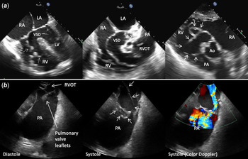
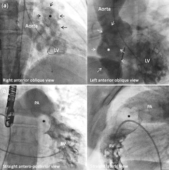
Membranous ventricular septal aneurysms are a rare condition with most being asymptomatic in nature though can have symptoms of arrhythmia, thrombosis or more rarely outflow tract obstruction.Reference Carcano, Kanne and Kirsch1 A 40-year-old female with trisomy 18 and unrepaired ventricular septal defect was referred to our centre because transthoracic echocardiography showed severe right ventricular outflow obstruction. Her large inlet ventricular septal defect extended into the perimembranous septum. The enormous aneurysmal tricuspid valve tissue not only closed off the ventricular septal defect with no residual shunt but also extended into the right ventricular outflow tract causing severe obstruction. Clinically she remained asymptomatic. She had past medical history of developmental delay, brain cyst and hypertension. Cardiac catheterisation showed severely elevated right ventricular systolic pressure (98 mmHg) with a gradient of 70 mmHg to the pulmonary artery. Pulmonary vascular resistance was 3 Woods Units. Transthoracic and intracardiac echocardiography showed to and fro flow through the ventricular septal defect from left ventricle to the blind pouch created by the membranous ventricular septal aneurysm (Fig 1, Video). This aneurysm crossed the pulmonary valve in systole and caused severe right ventricular outflow obstruction. Angiography in the left and right ventricles showed a large membranous ventricular septal aneurysm which caused dynamic obstruction in the right ventricular outflow obstruction (Fig 2). She was then referred for surgical repair.
(a ) Transesophageal echocardiography showing large inlet ventricular septal defect (VSD) and enormous membranous septal aneurysm (arrow) extending into the right ventricular outflow tract (RVOT). (b ) Intracardiac echocardiography showing the membranous septal aneurysm (arrow) crossing the pulmonary valve leaflets in systole and causing severe RVOT obstruction. RA = right atrium; RV = right ventricle; LA = left atrium; LV = left ventricle; Ao = aorta; pulmonary artery.

(a ) Left ventricular (LV) angiography shows the large membranous septal aneurysm (*) protruding into the right ventricular (RV) outflow tract. (b ) RV angiography showing the membranous septal aneurysm (*). Pulmonary artery (PA) is dilated.

Supplementary material
To view supplementary material for this article, please visit https://doi.org/10.1017/S1047951121001931
Acknowledgements
None.
Financial support
This research received no specific grant from any funding agency, commercial or not-for-profit sectors.
Conflicts of interest
None.
Ethical standards
All procedures performed in studies involving human participants were in accordance with the ethical standards of the institutional and/or national research committee and with the 1964 Helsinki declaration and its later amendments or comparable ethical standards. Informed consent was obtained from all individual participants included in this case report.