Introduction
Ecological immunology is a rapidly expanding, young discipline (Rolff & Reynolds, Reference Rolff and Reynolds2009). Within this field, immune response is seen as a set of traits that involve costs (Sheldon & Verhulst, Reference Sheldon and Verhulst1996). Resources must be allocated between an organism's maintenance and proper functioning and the expression of other traits linked with survival and reproduction, resulting in ecological trade-offs among trait types. At the moment, there is growing empirical evidence of trade-offs associated between life-history traits (size at birth, growth and mortality rates, size and age at maturity, longevity and survival, clutch size and reproductive effort: reviewed in Stearns, Reference Stearns1992) and immune defense (e.g. Boots & Begon, Reference Boots and Begon1993; Kraaijeveld & Godfray, Reference Kraaijeveld and Godfray1997; Fellowes et al., Reference Fellowes, Kraaijeveld and Godfray1998; Moret & Schmid-Hempel, Reference Moret and Schmid-Hempel2000; Rigby & Jokela, Reference Rigby and Jokela2000; Hoang, Reference Hoang2001).
The ecological immunity framework has been applied to a number of disciplines, including sexual selection (i.e. differential mating and fertilization success: Darwin, Reference Darwin1871) (e.g. Siva-Jothy, Reference Siva-Jothy2000; McKean & Nunney, Reference McKean and Nunney2001; Roberts et al., Reference Roberts, Buchanan and Evans2004) and, to a lesser extent, social evolution (e.g. Calleri et al., Reference Calleri, Rosengars and Traniello2007; Bocher et al., Reference Bocher, Doums, Millot and Tirard2008), predator-prey relationships (e.g. Packer et al., Reference Packer, Holt, Hudson, Lafferty and Dobson2003; Roy & Holt, Reference Roy and Holt2008) and parental investment (e.g. Hoi-Leitner et al., Reference Hoi-Leitner, Romero-Pujante, Hoy and Pavlova2001; Brzek & Konarzewski, Reference Brzek and Konarzewski2007). The influence of ecological immunity has also extended to broader fields, such as life-history theory (Schmid-Hempel, Reference Schmid-Hempel2005a), parasite-host coevolution (e.g. Day et al., Reference Day, Graham and Read2007; Duffy & Sivars-Becker, Reference Duffy and Sivars-Becker2007), conservation biology (e.g. Stevenson, Reference Stevenson2006; Tarlow & Blumstein, Reference Tarlow and Blumsteid2007) and learning (e.g. Barnard et al., Reference Barnard, Collins, Daisley and Behnke2006), among others.
One animal group where ecological immunology has been extensively investigated is insects (see, for example, Lawniczak et al., Reference Lawniczak, Barnes, Linklater, Boone, Wigby and Chapman2006). Insects have properties (e.g. small size, large sample sizes, short development times) that allow a wide range of experiments. Insect studies have measured a number of immune effectors related to both cellular and humoral defence reactions (hereafter referred to as immune markers), in which a cost of such immune response, measured through these markers, is assumed (see also Adamo, Reference Adamo2004). The logic of choosing such markers has been based on the assumption that they protect the insect, but their production negatively correlates with other life history or intermediate traits (e.g. colored traits, behavioral traits, etc.). Many feasible and highly sensitive techniques in insect immunology (e.g. proteomic, transcriptomic, liquid chromatography/mass spectrometry) have been employed at the individual-level, but costs confine these approaches to exploratory or qualitative studies at the population level. This turns into a restriction when the objects of study are primarily numerous individuals in natural populations, because differences among organisms in immune responses (and their trade-offs among other behavioural or reproductive traits) could be an important cause of adaptive change of the population.
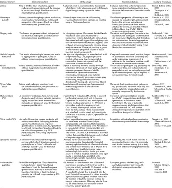
Here, we review each of the most common immune markers used in ecological and evolutionary studies that can be utilized across large sample sizes. Our first aim is to identify, when possible, the physiological processes behind each marker. This may allow the inference of trade-offs with other functions through individual data. Second, we briefly outline the methodological inconveniences in the assessment of such markers and suggest solutions. Our aim is to provide a set of more robust methodological practices. An outline of these two aims appears in table 1; we present each in greater detail in the text below. First, however, we present a basic short review of how insect immunity works, which serves as the framework for discussing the trade-offs and methodological issues.
Immune markers used in insect ecological immunity studies (see text for full discussion).

* Before inoculation, minimal lethal and/or sub-lethal doses must be determined. Overdoses can promote septic shock, rather than a measurable immunological response.
** Because antimicrobial activity can be due to lysozymes, reactive oxygen species and/or antimicrobial peptides, immune response measurement is commonly referred to as hemolytic, lysozyme-like or antibacterial activity.
Insect immune mechanisms
The insect immune system is formed by a set of cells, molecules and reactions. All of these features are continuously evolving to resist (attack and eliminate) pathogen invasion and to limit the negative consequences of the infection (Hoffman & Reichhart, Reference Hoffmann and Reichhart2002; Schmid-Hempel, Reference Schmid-Hempel2005a; Ayres & Schneider, Reference Ayres and Schneider2009). The first lines of defence include the exoskeleton cuticle, physical and chemical properties (e.g. pH) of the epidermis, gut epithelium, and male and female reproductive accessory glands (Gillespie et al., Reference Gillespie, Kanost and Trenzeck1997; Casteels, Reference Casteels, Brey and Hultmark1998). These tissues also secrete lysozymes and cytotoxic molecules such as reactive oxygen species (ROS: e.g. superoxide anions, peroxides, hydroxyl radicals) (Schmid-Hempel, Reference Schmid-Hempel2005a). Pathogens are mainly recognized by the membrane of haemocytes (which occur free in the haemolymph) or by the membranes of epithelial cells. These membranes bear proteins called pattern recognition receptors (PRRs) that recognize conserved molecular features of pathogens called pathogen-associated molecular patterns (PAMPs: LPS, mannoses, β-1, 3 glucans and peptidoglycans) (Gillespie et al., Reference Gillespie, Kanost and Trenzeck1997). Once pathogens are recognized as non-self, cellular and humoral immune mechanisms are activated. Cellular responses include phagocytosis, nodulation and encapsulation. During encapsulation, small and large pathogens are surrounded and bound by haemocytes (Gillespie et al., Reference Gillespie, Kanost and Trenzeck1997). During nodulation and encapsulation, a melanin layer is constructed (frequently referred to as melanotic nodulation/encapsulation) to cover foreign agents, which ultimately die by anoxia, toxic ROS or starvation (Nappi & Ottoviani, Reference Nappi and Ottaviani2000; Narayanan, Reference Narayanan2004).
Melanin production is a consequence of the phenoloxidase (PO) cascade (Söderhäll & Cerenius, Reference Söderhäll and Cerenius1998). During the cascade, opsonic factors, ROS and cytotoxins, such as quinones and semiquinones, are produced. These important intermediate molecules are highly reactive and toxic to pathogens, and serve to amplify the immune response (Nappi & Ottovianni, Reference Nappi and Ottaviani2000; Cerenius & Söderhäll, Reference Cerenius and Söderhäll2004). PO is involved in various physiological processes, including cuticular sclerotization and immune defense (such as melanotic encapsulation and wound healing). Three kinds of PO exist (Sugumaran & Kanost, Reference Sugumaran, Kanost, Beckage, Thompson and Federico1993): monophenol monooxigenase (also referred to as tyrosinase-type PO: Ashida & Brey, Reference Ashida, Brey, Brey and Hultmark1997), ο-diphenoloxidase (also referred to as cathecoloxidase-type PO: Decker & Jaenicke, Reference Decker and Jaenicke2004) and ρ-diphenoloxidase (also referred to as laccase-type PO: Sugumaran & Kanost, Reference Sugumaran, Kanost, Beckage, Thompson and Federico1993). These three kinds of PO and their activating systems are structurally almost indistinguishable; hence, the term phenoloxidase is often used in the literature without distinguishing among them. PO associated with sclerotization and pigmentation of cuticle (monophenol monooxigenase and ρ-diphenoloxidase) appears to be under hormonal control (Ashida & Brey, Reference Ashida, Brey, Brey and Hultmark1997), while ο-diphenoloxidase is triggered by recognition of non-self particles (Ashida & Brey, Reference Ashida, Brey, Brey and Hultmark1997). Haemolymph PO is mainly kept inside or near haemocytes; nevertheless, it can be a feature of the humoral response. The precursor of PO, pro-phenoloxidase (proPO) is primarily released from haemocytes into the haemolymph after contact with non-self particles (Ling & Yu, Reference Ling and Yu2006). It also contributes to humoral melanization of pathogens. The general PO activation pathway is initiated when phenylalanine is hydroxylated and converted into tyrosine. Then, proPO is converted by specific protease cleavage into active form PO. PO catalyzes both the hydroxylation of tyrosine to dopa and the oxidation of dopa to dopaquinone. Finally, dopaquinone is converted to melanin, which is used for wrapping pathogens and wound clotting (Söderhäll & Cerenius, Reference Söderhäll and Cerenius1998; Christensen et al., Reference Christensen, Li, Ch.-Ch. and Nappi2005; Nappi & Christensen, Reference Nappi and Christensen2005).
Other humoral responses include antimicrobial systemic molecules, which are also synthesized mainly in the fat body and reproductive accessory glands, gut cells and haemocytes (Manetti et al., Reference Manetti, Rosetto, Marchini, Brey and Hultmark1998; Schmid-Hempel, Reference Schmid-Hempel2005a). Most of these molecules are secreted close to the cuticle or into the haemolymph, gut tract or malpighian tubes. Antimicrobial peptide (AMP) molecules induce a number of negative effects on pathogen membranes including membrane collapse, prevention of cell division and permeability disruption (Otvos Jr, Reference Otvos2000; Bulet et al., Reference Bulet, Charlet, Hetru, Ezekowitz and Hoffmann2003). Nitric oxide (NO) is another immune-relevant molecule; it is a highly reactive and unstable free radical gas that crosses cell membranes to act on nearby targets (Müller, Reference Müller1997). NO also inhibits protein catalytic activity and has damaging effects on pathogen protein and DNA (reviewed in Rivero, Reference Rivero2006). Molecules such as ROS can damage pathogen nucleic acids, proteins and cell membrane (Nappi et al., Reference Nappi, Vass, Frey and Carton2000; Herrera-Ortiz et al., Reference Herrera-Ortiz, Lanz-Mendoza, Martínez-Barnetche, Hernández-Martínez, Villarreal-Treviño, Aguilar-Marcelino and Rodríguez2004). Lysozymes are hydrolytic enzymes that cleave the glycosidic bond between N-acetylmuramic acid and N-acetylglucosamine in peptidoglycan, a major component of the Gram-wall polymer (Jollès, Reference Jollès1996). It must be mentioned that most of the information regarding the humoral response has been obtained under laboratory conditions. Further investigation/characterization of these molecules must be done in the wild including the natural pathogens of each host.
Trade-offs among immune responses
Trade-offs within immune system effectors are also possible. In an individual, different traits are frequently found to be genetically or phenotypically correlated. Genetic correlation arises because a single gene can influence multiple traits in positive or negative fashions (pleiotropy and antagonistic pleiotropy, respectively) or because of linkage disequilibrium between genes affecting different characters (Falconer & Mackay, Reference Falconer and Mackay1996). Phenotypic correlations include the positive or negative influences of environmental factors on traits (Roff, Reference Roff1992). Reactions and products of immune response are interconnected and the kind of response could be related to the type of attacking pathogen. A number of studies have examined genetic and phenotypic correlations among encapsulation, lytic activity, cuticular darkness, PO activity and haemocyte load. In summary, these studies report positive and negative correlations, resulting in a lack of a clear pattern across species or even populations (see Ryder & Siva-Jothy, Reference Ryder and Siva-Jothy2001; Rantala & Kortet, Reference Rantala and Kortet2003; Cotter et al., Reference Cotter, Hails, Cory and Wilson2004; Fedorka et al., Reference Fedorka, Zuk and Mousseau2004; Rantala & Roff, Reference Rantala and Roff2005; Roff et al., Reference Rolff, Armitage and Coltman2005; Moreno-García et al., Reference Moreno-García, Lanz-Mendoza and Córdoba-Aguilar2010). This point will be reconsidered later.
Immune markers used in evolutionary ecology research
Cuticle darkness
The main components of insect cuticle – quinones and melanin (see Phenoloxidase activity) – are also used for other functions, thus strongly suggesting a trade-off (e.g. Barnes & Siva-Jothy, Reference Barnes and Siva-Jothy2000; Armitage & Siva-Jothy, Reference Armitage and Siva-Jothy2005). Quinone compounds determine cuticle pigmentation (via sclerotization) by making covalent links with proteins, resulting in coloured products, a process called quinone tanning (Riddiford & Hiruma, Reference Riddiford and Hiruma1988). Quinones also form methide derivatives that crosslink cuticle proteins through the quinone side chain (β-sclerotization reaction) and are responsible for cuticle hardening (Saul & Sugumaran, Reference Saul and Sugumaran1988). Cuticle darkness, thus, is related to how much quinone is deposited in every shed or wound repair site.
Methodology
Cuticular colour is frequently assessed under a fluorescent white light and the emerging colour classes are analyzed using a greyscale value (e.g. Barnes & Siva-Jothy, Reference Barnes and Siva-Jothy2000; Thompson et al., Reference Thompson, Armitage and Siva-Jothy2002; Rolff et al., Reference Rolff, Armitage and Coltman2005). However, since the cuticle structure is composed of hundreds of proteins (Andersen et al., Reference Andersen, Hojrup and Roepstorff1995), chitin and lipids (Chapman, Reference Chapman1998), and such protein composition greatly influences cuticle mechanical properties (e.g. flexibility: Haas et al., Reference Haas, Gorb and Blickhan2000; Gosline et al., Reference Gosline, Lillie, Carrington, Guerette, Ortlepp and Savage2002); there could be a possible overestimation of quinine and melanin tanning. Furthermore, since cuticles among insect taxa vary considerably in stiffness, hardness and pigmentation, it is possible that the dark aspect of a particular tissue may not necessarily reflect or be related to a reduced or heightened immune ability. In combination with cuticular colour analysis, we recommend the use of transverse section preparations (using a microtome) from different representative regions of the insect body and cuticle width measurement. This would reflect how much the insect has invested in producing a thick cuticle and, presumably, quinines and melanin. Note, however, that cuticular melanization is not always associated with greater immunogenic response (see Robb et al., Reference Robb, Forbes and Jamieson2003).
Cellular responses
Total number of haemocytes (haemocyte density)
Haemocyte counting and/or activity is a straightforward way of assessing cellular immune ability (e.g. Rantala et al., Reference Rantala, Koskimäki, Taskinen, Tynkkynen and Suhonen2000; Kraaijeveld et al., Reference Kraaijeveld, Limentani and Godfray2001; Cotter et al., Reference Cotter, Hails, Cory and Wilson2004). Haemocytes are generally responsible for immune activities, such as engulfing, surrounding and destroying infectious agents. After bacteria have penetrated the haemocoel, haemocytes are the first and most efficient bacteria-clearing mechanism, (Hillyer et al., Reference Hillyer, Schmidt and Christensen2003). However, there are different haemocyte types, each playing a different function (recognition, phagocytosis, nodulation, encapsulation or melanization) (see reviews by Lavine & Strand, Reference Lavine and Strand2002; Ribeiro & Brehélin, Reference Ribeiro and Brehélin2006). Differences in pathogen type (e.g. bacteria, virus, nematodes, etc.) among individuals, sexes or species must be considered, for example, for example, the mosquito Aedes aegypti mainly phagocyte Escherichia coli. In contrast, other types of bacteria (e.g. Micrococcus luteus) are melanized (Hillyer et al., Reference Hillyer, Schmidt and Christensen2003). However, the factors eliciting phagocytic vs. melanization responses against bacteria are independent of Gram type (Hillyer et al., Reference Hillyer, Schmidt and Christensen2004).
Methodology
For total and viable counts, haemolymph is collected and incubated in humidity chamber, then counted using an hemocytometer using (phase contrast) microscopy (e.g. Rantala et al., Reference Rantala, Koskimäki, Taskinen, Tynkkynen and Suhonen2000; Kraaijeveld et al., Reference Kraaijeveld, Limentani and Godfray2001). For collection and incubation of haemolymph, we suggest the use of cell culture media, such as RPMI, Schneider or Grace rather than PBS buffers. This media may allow a better viability of cells. Trypan blue can be used to differentiate between dead and viable cells. Haemolymph extraction may be accomplished by perfusion-bleed; a droplet can be collected from a puncture/cut on the thorax or by a removed leg. Although this may appear an easy task, haemocytes can adhere to tissues such as the fat body. One solution to this problem is to use anticoagulant buffers of low pH (Pech et al., Reference Pech, Trudeau and Strand1994). An accurate extraction of these cells can also be carried out by injecting a protease inhibitor blend (e.g. PMSF, TLCK, leupeptine). However, optimal conditions for obtaining insect haemocytes cannot be generalized; and, in each procedure of extraction, the use (or non-use) of anticoagulant must be adjusted.
Phagocytosis
Phagocytic activities require the internalization of the surface membrane and action of several organelles, including endosomes and lysosomes, which fuse with the plasma membrane and provide the membrane for phagosome formation (Desjardins et al., Reference Desjardins, Houde and Gagnon2005). In mosquitoes, the synthesis of lipids for the membranes of the phagosome, plasma membrane and for the production of cytotoxic proteins for the degradation of phagocytised material decreases their reproductive output (Ferdig et al., Reference Ferdig, Beerntsen, Spray, Li and Christensen1993; Hogg & Hurd, Reference Hogg and Hurd1995; Hillyer et al., Reference Hillyer, Schmidt and Christensen2003). For these reasons, trade-offs are expected among phagocytic activities and fitness traits. Furthermore, it is also probable that haemocytes undergo apoptosis after phagocytosis, which reduces the number of circulating haemocytes (as occurs in Ae. aegypti: Hillyer et al., Reference Hillyer, Schmidt, Fuchs, Boyle and Christensen2005). This would lead to another trade-off, as it would generate a decrease in the homeostasis and remodelling of tissues, as well as the capacity for fighting pathogens. Phagocytosis is an important mechanism to remove dead or damaged cells, as well as to remodel organs during metamorphosis (Abrams et al., Reference Abrams, White, Fessler and Steller1993; Franc et al., Reference Franc, Dimarcq, Lagueux, Hoffmann and Ezekowitz1996). Thus, a delay in growth seems necessary for pathogen clearing, via releasing haemocytes from their immune activity and facilitating the wound repairing process before cuticle shedding (Lavine & Strand, Reference Lavine and Strand2002).
Methodology
Phagocytic activity has been measured by exposing the host to microbial pathogens, such as bacteria and yeast cells or polystyrene beads, and assessing in vitro phagocytosis (Kurtz & Sauer, Reference Kurtz and Sauer1999; Kurtz et al., Reference Kurtz, Wiesner, Götz and Sauer2000; Kurtz, Reference Kurtz2002). One way to assess this process is by inoculating fluorescent, labelled beads or bacteria in vivo (Kurtz, Reference Kurtz2002). In either process, the phagocytised intruders are counted manually or by using image analysis software. Care must be taken to ensure the beads do not become obstructed by melanin. Recovery of remaining beads (if possible) from the haemolymph is a useful method to estimate phagocytosis activity. Trypan blue also can further assess the viability of labelled cells or molecules. The effect of this colorant is that only the ingested particles retain their fluorescence, while non-ingested particles become coated with trypan blue dye (see Kurtz et al., Reference Kurtz, Wiesner, Götz and Sauer2000). Another method is to use pHrodo dye conjugated bacteria. Bacteria become fluorescent only in the acidic environment of the haemocyte phagosome (see Luce-Fedrow et al., Reference Luce-Fedrow, Von Ohlen and Chapes2009), while extracellular non-phagocytosed bacteria do not fluoresce. When using live bacteria to evaluate the efficiency of phagocytosis, or any of the immune parameters referred to in this paper, we recommend first to assess the bacterial clearance of the infected host. To do this, supernatant of haemolymph (or homogenized/macerated individuals) can be plated (e.g. LB-agar, blood-agar) and colony forming units counted (see Pham et al., Reference Pham, Dionne, Shirasu-Hiza and Schneider2007).
Capacity to attach to non-self surfaces (nodule/capsule formation)
Quite frequently, researchers have introduced pathogens or microbial cell wall components to insect hosts to assess cellular immune response (e.g. Howard et al., Reference Howard, Miller and Stanley1998). This induces the production of nodules/capsules, which are then counted when haemolymph is extracted after the challenge or directly in the haemocoel (e.g. Miller et al., Reference Miller, Howard, Nguyen, Rosario and Stanley-Samuelson1996; Goldsworthy et al., Reference Goldsworthy, Mullen, Opoku-Ware and Chandrakant2003). Nodules can persist in insects for long periods of time (Carton & Nappi, Reference Carton and Nappi1997), which means that the haemocytes used for nodule formation remain attached, thereby decreasing haemocyte density during insect lifetime. This reduction in haemocyte number could in turn limit the availability of haemocytes that become relevant during moulting and/or metamorphosis. Not all adult insects produce haemocytes since haematopoietic organs are not always present at this stage (Chapman, Reference Chapman1998). In lepidopterans and dipterans, hematopoietic organs have been identified exclusively in the embryonic, larval and pupal stages (Gardiner & Strand, Reference Gardiner and Strand2000; Holz et al., Reference Holz, Bossinger, Strasser, Janning and Klapper2003; Nardi et al., Reference Nardi, Pilas, Ujhelyi, Garsha and Kanost2003; Williams, Reference Williams2007). This means that any haemocytes present in the adult stage were produced during embryogenesis, larval stages and metamorphosis. However, the presence of hematopoietic organs in adult grasshoppers has been reported (Hoffman, Reference Hoffmann1973; Hoffman et al., Reference Hoffmann, Brehelin and Hoffmann1974). Natural history differences among insect groups (e.g. the presence of metamorphosis) likely leads to variation in haemocyte production (consequently cell number).
Methodology
Pathogens such as bacteria or yeast cells are inoculated directly to haemocoel (Brookman et al., Reference Brookman, Rowley and Ratcliffe1989). Also, injected parasites can be used to assess nodule/encapsulation formation (see Eleftherianos et al., Reference Eleftherianos, Baldwin, ffrench-Constant and Reynolds2008). Haemolymph is collected and incubated in a humidity chamber; nodules are observed using a microscope and counted. Care must be taken since pathogens can be destroyed before nodulation takes place, for example, if pathogens enter the midgut (Luckhart et al., Reference Luckhart, Vodovotz, Cui and Rosenberg1998). Furthermore, nodules must be counted even when their size varies, which can occur even within the same individual (see Howard et al., Reference Howard, Miller and Stanley1998). Enumeration of different sizes can provide further information on differential immune ability among hosts. We recommend directly assessing the size of nodules either qualitatively (by categorizing the different sizes and counting how many of these fall into the distinct categories) or quantitatively with the use of image analysis software.
Melanotic nodulation/encapsulation (nylon monofilament and beads insertion)
For encapsulation/melanotic encapsulation assessment, inoculation with sephadex or polystyrene beads or insertion of a nylon monofilament into the haemocoel is frequently used. In this method, nylon and beads are thought to mimic natural infection (like that of a parasite/parasitoid) while avoiding the damaging effects of pathogens (e.g. Siva-Jothy, Reference Siva-Jothy2000; Koskimäki et al., Reference Koskimaki, Rantala, Taskinen, Tynkkynen and Suhonen2004; Simmons et al., Reference Simmons, Zuk and Rotenberry2005; Rantala & Roff, Reference Rantala and Roff2007). Cellular and humoral responses are activated, which could lead to trade-offs similar to when nodule formation and phagocytosis are elicited. In the melanin layer, which is frequently produced during nodulation/encapsulation, synthesis of melanin is required. Melanin is costly to produce, not only because of the resources used (see Phenoloxidase Activity below), but because melanin is required for cuticle formation, humoral toxic molecule production, and colouring and hardening of other structural traits (e.g. wings, eggs, spermatheca: Li & Christensen, Reference Li and Christensen1993; Ilango, Reference Ilango2005).
Methodology
After insertion, filament/beads are retrieved and the volume of melanized and non-melanized cell masses that encapsulate the filament are measured (Siva-Jothy, Reference Siva-Jothy2000). In vitro encapsulation assays can be also be performed. For this, haemolymph is collected from each individual and mixed with beads. Encapsulation and melanization are observed after incubation by microscopy. Three methods have been developed for measuring such masses. One method observes the mean gray scale of the covered area using image analysis software (Simmons et al., Reference Simmons, Zuk and Rotenberry2005), while a second uses images software filters to measure the density of the covered area (Koskimäki et al., Reference Koskimaki, Rantala, Taskinen, Tynkkynen and Suhonen2004). Another approach measures the encapsulated/melanized area (Contreras-Garduño et al., Reference Contreras-Garduño, Canales-Lazcano and Córdoba-Aguilar2006). All measures, however, are unable to differentiate the functional aspects of haemocytes, as cellular nodulation/encapsulation and humoral melanization are two separate but related processes (Ling & Yu, Reference Ling and Yu2006). Haemocytes release or contain surface proPO that, when activated to PO, causes melanization. This cellular encapsulation also enhances interactions between haemolymph and PO. Ling & Yu (Reference Ling and Yu2006) noticed that melanization did not occur when isolated haemocytes were used. Rather, melanization occurred only in the presence of plasma. This suggests that factors in haemolymph are required for PO cascade activation and, hence, melanization. Therefore, cellular encapsulation could be implicated in both cellular and humoral immune responses. A possible adverse effect of melanization is the production of toxic molecules that could affect self tissues or cells. Also, melanotic capsules consists of an inner area of necrotic hemocytes with melanin formation at the interface with the foreign object (Hernández-Martínez et al., Reference Hernández-Martínez, Lanz, Rodrguez, González-Ceron and Tsutsumi2002); this loss of haemocytes could be detrimental for the host.
Trade-offs among immunity and other traits (e.g. developmental time) using implants have been found (see Rantala & Rolff, Reference Rantala and Roff2005). The use of non-natural infections seems to be a suitable index of host immune activation-recognition and its ecological costs (e.g. Rantala & Roff, Reference Rantala and Roff2007). For the methods discussed above, unfortunately, the distribution of melanized/encapsulated areas is often so irregular that it impedes thorough assessment (i.e. it depends on the position of the artificial object during area measurement). We recommend several measures of the same implant. Additionally, inserting a nylon piece is a difficult task for small insects. In this case, we recommend the use of inoculated beads. We also recommend using live pathogens that induce melanotic encapsulation or other immune responses. This can be useful, for example, when testing for immune tolerance and behaviour (see Ayres & Schneider, Reference Ayres and Schneider2009). Tolerance theory predicts a possible increase in the pathogen burden, but the immune response will limit the health or fitness consequences of this pathogen load (sensu Read et al., Reference Read, Graham and Råberg2008; Schneider & Ayres, Reference Schneider and Ayres2008). Only live pathogens can replicate and continuously damage a host. It is also possible that live pathogens activate more immune pathways (compared with inert implants) and, therefore, give a more complete picture of an immune response. However, when using live pathogens, it is important to first estimate the dosage of pathogen needed for accurate measurement of a particular immune response. It has been observed that in some insects, low to medium doses elicit phagocytosis; moderate to high doses elicit nodulation; and overdoses can promote septic shock (S. Hernández-Martínez, personal communication). Before a challenge, we recommend discovering the immune response that is elicited in relation to the type and dose of infectious agent (natural or artificial) model.
Humoral responses
Phenoloxidase activity
PO is the most widely used immune marker in ecological immunology studies (e.g. Goldsworthy et al., Reference Goldsworthy, Mullen, Opoku-Ware and Chandrakant2003; Wilson et al., Reference Wilson, Knell, Boots and Koch-Osborne2003; Jacot et al., Reference Jacot, Scheuber, Kurtz and Brinkhof2005). The PO enzyme is used for different physiological processes (wounding, clotting, cuticle composition, melanotic encapsulation, production of cytotoxic molecules) and probably spermatheca formation (Ilango, Reference Ilango2005). Due to these different functions, PO is constantly synthesized; therefore, it could be costly to produce for the organism (see González-Santoyo & Córdoba-Aguilar, Reference González-Santoyo and Córdoba-Aguilar2012). Recall that proPO molecules are produced mainly by haemocytes (other tissues, such as fat body and midgut epithelium, may also produce proPO in smaller quantities), and that some of these molecules remain attached to haemocytes, while some proPO might end up in several other tissues (i.e. cuticle, midgut, salivary glands, eggs: Ashida & Brey, Reference Ashida, Brey, Brey and Hultmark1997). If pathogens are detected during proPO transportation from haemocytes to other tissues, it is possible that this proPO gets activated in the haemocele for clearing pathogens, consequently leaving target tissues with a proPO deficiency. This means that a trade-off may arise because of a proPO deficiency (but not of other components, such as PO substrates; see below), as has been assumed in general studies where PO and melanin have been involved (Siva-Jothy, Reference Siva-Jothy2000). This different perspective of a possible trade-off when measuring PO activity has not been put forward nor examined previously and needs further empirical study.
The PO substrate tyrosine (TYR) and tyrosine subproducts are used for the expression and manufacture of other traits. TYR is obtained through food and is naturally synthesized by organisms or by the hydroxylation of phenylalanine (PHE) (Christensen et al., Reference Christensen, Li, Ch.-Ch. and Nappi2005). Since PHE is an essential amino acid for protein synthesis, its importance lies in the cost of acquiring this resource. TYR is also involved in a variety of functions. One of these, for example, is the colouring of the egg chorion in some insects (Li & Christensen, Reference Li and Christensen1993). In mosquitoes, melanization responses against worms generate a delay in TYR accumulation in the ovaries and, therefore, a decrease in the number of eggs produced after an immune challenge (Li & Christensen, Reference Li and Christensen1993) and a delay in oviposition (Ferdig et al., Reference Ferdig, Beerntsen, Spray, Li and Christensen1993). Examples like this are suggestive of substrates of PO and melanin – PHE and TYR – being restrictive resources that may lead to trade-offs between the immune response and other key functions, including moulting, basal metabolic rates and protein synthesis. Although TYR can be stored (as tyrosine-O-phosphate, beta-alanyl-L-tyrosine, tyrosine glucoside: Mitchell & Lunan, Reference Mitchell and Lunan1964; Levenbook et al., Reference Levenbook, Bodnaryk and Spande1969; Chen et al., Reference Chen, Mitchell and Neuweg1978), trade-offs could be detected if not enough TYR is gathered in order to provide this amino acid to all the metabolic requirements through the insect lifetime. However, not all insects necessarily use TYR or PHE in the same way, PO measures need to be carefully interpreted in every insect model. Melanin, the final product of the PO cascade, is also involved in other morphological traits, such as cuticle formation and pigmentation, which may affect functions related to aposematism and crypsis, thermoregulation, resistance to UV radiation and coloured sexual traits. Many of these functions and their supposed trade-offs have not been adequately investigated.
Methodology
PO activity is assayed by thawing haemolymph and o-diphenols (dopa or dopamine) as substrate into a microplate well. This assay will not distinguish the three different PO activities, since it is inferred that the ο-diphenoloxidase activity of all three enzymes will be detected (Sugumaran & Kanost, Reference Sugumaran, Kanost, Beckage, Thompson and Federico1993). Care must be taken when using o-diphenols, since there could also be a peroxidase-mediated melanin formation (Christensen et al., Reference Christensen, Li, Ch.-Ch. and Nappi2005), which can result in an overestimation of PO activity. We recommend the use of a peroxidase suppressor (e.g. hydrogen peroxide in methanol or commercially available). Another current problem with assessing PO is that protein load standardization is needed before measurement, using a standard protein assay (Contreras-Garduño et al., Reference Contreras-Garduño, Lanz-Mendoza and Córdoba-Aguilar2007), a practice that is rarely carried out and without which there may result disparate and unreliable PO readings. Since, occasionally, real protein load can be masked if there are differences among individuals (e.g. bigger size of females than males, inaccurate extract of total amount of haemolymph). We recommend using the protein load practice mentioned above. Furthermore, there might be a rapid degradation of PO if stored for a relatively long time. This issue can be resolved by using a protease inhibitor cocktail (e.g. PMSF, Leupeptin). Another problem is that small-sized insects may not provide PO readings, since PO quantities are so small. This problem can be solved by using more than one individual for a single sample to obtain PO readings. Also, the total PO activity can be estimated using the enzyme α-chymotrypsin to activate all pro-PO present in the haemolymph (see Bailey & Zuk, Reference Bailey and Zuk2010).
Nitric oxide
NO quantification is a relatively new immune marker in ecological immunity studies (see, for example, Moreno-García et al., Reference Moreno-García, Lanz-Mendoza and Córdoba-Aguilar2010). NO damages pathogens and is also used as a signalling molecule. In Drosophila, NO is indispensable for activation of the immune deficiency (Imd) pathway (Foley & O'Farrel, Reference Foley and O'Farrell2003). This is one of the three pathways (the other two being Toll and JAK/STAT; see Ferrandon et al., Reference Ferrandon, Imler, Hetru and Hoffman2007) that produce transcriptional factors in the fat body, leading to the synthesis of antimicrobial peptides and other factors that prevent self tissue damage and control haemocyte proliferation and differentiation (Foley & O'Farrell, Reference Foley and O'Farrell2003; Agaisse & Perrimon, Reference Agaisse and Perrimon2004). Another interesting point is the fact that the NO synthase (NOS) (an enzyme that converts L-Arginine to L-Citrulline and generates NO) is constitutive and inducible (Müller, Reference Müller1997). NO is also a signalling molecule, so a constant production of NOS is necessary for homeostatic purposes, while inducible NOS is only synthesized after an immune challenge (Nappi et al., Reference Nappi, Vass, Frey and Carton2000). It is, therefore, expected that NO production increases after an immune challenge. Nevertheless, Krishnana et al. (Reference Krishnana, Hyršl and Šimek2006) found that NO production was similar in both non-stimulated and stimulated haemocytes in lepidopteran larvae. In this particular case, the authors indicate that NO can be continuously synthesized by the induced NOS for long periods of time (hours to days). This means that the effectiveness for killing pathogens with NO may not only reside on NO concentration but in the duration of NO synthesis (see Laurent et al., Reference Laurent, Lepoivre and Tenu1996).
NO is produced during the oxidation of L-Arginine (Müller, Reference Müller1997). Arginine is an amino acid, which must be obtained from the diet (Rivero, Reference Rivero2006). Arginine is also important for sperm maturation (Osanai & Chen, Reference Osanai and Chen1993), egg production (Uchida, Reference Uchida1993), long term memory, chemosensory (antennal lobes, olfaction) and visual information processing (Müller, Reference Müller1997). Trade-offs are thus expected among these traits and the immune response.
Methodology
Basal levels of NO are normally used, which provides an incomplete picture of host immune ability. A better alternative is to assess NO production after host injection or ingestion of pathogens. The standard method to measure NO is to use the Griess reaction that measures nitrite production, an indirect measure of NO (Bredt & Snyder, Reference Bredt and Snyder1994). The use of controls such as an L-NAME (a NOS-inhibitory arginine analogue; see Rivero, Reference Rivero2006) and its inactive enantiomer D-NAME is recommended. Controls are needed because nitrites can also be sub-products of other reactions not related with immunity. For this method, however, it is unknown whether haemolymph protein load standardization is needed before measurement. This is a technical problem that must be resolved in the near future. Similar to PO, more than a single individual may be needed if an individual host sample is too small.
Lysozyme activity and antimicrobial peptides
Lysozyme is an enzyme with hydrolytic action, mainly against the peptidoglycan of Gram+ cell walls. It can be induced or constitutively expressed in the gut tract (Hetru et al., Reference Hetru, Hoffman, Bulet, Brey and Hultmark1997), haemocytes and fat body (Gillispie et al., Reference Gillespie, Kanost and Trenzeck1997). AMPs show great structural diversity (more than 170 isoforms have been found in insects: Bulet et al., Reference Bulet, Hetru, Dimarq and Hoffmann1999) and are produced soon after foreign recognition (1–4 h) with an efficient pathogen-specific killing action (Schmid-Hempel, Reference Schmid-Hempel2005b). Some AMPs may remain in haemolymph for up to three weeks (Schmid-Hempel, Reference Schmid-Hempel2005b), which is convenient during subsequent pathogen encounters. AMPs are generally short peptides, containing fewer than 150–200 amino acids (Bulet et al., Reference Bulet, Stöcklin and Menin2004), so they are considered energy efficient, quick and economical to produce (Otvos Jr, Reference Otvos2000). However, the overall AMPs concentration in Drosophila haemolymph can reach 200 μM (Otvos Jr, Reference Otvos2000). At this concentration, antimicrobial peptides do seem costly to produce. It is possible that resources (proteins) gathered through ontogeny are indispensable for antimicrobial peptide generation as well as for the synthesis of other molecules. This fact could potentially lead to trade-offs between immunity and synthesis of other products (e.g. spermatophalyx peptides) and tissue conformation.
Methodology
Lysozyme activity is commonly assayed via the clearance rate of bacterial suspension using haemolymph. The method is relatively simple; after the haemolymph is extracted and mixed with a bacterial solution, a turbidity assay is then performed (e.g. Adamo, Reference Adamo2004). Small absorbance values indicate high lytic activity. Another way is to add small drops of haemolymph to bacterial culture, measuring the area of lytic activity after some time (12 h). The lytic activity appears as clear circular zones in the culture, which indicates that bacteria have been cleared. The diameter of the hole is measured and taken as the indicator of lytic effectiveness.
AMPs have been measured by injection or ingestion of complete bacteria, fungi, parasites or their fractions (e.g. LPS, peptidoglycane). AMP activity is assessed in three ways: (i) measurement of the clearance rate of a bacterial suspension (via a turbidity assay, similar to the lysozyme protocol described above); (ii) measurement of the rate of bacterial growth on a plate, after the same bacteria have been injected into the host and the haemolymph has been extracted after some time; and (iii) the antibacterial zone assay, via measurement of the hole area, similar to the lysozyme protocol described above. A problem with these different assays is that antimicrobial activity can be due to other molecules and not necessarily AMPs. Since lysozyme needs a pH 8 to 6.5 for optimal activity, this can be used to distinguish between other AMP activities (Rantala & Kortet, Reference Rantala and Kortet2004; Ahtiainen et al., Reference Ahtiainen, Alatalo, Kortet and Rantala2005; but see da Silva et al., Reference da Silva, Dunphy and Rau2000; Adamo & Parsons, Reference Adamo and Parsons2006).
Some concluding remarks
One intention of this review is to highlight the ecological and physiological context of insect immune markers and their value for estimating trade-offs between immunity and other traits. Thus, current (and new) knowledge of immune physiological mechanisms must be incorporated into ecological data to allow for a deeper understanding of the reasons for evolutionary correlations and constraints. The use of each method mentioned here must be determined by each researcher in terms of its overall cost (in terms of time-consumption or price), differences in the kind of pathogens or elicitors used to induce immune response, and the immune marker that can address questions via an appropriate experimental design. Another important consideration for each researcher is the potential to make repeated immune measurements on the same sample. Unfortunately, some of the measurement methods referred to here are not suitable to be repeated more than once. For example, PO and NO contained in haemolymph extraction gradually degrade even when stored in an ultrafreezer (–70 °C). Also, unfreezing and re-freezing samples accelerates degradation. Occasionally, haemocyte membranes can become disrupted even in a buffer solution or culture medium if stored for long periods of time (e.g. weeks to months). We recommend extracting enough samples to do all measurements required for the experimental design. If a large number of individuals are required, they (or extractions) can be stored and measurements should only be done for the exact number of tests that can be processed and quantified in a single day.
Finally, there are two related points that we would like to highlight: (i) some immune markers mentioned here are usually seen as responses to attack and kill pathogens; and (ii) the existing correlations among immune responses. As mentioned before, negative and positive correlations have been found among immune traits, but correlations seem to be host-specific. Commonly, resource limitation has been used to explain the negative correlations; however, the non-expression of an immune effector does not necessarily mean that there is no response. Reactions and products of immune responses are interconnected; and the type of response is related to the pathogens virulence. As mentioned before, not all pathogens will induce the same reaction; or, at least, the magnitude of a response can differ depending on the host-pathogen interaction type. Occasionally, tolerance could be the best strategy to cope with infections. The resistance or tolerance strategies adopted by an individual will depend on the force and course of the infection. There could be a critical host damage threshold; thus, when the intensity of infection is below this threshold (i.e. no significant damage to the host), the insect could tolerate it. Above the threshold, it then pays to allocate more resources into resisting the pathogen. For example, phagocytic activity could be turned on, while PO activity could remain down regulated; in this case, probably the host is avoiding autoreactivity molecules (generated through the PO cascade). In this example, the immune strategy is enough in order to limit pathogen and self damage; nevertheless, the pathogen is not eliminated. Since the immune system is highly complex, several aspects of immune response must be measured to comprehend the insect immunity strategies. Also, occasionally, some immune responses are not suitable to test some hypothesis; for example, pro-PO cascade and PO activity occasionally are not affected by the nutritional condition, but other responses, such as lysozyme, are affected (see Jacot et al., Reference Jacot, Scheuber, Kurtz and Brinkhof2005; Moreno-García et al., Reference Moreno-García, Lanz-Mendoza and Córdoba-Aguilar2010). Therefore, it is recommended to use more than one immune marker when possible. Finally, the starting point in insect immunoecology is that it must incorporate the great range of environmental factors involved using a trade-off framework. Resource allocation and investment strategies should be studied by taking into account evidence of the real cost in natural conditions and their effects in the expression of immune response and generational changes.
Ideally, an ecological study of insect immunity should start with thoughtful design regarding appropriate immune markers. Regarding methodological issues, in many cases, methodological information is available, but it is possible that the status quo of how other evolutionary ecology researchers have used such markers leads others not to think twice about the best way to proceed. Lastly, we encourage ecological and evolutionary biologists alike to incorporate useful molecular techniques (e.g. microassays, proteomics, quantitative Reverse Transcriptase-PCR, etc.) to identify the possible mechanisms and the genetic basis underlying the trade-offs between immunity and life history traits. It is hoped that this information proves constructive in paving the way to a more robust field of ecological immunology.
Acknowledgements
We thank Salvador Hernández-Martínez for providing assistance with protocols and for helpful suggestions. We thank Gloria Tavera and Chris Anderson for revising this paper in English, and providing thoughtful comments. MM-G was supported by the Posgrado en Ciencias Biomédicas (CONACYT: grant no. 172947), Instituto de Ecología, (Universidad Nacional Autónoma de México), and the Centro de Investigación Sobre Enfermedades Infecciosas, Instituto Nacional de Salud Pública, México. AC-A was supported by a grant provided by PAPIIT-UNAM (Project No. 211506). An anonymous reviewer provided corrections to the manuscript.