Parkinson disease (PD) is one of the most common neurodegenerative disorders, second in prevalence only to Alzheimer disease, and affects ~1–2% of individuals over 60 years of age. It is estimated that in the European Union alone there are more than 500 000 people with PD. The motor syndrome of PD is characterised by progressive akinesia, rigidity, rest tremor and postural disturbances, but it is also increasingly recognised that nonmotor symptoms, including autonomic and cognitive disturbances, are an integral part of the disease. The motor syndrome is caused by the predominant degeneration of dopaminergic neurons of the substantia nigra, resulting in a loss of dopamine in the striatal projection areas of these neurons. Dopamine-replacement treatment by levodopa (l-dopa) greatly ameliorates motor symptoms. However, nonmotor symptoms are likely to have a different pathological basis and are therefore largely refractory to dopaminergic drugs (Ref. Reference Poewe1). Pathologically, PD is characterised by intraneuronal inclusions of various shapes and subcellular locations consisting mainly of fibrillar α-synuclein (Lewy bodies and Lewy neurites).
It is assumed that in most cases PD is caused by an interaction of multiple genetic susceptibilities and environmental factors on the background of an ageing brain. In fact, there is rapidly accumulating evidence that genetic risk factors are of major importance in the sporadic form of the disease, while evidence for environmental causes is still sparse, despite decades of epidemiological research. In a way, ageing is probably the single most important and arguably the only definite ‘environmental’ risk factor, if environment for this purpose is defined as the sum of factors influencing the ‘cellular milieu’, but the specific age-dependent changes that eventually initiate and/or maintain the process of neurodegeneration are very poorly understood.
It is only in the past 20 years that significant advances have been made in the understanding of the molecular pathogenesis of PD, made possible by the identification of genes for rare forms of the disease with a clear ‘mendelian’, autosomal dominant or autosomal recessive, pattern of inheritance. Initially, progress in the genetics of PD was met with considerable scepticism, as many experts felt that the rare monogenic forms might be of little relevance for the aetiology of the common sporadic disorder. Differences in clinical presentation and pathology were emphasised (Ref. Reference Duvoisin2). More recently, however, evidence has emerged that molecular pathways unveiled by genetic studies do play a role in the common sporadic disorder and in addition genetic variability in those genes directly influences disease risk (Ref. Reference Gasser3). Furthermore, many recent discoveries indicate that there is no clear distinction between high-penetrance bona fide ‘PD genes’ and risk-modifying susceptibility variants, but rather that we are confronted with a continuum of effect strengths operating in several crucial cellular pathways (Ref. Reference Farrer4).
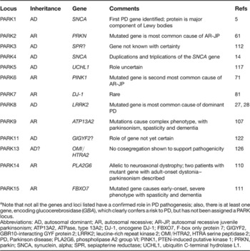
This article first reviews the different autosomal dominant and recessive PD genes (Table 1) – discussing the clinical and pathological features associated with their mutations, and the molecular pathways thought to be relevant for their pathogenic effect. Later, the evidence for a role of these genes in the sporadic disorder is reviewed. Interestingly, as more and more genes causing mendelian forms of parkinsonism are discovered, the dominant forms more closely seem to mimic idiopathic PD (or ‘pure’ forms of parkinsonism), while the recessive forms fall into two classes: those with pure parkinsonism and those that differ clinically and pathologically from the sporadic disease (‘complex’ forms of parkinsonism, often including spasticity, dystonia and other atypical features). This pattern is also seen in other complex diseases that are caused by several different dominant or recessive mutations, such as the spastic paraplegias or the ataxias.
Currently listed genetic loci and genes associated with monogenic forms of Parkinson disease (PARK loci)a

a Note that not all the genes and loci listed have a confirmed role in PD pathogenesis; also, there is at least one gene, encoding glucocerebrosidase (GBA), which clearly confers a risk to PD, but has not been assigned a PARK locus.
Abbreviations: AD, autosomal dominant; AR, autosomal recessive; AR-JP, autosomal recessive juvenile parkinsonism; ATP13A2, ATPase, type 13A2; DJ-1, oncogene DJ-1; FBXO7, F-box only protein 7; GIGYF2, GBR10-interacting GYF protein 2; LRRK2; leucine-rich repeat kinase 2; OMI/HTRA2, HTRA serine peptidase 2; PD, Parkinson disease; PLA2G6, phospholipase A2 group VI; PINK1, PTEN-induced putative kinase 1; PRKN, parkin; SNCA, synuclein, alpha; SPR, sepiapterine reductase; UCHL1, ubiquitin C-terminal hydrolase L1.
Mutations in autosomal dominant PD genes
The PARK1 locus: SNCA (α-synuclein)
Clinicogenetics
A missense mutation (A53T) in the gene for α-synuclein (SNCA) was the first PD-causing mutation to be identified – in a PD family with autosomal dominant inheritance (Ref. Reference Polymeropoulos5). Two additional different point mutations in the SNCA gene have subsequently been identified in families with dominantly inherited PD: A30P in a German kindred (Ref. Reference Krüger6), and E46K in a family from Spain (Ref. Reference Zarranz7). SNCA point mutations have so far been found only in large, multigenerational PD families – never in sporadic PD (Ref. Reference Berg8). The phenotype of patients with SNCA point mutations is that of l-dopa-responsive parkinsonism with a relatively early age of onset, rapid progression, and high prevalence of dementia, and psychiatric and autonomic disturbances, reminiscent of Lewy-body dementia (Ref. Reference Golbe9). Several family members from these kindreds have come to autopsy and they invariably showed cell loss of dopaminergic neurons of the substantia nigra, and severe and widespread Lewy pathology, particularly in the form of Lewy neurites (Ref. Reference Duda10).
The identification of SNCA point mutations as a cause of PD soon led to the discovery that the encoded protein, α-synuclein, is the major fibrillar component of Lewy bodies and Lewy neurites (Ref. Reference Spillantini11) in familial as well as sporadic cases. The currently favoured hypothesis states that the amino acid changes in α-synuclein lead to an increased tendency of the protein to form oligomers and later on fibrillar aggregates (Ref. Reference Goedert, Spillantini and Davies12), representing a ‘toxic gain of function’. However, the precise sequence of events and the causal links leading from aggregation to cellular dysfunction and cell death are still unknown. Some studies favour the hypothesis that the mature aggregates (Lewy bodies) are not themselves the toxic moiety, but rather an attempt of the cell to clear small toxic oligomers (Ref. Reference Cookson and van der Brug13).
A direct link between α-synuclein and PD was further supported by the discovery that multiplications of the wild-type sequence of SNCA (duplications and triplications) cause PD with or without dementia in some families (Refs Reference Singleton14, Reference Ibanez15). This finding was of major mechanistic importance because it indicates that an increase in wild-type α-synuclein protein expression appears to be toxic to neurons (Ref. Reference Miller16). A dose dependency of this effect is demonstrated by the fact that patients with SNCA triplications have an early age of onset (mean of around 35 years) and a high prevalence of dementia, while patients with SNCA duplications have a more typical late-onset PD phenotype (Ref. Reference Chartier-Harlin17). This could even be confirmed in a single family in whom a duplication and a triplication of the identical genomic fragment was found to cosegregate with early and late age of onset, respectively.
Possible pathogenic pathways
α-Synuclein is a relatively small protein that is abundantly expressed in many parts of the brain and localised mostly to presynaptic nerve terminals. Many aspects of the normal function of α-synuclein are still unknown. The protein has been shown to bind to brain vesicles (Ref. Reference Jensen18) and may be functionally involved in brain plasticity (Ref. Reference Abeliovich19).
There is still no good explanation for the selectivity of neuronal damage in PD, which is prominent in dopaminergic cells whereas α-synuclein is widely expressed in many areas of the brain. The identification of proteins that specifically bind α-synuclein, such as synphilin-1 (Ref. Reference Engelender20), may shed new light on this important question. In addition, several relevant animal models have recently been described. Transgenic mice overexpressing α-synuclein – both the normal and the mutated human sequence, under different promoters (Refs Reference Masliah21, Reference Kahle22) – show accumulation of the protein but do not mimic the full degenerative process in the substantia nigra. A transgenic Drosophila model may be more true to the human disease (Ref. Reference Feany and Bender23). Interestingly, in this model, pathology appears to be mediated by mitochondrial dysfunction, at least in muscle tissue (Ref. Reference Greene24), and can be partially rescued by coexpression of molecular chaperones (Ref. Reference Auluck25).
The PARK8 locus: LRRK2 (leucine-rich repeat kinase 2)
Clinicogenetics
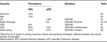
The PARK8 locus was mapped to chromosome 12 (Ref. Reference Funayama26). Missense mutations in leucine-rich repeat kinase 2 (LRRK2) (Refs Reference Zimprich27, Reference Paisan-Ruiz28) – a large gene in this region spanning 144 kb and encoding 2527 amino acids – were found to cosegregate with the disease in several families. Mutations in the LRRK2 gene are the most common cause of mendelian PD identified so far. In studies across several populations, 5–15% of autosomal dominant PD families carried mutations in LRRK2 (Refs Reference Di Fonzo29, Reference Berg30). One particularly common mutation, a base pair change at position 6055, better known as the ‘G2019S-mutation’, is responsible for familial PD in up to 7% of cases in different Caucasian populations (Refs Reference Nichols31, Reference Di Fonzo32, Reference Kachergus33), but was found, somewhat surprisingly, also in 1–2% of sporadic patients (Ref. Reference Gilks34). Even higher G2019S prevalence rates of up to 40% were found in genetically isolated populations, such as the Ashkenazi Jewish and the North-African Arab populations, both in sporadic and familial cases (Refs Reference Ozelius35, Reference Lesage36) (Table 2). The high prevalence in sporadic cases suggests a reduced penetrance of this mutation (Ref. Reference Goldwurm37), which must be taken into account when counselling patients and families. Another, even more common, LRRK2 variant, G2385R, has been found in the Asian population in ~6–10% of sporadic PD patients, as opposed to 3–5% of controls (Refs Reference Di Fonzo38, Reference Tan39, Reference Funayama40). Consequently, this variant confers a relative risk of about 2 to 3 of developing PD, suggesting that different alterations in one and the same gene may act as a high-penetrance disease-causing mutation or as a genetic risk variant in sporadic populations.
Selected studies on the prevalence of specific LRRK2 mutations in different populationsa

a Note that as a result of varying inclusion criteria and sampling strategies, different studies may not be directly comparable.
Abbreviations: fPD, familial Parkinson disease; sPD, sporadic Parkinson disease.
To date, more than 20 potentially pathogenic mutations in LRRK2 have been identified, but in only six of them can pathogenicity be considered to be highly likely (R1441C, R1441G, R1441H, Y1699C, G2019S and I2020T), because of firm evidence of cosegregation in affected families and functional data suggesting an alteration of kinase activity (Refs Reference Gloeckner41, Reference Greggio42).
The most extensive clinicogenetic study so far estimated the overall frequency of LRRK2 mutations in the European population to be ~1.5% in sporadic and ~4% in familial cases, with a geographic gradient decreasing from Mediterranean countries (Spain, Portugal, Italy) (Refs Reference Mata43, Reference Bras44) to northern countries (Ref. Reference Healy45). The average age of onset, based on more than 350 patients, was 58 years, with a wide range from the mid-20s (rarely) to over 90 years. The clinical picture was that of typical asymmetric l-dopa-responsive parkinsonism that was indistinguishable by any single criterion from PD in individuals without LRRK2 mutations. As a group, the disease appeared to be somewhat more benign in patients with mutations in LRRK2, with slower progression and a lower frequency of dementia and psychiatric complications.
Due to its relatively high frequency, the G2019S mutation offers for the first time the possibility of looking at gene–gene interactions. Three Spanish patients simultaneously harbouring heterozygous mutations in LRRK2 and in the parkin gene (see below) did not present with an earlier age at onset or a more severe disease (Ref. Reference Dachsel46). The G2019S mutation also seems to be fully dominant, as homozygous mutation carriers have been identified who also do not differ from heterozygotes with respect to disease severity or age of onset (Ref. Reference Nichols31).
Possible pathogenic pathways
LRRK2 is widely expressed in the brain, with highest levels in the striatum and the hippocampus but a relatively low abundance in the substantia nigra (Ref. Reference Higashi149); it can also be detected in other organs, such as the spleen, the lung and the liver. It has been found in the cytoplasm as well as associated with membranes. Its function and the mechanism by which LRRK2 mutations cause neuronal degeneration are unknown. By sequence homology, LRRK2 can be assigned to the group of recently identified ROCO proteins (Ref. Reference Bosgraaf and Van Haastert47) and contains a protein kinase domain of the mitogen-activated kinase kinase kinase class, suggesting a role in intracellular signalling pathways. Although the natural substrate of LRRK2 is unknown, cell culture studies using generic substrates or LRRK2 autophosphorylation paradigms suggested that at least some pathogenic mutations seem to be associated with an increase, rather than a loss, of kinase activity (Refs Reference Gloeckner41, Reference Smith48, Reference West49), and that kinase activity appears to be necessary for neurotoxicity in vitro (Ref. Reference Greggio50), raising the interesting possibility that kinase inhibition may be a potential therapeutic strategy.
A puzzling aspect of LRRK2-associated PD is its pathology. Post-mortem changes in patients with LRRK2 mutations are those of typical Lewy-body PD in most cases (Ref. Reference Ross51), but also include diffuse Lewy-body disease, nigral degeneration without distinctive histopathology and, rarely, even aggregates of the microtubule-associated protein tau (Maptau) suggestive of progressive supranuclear palsy or frontotemporal dementia (Ref. Reference Dachsel52). Different pathological findings were even reported in a single family with an R1441C mutation (Ref. Reference Zimprich27). LRRK2 mutations may therefore be an upstream event in the cascade leading to neurodegeneration with different pathologies. However, the vast majority of patients with a G2019S mutation, in whom pathology has been reported, seem to conform to the typical α-synuclein Lewy-body type of PD (Ref. Reference Ross51). No direct link between LRRK2 and α-synuclein has so far been demonstrated. α-Synuclein does not seem to be phosphorylated by LRRK2.
Despite its still poorly understood role in pathogenesis, LRRK2-associated PD is of particular interest since it is the first example of a mendelian form of PD that is common enough to provide the opportunity to study the development of the disease in a sizeable population. Longitudinal studies in presymptomatic mutation carriers may reveal premotor changes by clinical, biochemical or imaging methods, indicating the very early phases of the neurodegenerative process. It is in this population that studies exploring neuroprotective or preventive measures, be they specific for the LRRK2-associated pathways or more general, are most promising to yield first results.
PD caused by mutations in the glucocerebrosidase gene
Clinicogenetics
Mutations in the gene for the glucocerebrosidase, GBA, cause Gaucher disease, a rare recessive neurometabolic disease characterised by accumulation of the glycolipid glucocerebroside in the lysosomal compartment of various cells. Patients and relatives of patients with Gaucher disease have been found to have a conspicuously high prevalence of PD. Screening of PD patients for GBA mutations identified a higher number of heterozygous mutation carriers than expected (Ref. Reference Aharon-Peretz, Rosenbaum and Gershoni-Baruch53). This has now been confirmed in several populations and in a large meta-analysis, with very similar odds ratios (Refs Reference Mata54, Reference De Marco55, Reference Ziegler56). Mutations have been found in ~4–6% of Caucasian PD patients, and <2% of controls. The relative risk of developing PD for heterozygous mutation carriers is therefore in the range of 2 to 3. GBA mutations as well as Gaucher disease are much more common in Ashkenazi Jews than in non-Jewish European populations. As many as 20–30% of Ashkenazi Jewish PD patients carry GBA mutations (about the same proportion carries the G2019S LRRK2 mutation). Depending on the type of mutation, the relative risk for mutation carriers can be 4 to 6 or even higher (Ref. Reference Goker-Alpan57).
Possible pathogenic pathways
GBA encodes a lysosomal enzyme that cleaves glucocerebroside, and homozygous or compound heterozygous mutations are associated with an accumulation of the uncleaved substrate in the lysosomal compartment (Gaucher cells). Patients with Gaucher disease with parkinsonism and also heterozygous mutation carriers have typical α-synuclein-positive Lewy-body pathology as in sporadic PD. However, cortical involvement is probably more common in GBA-mutation carriers compared with PD patients without mutations in GBA, and the rate of clinical dementia may also be higher. GBA mutations have also been described in a high proportion of patients with Lewy-body dementia (Ref. Reference Goker-Alpan57).
As α-synuclein is at least in part degraded by a chaperone-mediated lysosomal pathway, it is conceivable that heterozygous GBA mutations, even if the residual enzyme activity is sufficient to avoid accumulation of glucocerebroside, may increase the risk for PD by altering cellular α-synuclein homeostasis. At present this remains an attractive speculation.
Recessive forms of PD and parkinsonism
So far, mutations in three genes have been identified in clinically ‘pure’ forms of autosomal recessive PD, which are characterised by an early age at onset and therefore referred to as autosomal recessive juvenile parkinsonism (AR-JP): parkin (PRKN, or PARK2), PINK1 (PARK6) and DJ-1 (PARK7). Three additional recessive genes have been added to the list more recently – ATP13A2, PLA2G6 and FBXO7 – which all also cause, when mutated, an early-onset disease with parkinsonism, but also with additional features such as dystonia, dementia, oculomotor disturbances and spasticity. Currently, mutations in PRKN and PINK1 are numerically clearly the most important causes for AR-JP.
AR-JP caused by mutations in PRKN, PINK1 and DJ-1
PD with early disease onset and recessive inheritance was first systematically studied as a clinical entity in Japan (Ref. Reference Ishikawa and Tsuji58). Patients suffered from l-dopa-responsive parkinsonism with onset in the second to fourth decade. Some showed diurnal fluctuations, with symptoms becoming worse later in the day, similar to that seen in patients with l-dopa-responsive dystonia (DRD), which is caused by mutations in the gene for GTP-cyclohydrolase (Ref. Reference Ichinose59). Recessive PD on average tends to have a more symmetrical distribution of symptoms, a slower progression and a lower prevalence of dementia, as compared with typical idiopathic PD. Despite the generally slow progression of symptoms, patients with AR-JP can develop early and severe l-dopa-induced motor fluctuations and dyskinesias.
PRKN
A first genetic locus for AR-JP was mapped to the long arm of chromosome 6 (Ref. Reference Matsumine60). Several homozygous deletions in consanguineous families with AR-JP were then found in a large gene in this region that was named parkin (PRKN) (Refs Reference Kitada61, Reference Hattori62). PRKN mutations are the most common cause of AR-JP, and many mutations have been reported (Refs Reference Lücking63, Reference Abbas64, Reference Lücking65, Reference Hedrich66). All types of mutations, including missense mutations leading to amino acid exchanges, nonsense mutations resulting in premature termination of translation, and exonic rearrangements (deletions, duplications and triplications) were identified (Ref. Reference Lücking67). Mutations have been found in several different ethnic populations. It is difficult to compare prevalence rates between these studies, as the frequency of PRKN mutations identified in a given cohort depends most strongly on the age of disease onset of the patients included (Refs Reference Lücking65, Reference Periquet68). As about a half to two-thirds of mutations are exonic rearangements and most of those are likely to have occurred as relatively recent new mutational events (Ref. Reference Periquet69), marked differences in population frequencies should not be expected.
By contrast to some of the more recently identified recessive forms of parkinsonism, the clinical picture in parkin-related PD is usually that of ‘pure’ PD, frequently indistinguishable from the sporadic disease with the notable exception of the earlier age at onset. The vast majority of patients manifest the disease before the age of 40; very few patients with onset in their 50s have been described (Ref. Reference Klein70). In the largest European series reported (Ref. Reference Lücking65), mean age at onset was 32 years, progression of the disease was slow but l-dopa-associated fluctuations and dyskinesias occurred frequently and early. Brisk reflexes of the lower limbs were present in 44%.
PINK1
In a large Italian family with autosomal recessive early-onset parkinsonism, a novel locus, PARK6, was identified on chromosome 1p36 (Ref. Reference Valente71). The phenotype was similar to that seen with PRKN mutations and characterised by early-onset parkinsonism (range 32 to 48 years), with slow progression and sustained response to l-dopa. In this and two other consanguineous PARK6-linked families, two different mutations in the gene PINK1 (encoding PTEN-induced putative kinase 1) were identified (Ref. Reference Valente71).
Several studies confirmed the presence of PINK1 mutations in patients with early-onset PD (Refs Reference Valente72, Reference Hatano73, Reference Bonifati74, Reference Hedrich75, Reference Prestel76). Most mutations were missense mutations in conserved regions, but whole-gene deletions have also been described (Ref. Reference Marongiu77). Almost all described patients with PINK1 mutations have slow disease progression and a good response to l-dopa. As in PRKN-related disease, except for the earlier average age of onset, no single feature can separate PINK1-related disease from idiopathic PD (Ref. Reference Albanese78). There are some indications that PINK1-mutated patients have a higher prevalence of psychiatric disturbances, particularly anxiety and depression, which is only relatively rarely observed in PRKN-related cases (Ref. Reference Ephraty79).
DJ-1
The third locus for AR-JP, PARK7, was mapped also to chromosome 1p36, in a Dutch family (Ref. Reference van Duijn80), and the gene was identified as the oncogene DJ-1 (Ref. Reference Bonifati81). Again, the phenotype closely resembles that found in patients with PRKN and PINK1 mutations, but this statement is based on a small number of identified patients (Refs Reference Abou-Sleiman82, Reference Hering83). However, one recessive family, which carries two homozygous mutations in the DJ-1 gene, has been described with early-onset parkinsonism, dementia and amyotrophic lateral sclerosis, suggesting that the clinical phenotype associated with mutations in this gene, although rare, may be rather wide (Ref. Reference Annesi84). The associated pathology is still unknown, as no autopsies have been reported.
Neuropathology
Even ten years after the identification of PRKN, there is still relatively little information available on the neuropathology of molecularly confirmed cases of AR-JP to shed light on the molecular pathogenesis. Only six autopsy cases of patients with PRKN mutations, and not a single one with a PINK1 or DJ-1 mutation, have been published. In the reported cases, severe and selective degeneration of dopaminergic neurons and gliosis in the substantia nigra pars compacta, and, to a somewhat lesser degree, of the locus coeruleus, have been described (Refs Reference Takahashi85, Reference van De Warrenburg86, Reference Mori87). Lewy bodies or other α-synuclein-containing Lewy pathology have not been seen in these studies, but have been found in two other individuals with PRKN mutations (Refs Reference Farrer88, Reference Pramstaller89). As in LRRK2-associated PD, α-synuclein pathology therefore does not seem to be a necessary feature in these forms of familial PD, suggesting that the disease process may differ in some important ways from that in typical idiopathic PD or from those cases caused by SNCA point mutations or multiplications.
Molecular pathogenesis of AR-JP
As autosomal recessive mutations are usually thought to act in a ‘loss-of-function’ way, it is important to understand the normal function of the gene products. The genes identified in early-onset recessive PD point to three important cellular pathways/components and their role in the molecular pathogenesis of PD: the ubiquitin–proteasome system, the mitochondria and the cellular response to oxidative stress.
The ubiquitin–proteasome system degrades damaged and/or misfolded proteins. This system came into focus in PD when it was discovered that parkin functions as an E3 ubiquitin ligase and thus is a component of the ubiquitin–proteasome system (Ref. Reference Shimura90). Initially, it was therefore assumed that the loss of E3 ubiquitin ligase function might lead to the accumulation of toxic misfolded proteins, causing damage to dopaminergic neurons. While several putative interaction partners and potential substrates of parkin have been discovered (Refs Reference Chung91, Reference Imai92, Reference Zhang93), none of them has been convincingly and reproducibly shown to accumulate to significant levels in PD. However, the classic polyubiquitination at lysine 48 (K48) of ubiquitin, which is the signal for proteasomal degradation, is not the only way in which parkin can modify proteins. Parkin has also been found to mediate monoubiquitination of certain proteins, which does not lead to protein degradation but rather modifies their function and therefore can be seen as a signalling process (Ref. Reference Joch94).
Recently, a potential role of the parkin protein in mitochondrial biogenesis, function and stability has received increased attention. Mitochondrial dysfunction has of course been suspected to contribute to the pathogenesis of PD for many years (Ref. Reference Schapira95). For example, parkin enhances mitochondrial biogenesis in proliferating cells through transcription and replication of mitochondrial DNA (Ref. Reference Kuroda96). Downregulation of several proteins involved in mitochondrial function or oxidative stress in the midbrain of prkn −/− mice has been demonstrated by two-dimensional gel electrophoresis (Ref. Reference Palacino97). Consistent with reductions in several subunits of complexes I and IV, functional assays showed reduced respiratory capacity of striatal mitochondria. Mice lacking parkin also exhibited decreased levels of proteins involved in protection from oxidative stress, decreased serum antioxidant capacity and increased protein and lipid peroxidation (Ref. Reference Palacino97). It has also been shown that loss of parkin worsens mitochondrial damage in mice overexpressing α-synuclein (Ref. Reference Stichel98).
Interestingly, mitochondrial dysfunction also appears to be central to another model of parkin-associated disease: the parkin-knockout model in Drosophila (Ref. Reference Greene24). Flies lacking parkin have a decreased lifespan and impaired locomotor activity, due to apoptotic degeneration of flight muscle cells, and male sterility. Mitochondrial pathology is the earliest manifestation of muscle degeneration in flies lacking parkin, raising the possibility that similar mitochondrial impairment triggers the selective cell loss observed in AR-JP (Ref. Reference Greene24).
That mitochondrial dysfunction may be central in PINK1-mutated cases has been suspected since the gene was first identified as a cause for AR-JP, as the protein has been located, at least in good part, to the mitochondrial compartment, where it may act as a protein kinase (Ref. Reference Valente72). Not unexpectedly, therefore, a deletion of the PINK1 gene in Drosophila leads to a similar muscle cell phenotype as parkin deficiency, with mitochondrial enlargement and disintegration. Interestingly, this phenotype can be largely rescued by the overexpression of the gene for parkin, suggesting that both proteins act in a common pathway, which is related to mitochondrial function and integrity (Ref. Reference Yang99).
A mitochondrial phenotype has also been confirmed in fibroblasts derived from patients with PRKN mutations (Ref. Reference Mortiboys100). PRKN-mutant cells had lower mitochondrial complex I activity and ATP production, which correlated with a greater degree of mitochondrial branching, suggesting that the functional and morphological effects of parkin are related. A similar study in primary fibroblasts and immortalised lymphoblasts from three patients homozygous for G309D-PINK1 showed increased oxidative stress in the form of increases in lipid peroxidation and in antioxidant defences by mitochondrial superoxide dismutase and glutathione (Ref. Reference Hoepken101). Also in fibroblasts, the morphological changes caused by mutant PINK1 could be rescued by overexpression of parkin.
A further step towards a mechanistic explanation of the mitochondrial phenotype seen in parkin- and PINK1-deficiency came from another Drosophila study. Manipulations that reduce mitochondrial fission enhance the PINK1- and parkin-mutant phenotypes, whereas perturbations that reduce mitochondrial fusion or increase mitochondrial fission suppress the PINK1- and parkin-mutant phenotypes, indicating that the PINK1/parkin pathway promotes mitochondrial fission (Ref. Reference Poole102).
The role of the protein encoded by the third gene for AR-JP, DJ-1, also seems to tie into the mitochondrial and oxidative stress pathways, but its precise relationship, if any, to the parkin- and PINK1-related processes is still unclear. DJ-1 is a multifunctional redox-sensitive protein that may mediate neuroprotection by dampening mitochondrial oxidative stress through regulation of anti-apoptotic and antioxidative gene expression, facilitation of the prosurvival kinase AKT and suppression of apoptosis signal-regulating kinase 1 (ASK1) pathways (Ref. Reference Lev103). It has been shown that wild-type DJ-1 translocates to the outer mitochondrial membrane upon oxidative stress, which is associated with neuroprotection (Ref. Reference Canet-Aviles104).
Very recently, a functional E3 ubiquitin ligase complex consisting of parkin, PINK1 and DJ-1 that promotes degradation of misfolded proteins was identified; thus, impairment of E3 ligase activity by PD-pathogenic mutations in these genes might constitute a mechanism underlying PD pathogenesis (Ref. Reference Xiong105).
In summary, there is now much converging evidence suggesting that all three recessive PD genes appear to promote cell survival by positively regulating mitochondrial function and helping the cell to deal better with oxidative stress. At least the proteins encoded by the two more commonly mutated genes, PRKN and PINK1, appear to be acting in the same pathway.
Other recessive forms of early-onset parkinsonism
More recently, three genes have been identified that cause early-onset forms of parkinsonism associated with a variety of other signs and symptoms, including, in variable combinations, dystonia, ataxia, spasticity and dementia.
ATP13A2
The first of these genes to be identified was ATP13A2, encoding a previously uncharacterised, predominantly neuronal P-type ATPase, in a recessively inherited early-onset parkinsonian syndrome described as ‘Kufor-Rakeb syndrome’ (Ref. Reference Najim al Din106). Patients with this disease have rapidly progressive parkinsonism, spasticity, vertical upgaze palsy and dementia. The substrate and function of the protein is unknown. The wild-type protein was found to be located in the lysosome of transiently transfected cells, while the unstable truncated mutant proteins were retained in the endoplasmic reticulum and degraded by the proteasome (Ref. Reference Ramirez107). It can be speculated that either overload of the proteasomal protein degradation machinery or lysosomal dysfunction due to the absence of sufficient levels of ATP13A2 protein might lead to neurodegeneration. In fact, there is increasing evidence for an important role of the lysosome in the aetiology of PD: as indicated above, α-synuclein is degraded by chaperone-mediated autophagy, and mutations in the gene encoding lysosomal glucocerebrosidase are an important cause of PD. Nevertheless, a direct involvement of the lysosome in the neurodegenerative process in Kufor-Rakeb syndrome and its potential bearing for PD remains speculative at this time (Ref. Reference Vilarino-Guell108).
PLA2G6
Another interesting addition to the recessive genes causing parkinsonian syndromes that may give important insight into underlying pathogenetic processes is the gene for phospholipase A2 group VI (PLA2G6). Mutations in this gene have been identified in two recessive childhood-onset disorders: infantile neuroaxonal dystrophy (INAD) and neurodegeneration with brain iron accumulation (NBIA). There is brain iron accumulation in some patients with INAD, so when gene mapping identified a common locus in families with these disorders on chromosome 22, it was reasonable to suspect that the disorders may be allelic. In fact, a large number of mutations were identified, including missense changes, small deletions with and without frameshift, nonsense mutations and large deletions (Ref. Reference Morgan109). As patients with two null mutations tended to have the most severe phenotype, a loss of function mechanism can be assumed. Interestingly, in addition to axonal swellings throughout the cortex, striatum, cerebellum, brainstem and spinal cord, the pathological picture also includes α-synuclein-positive Lewy bodies, and thus these disorders share this important pathological feature with PD. Lewy bodies are also found in another form of neurodegeneration with brain iron accumulation (NBIA type 1; formerly called Hallervorden–Spatz disease), caused by mutations in the gene for pantothenate kinase 2 (PANK2). Therefore, there seems to be an interesting but still little understood link between iron accumulation (which is also found in PD proper), α-synuclein aggregation, and neurodegeneration with parkinsonian symptoms. While the ‘classic’ phenotype of NBIA types 1 and 2 is that of a young-onset progressive extrapyramidal–pyramidal syndrome with visual disturbance through optic atrophy or pigmentary retinopathy, mutations in both genes can be associated with a parkinsonian syndrome of later onset. Recently, PLA2G6 mutations have been identified in patients with adult-onset l-dopa-responsive dystonia–parkinsonism, pyramidal signs and cognitive/psychiatric features, and cerebral and cerebellar atrophy on magnetic resonance imaging but lack of iron in the basal ganglia (Ref. Reference Paisan-Ruiz110).
FBXO7
Finally, mutations in a novel and still poorly characterised gene, FBXO7, have been found in members of two families with early-onset, progressive parkinsonism and pyramidal tract signs, a phenotype that had been described clinically as the pallidopyramidal syndrome (Ref. Reference Di Fonzo111). Fbxo7 is a member of the F-box-containing protein (FBP) family, characterised by a 40 amino acid domain, the F-box. FBPs serve as molecular scaffolds in the formation of protein complexes, and have been implicated in a range of processes, such as cell cycle regulation, genome stability, and development.
Other potential genes and loci
Several other loci have been mapped in families with PD showing mendelian inheritance, but either the genes have not yet been identified, or their role is still controversial. A dominant locus has been described on chromosome 2p13 (PARK3) (Ref. Reference Gasser112). Clinical features are similar to those of sporadic PD. So far, however, the gene has not been identified in these families. Nevertheless, several independent recent reports implicate PARK3 as a disease-modifying locus influencing age at onset in two sib-pair cohorts with PD (Refs Reference DeStefano113, Reference Pankratz114). A further study refined this association to a region containing the sepiapterine reductase (SPR) gene (Ref. Reference Karamohamed115), which was confirmed in another study (Ref. Reference Sharma116). Sepiapterine reductase is involved in dopamine synthesis. This finding may indicate that variants in SPR modify age of onset of PD, but the potentially causative sequence changes have not been identified.
A heterozygous missense mutation in the gene for ubiquitin C-terminal hydrolase L1 (UCHL1), which is located on chromosome 4p, has been identified in two individuals with PD in a single family of German ancestry (Ref. Reference Leroy117). To date, no other pathogenic mutations of this gene have been identified, and the segregation of the mutation in the original family does not sufficiently support the contention of UCHL1 being a dominant PD gene. Nevertheless, a common polymorphism in the UCHL1 gene, the S18Y variant, has been suggested to be associated with sporadic PD in several studies, including a large meta-analysis (Ref. Reference Maraganore118). Still, whether UCHL1 is really a PD gene is not clear yet.
Two other loci have been mapped: to chromosome 1 in an Icelandic population (PARK10) (Ref. Reference Hicks119), and to chromosome 2q in a mixed Caucasian population from the USA (PARK11) (Ref. Reference Pankratz120). The latter has not been confirmed in another cohort of European families (Ref. Reference Prestel121). Nevertheless a recent report provided evidence for mutations in a gene in this region, GIGYF2 (GBR10-interacting GYF protein 2), which encodes a component of the insulin signalling pathway (Ref. Reference Lautier122). However, several studies failed to confirm the association of mutations in the GIGYF2 gene with PD (Refs Reference Bras123, Reference Nichols124, Reference Zimprich125) so their pathogenic role is still uncertain.
Finally, functional studies in cell culture and a mouse model showing neurodegeneration in the basal ganglia support a possible pathogenic effect of rare variants identified in OMI/HTRA2, encoding a mitochondrial protease (Ref. Reference Strauss126).
The role of genes for mendelian forms of PD in the sporadic disease
Mendelian forms of PD account for less than 10% of all PD in most populations. A notable exception is the Ashkenazi Jewish population, in whom mutations in LRRK2 and GBA are found in a much higher proportion of cases compared with other ethnic groups. However, there is accumulating evidence that mutations or common genetic variants in the genes identified in the rare monogenic forms of PD also play an important role in the aetiology of the sporadic disorder. This became particularly evident for the SNCA gene encoding α-synuclein.
As the discovery of multiplications of the SNCA locus as a cause for dominantly inherited PD clearly indicates that an increase in expression of wild-type SNCA can be pathogenic, there has been considerable interest in studies looking at polymorphic variants in the SNCA gene that might influence expression levels as potential risk factors in the sporadic disease. Early studies looking at a complex polymorphic dinucleotide repeat polymorphism (NACP-REP1) located ~10 kb upstream of the transcriptional start site of SNCA initially produced controversial results, but a meta-analysis summarising data from over 2500 individuals appeared to confirm initial findings of an association with sporadic PD (Ref. Reference Maraganore127).
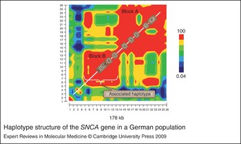
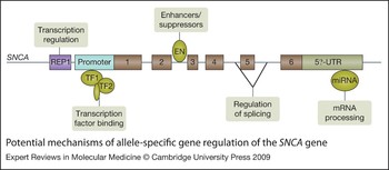
The first thorough analysis of the haplotype structure of the entire SNCA gene, by genotyping more than 50 SNPs across the gene, revealed a strong association of a haplotype block comprising exons 5 and 6 and the 3′ untranslated region (UTR) of SNCA with PD, conferring a relative risk of ~1.4 in heterozygous carriers of the risk haplotype and ~2 in homozygotes (Ref. Reference Mueller128) (Fig. 1). The strong association of the 3′-haplotype block with a similar relative risk has since been replicated in numerous populations from Japan (Ref. Reference Mizuta129), the UK (Ref. Reference Goris130), Germany and Serbia (Ref. Reference Winkler131), Ireland (Ref. Reference Ross132) and Norway (Ref. Reference Myhre133), and has recently also been detected in two whole-genome association studies (GWASs) with cohorts of patients with familial PD (Ref. Reference Pankratz134) as well as sporadic PD [A. Singleton (NIH, Bethesda, USA), pers. commun.; and T. Gasser, unpublished]. The mechanism by which genetic variants in SNCA influence the risk for PD is still unclear. Allele-specific binding of transcription factors to regulatory regions of the gene (Ref. Reference Fuchs135), or the binding of microRNAs to the 3′-UTR to modify translational processes (Ref. Reference Fuchs135), could be potential mechanisms (Fig. 2).
Haplotype structure of the SNCA gene in a German population. The SNCA gene (encoding α-synuclein) consists of two haplotype blocks (genetic regions usually inherited as a single block, with a high degree of so-called linkage disequilibrium) encoded in red. The numbers on the axes represent analysed SNPs; the colour indicates the degree of correlation (linkage disequilibrium) between any two given SNPs. The first block (Block A) ranges from the promoter region to intron 4; the second block comprises exons 5 and 6 and the 3′-untranslated region (Block B). The second block gives the strongest association signal with Parkinson disease. Associated SNPs in the first block may be derived from an independent association or due to some degree of linkage disequilibrium between the two linkage blocks. For further details see Ref. Reference Mueller128. Abbreviations: P, promoter; SNP, single-nucleotide polymorphism; UTR, untranslated region.

Potential mechanisms of allele-specific gene regulation of the SNCA gene. The mechanisms by which genetic variants in the SNCA gene (encoding α-synuclein) influence the risk for Parkinson disease is unclear, but might include differential binding of transcription factors (e.g. TF1) to the promoter, or of enhancers (ENs) or suppressors to intronic or exonic regulatory sequences, alternative splicing, or binding of microRNAs (miRNAs) to the 3′ untranslated region (UTR). REP1 (NACP-REP1) is a polymorphic dinucleotide repeat polymorphism that has been shown to influence SNCA expression, at least under in vitro conditions (Ref. Reference Chiba-Falek148).

Interestingly, the above-mentioned GWASs confirmed several earlier candidate gene association studies that implicated the gene MAPT (encoding microtubule-associated tau) in the aetiology of PD (e.g. (Refs Reference Healy136, Reference Martin137) (Ref. Reference Skipper138), although these findings had been controversial (Ref. Reference de Silva139). This finding is mechanistically interesting, since neurodegenerative disorders with parkinsonism have usually been considered to fall into two classes, depending on the aggregating protein: the synucleinopathies, such as PD, dementia with Lewy bodies, and multiple system atrophy; and the tauopathies, which include progressive supranuclear palsy, corticobasal degeneration, and frontotemporal lobar degeneration (Ref. Reference Wenning and Jellinger140). However, it has long been known that these pathologies are not mutually exclusive, as, for example, antibodies to tau stain Lewy bodies in sporadic (Ref. Reference Arima141) and familial (Ref. Reference Duda10) Lewy-body diseases, and interplay of tau and α-synuclein has been described on many levels (Ref. Reference Galpern and Lang142).
Another interesting link between mendelian forms of PD and the sporadic disorder has been raised by the observation of heterozygous mutations in AR-JP genes in patients with sporadic late-onset PD. The question whether (specific?) heterozygous PRKN mutations may cause or increase the susceptibility to late-onset typical PD is still a matter of debate. It is well established that heterozygous carriers of PRKN mutations have reduced [18F]fluorodopa uptake by positron emission tomography (Refs Reference Khan143, Reference Hilker144), even in the absence of clinical symptoms, which has been interpreted as a ‘first hit’ by the genetic abnormality to the dopaminergic system. Some larger multicase pedigrees have been ascertained on the basis of typical early-onset parkin-related cases. Heterozygous carriers of PRKN mutations in these families are obviously common and have been found to exhibit either minor signs of basal ganglia dysfunction (Ref. Reference Pramstaller145) or present with late-onset parkinsonism (Refs Reference Klein70, Reference Farrer88). In at least two of these cases, typical Lewy bodies have been described at autopsy, suggesting that a hetero- or hemizygous PRKN mutation may be a risk factor for typical Lewy-body-positive PD. However, PD is a common condition, and an occasional occurrence of PD due to different causes in a single family is a well-documented possibility. Therefore, the causal relationship between heterozygous PRKN mutations and disease status is still not completely clear. Studies screening late-onset PD cases and healthy, age-matched controls for PRKN mutations found similar frequencies of genetic variants, of the order of about 3%, suggesting that heterozygous PRKN mutations generally do not confer a significantly increased risk for late-onset PD (Refs Reference Lincoln146, Reference Kay147). However, ‘healthy’ controls in these studies have usually not been examined carefully for minor signs of parkinsonism.
Clinical implications
The identification and characterisation of mendelian forms of PD has sparked an extremely fruitful line of research, and has changed the way we think about the disease. Although in most populations monogenic forms of PD are rare, there are some, such as the Ashkenazi Jewish population, where genetic testing and counselling is now routine for most patients with PD and their families. In most other populations, genetic testing should still be restricted to those relatively small groups of patients with a clear positive family history (testing for the dominant genes SNCA and LRRK2) or exceptionally early onset (PRKN, PINK1 and DJ-1). Nevertheless, thinking about the possibility of a genetic aetiology and asking for a family history of the disease has increasingly entered medical thinking. As, undoubtedly, more PD genes are discovered, technologies develop and the price for large-scale genotyping continues to drop, genetic testing and counselling will have a firm place in the diagnostic evaluation of PD patients.
Research in progress and outstanding research questions
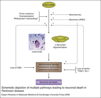
While many pieces of the puzzle depicting the molecular pathogenesis of PD have been discovered, we are still not able to see a complete picture. The PD genes discovered so far point to molecular pathways of α-synuclein aggregation, mitochondrial dysfunction and oxidative stress, dysfunction of proteasomal and lysosomal protein degradation and alterations in signalling cascades, but how all these components tie together is still enigmatic (Fig. 3). Better cellular and animal models are needed and disease-relevant interactions and protein networks will have to be identified. One possibility that has recently emerged is the generation of patient-specific induced pluripotent stem cells (iPS), which can be differentiated, in vitro, into neuronal-like cells. It is hoped that these cells will be closer to the human pathological situation as compared with immortalised cell lines or cellular models from other species.
Schematic depiction of multiple pathways leading to neuronal death in Parkinson disease. Point mutations in the SNCA gene (encoding α-synuclein), but also overexpression of the wild-type SNCA, in familial Parkinson disease, and potentially mislocalisation or mishandling in sporadic Parkinson disease, leads to α-synuclein oligomerisation, which in turn may damage multiple essential cellular processes. α-Synuclein is the major fibrillar component of Lewy bodies and Lewy neurites in neurons, the pathological hallmark of Parkinson disease. Abnormal tau species may induce α-synuclein oligomerisation by cross-seeding. The disease-relevant function of LRRK2 (leucine-rich repeat kinase 2) is still poorly understood. The mutated protein might cause mishandling of α-synuclein, or disturb essential cellular functions, which might lead to secondary accumulation of α-synuclein aggregates. Loss of parkin, PINK1 (PTEN-induced putative kinase 1) and DJ-1 function results in damage to mitochondria as well as dysfunction of proteasomal and lysosomal protein degradation, which may in turn enhance deleterious actions of α-synuclein oligomers. Lysosomal dysfunction is also likely to arise from loss of function mutations in GBA (encoding the lysosomal enzyme glucocerebrosidase). Solid lines indicate interactions supported by substantial experimental evidence, while dashed lines represent more speculative relationships.

It is also becoming increasingly clear that pathways identified in rare mendelian forms of parkinsonism are also relevant to the PD population as a whole. However, it is now necessary to determine how genetic associations translate into population and individual risks for disease, and how to translate this knowledge into epidemiological studies and intervention trials. It is conceivable that knowledge about genetic risks will allow us to stratify patient populations and thus make neuroprotective or preventive studies more powerful. What is still needed, however, are valid biomarkers that will allow early detection of the neurodegenerative process and noninvasive following of its course. Longitudinal studies of presymptomatic carriers of mutations in the mendelian PD genes may be able to provide these urgently needed biomarkers.
Acknowledgements and funding
The author has received funding from the Charitable Hertie Foundation, the German Ministery for Education and Research (BMBF), the German National Genome Network (NGFNplus #01GS08134) and the Helmholtz Alliance for Mental Health in an Ageing Society (HA 125). The help of Dr Angelika Oehmig in manuscript preparation is gratefully acknowledged. The author also thanks the reviewers for their helpful and benevolent comments.